Magnetic Field and Hydrogels Could Be Used to Grow New Cartilage

by Frank Otto

MRI Knee joint or Magnetic resonance imaging sagittal view for detect tear or sprain of the anterior cruciate ligament (ACL).

Using a magnetic field and hydrogels, a team of researchers in the Perelman School of Medicine have demonstrated a new possible way to rebuild complex body tissues, which could result in more lasting fixes to common injuries, such as cartilage degeneration. This research was published in Advanced Materials.

“We found that we were able to arrange objects, such as cells, in ways that could generate new, complex tissues without having to alter the cells themselves,” says the study’s first author, Hannah Zlotnick, a graduate student in bioengineering who works in the McKay Orthopaedic Research Laboratory at Penn Medicine. “Others have had to add magnetic particles to the cells so that they respond to a magnetic field, but that approach can have unwanted long-term effects on cell health. Instead, we manipulated the magnetic character of the environment surrounding the cells, allowing us to arrange the objects with magnets.”

In humans, tissues like cartilage can often break down, causing joint instability or pain. Often, the breakdown isn’t in total, but covers an area, forming a hole. Current fixes are to fill those holes in with synthetic or biologic materials, which can work but often wear away because they are not the same exact material as what was there before. It’s similar to fixing a pothole in a road by filling it with gravel and making a tar patch: The hole will be smoothed out but eventually wear away with use because it’s not the same material and can’t bond the same way.

What complicates fixing cartilage or other similar tissues is that their makeup is complex.

“There is a natural gradient from the top of cartilage to the bottom, where it contacts the bone,” Zlotnick explains. “Superficially, or at the surface, cartilage has a high cellularity, meaning there is a higher number of cells. But where cartilage attaches to the bone, deeper inside, its cellularity is low.”

So the researchers, which included senior author Robert Mauck, PhD, director of the McKay Lab and a professor of Orthopaedic Surgery and Bioengineering, sought to find a way to fix the potholes by repaving them instead of filling them in. With that in mind, the research team found that if they added a magnetic liquid to a three-dimensional hydrogel solution, cells, and other non-magnetic objects including drug delivery microcapsules, could be arranged into specific patterns that mimicked natural tissue through the use of an external magnetic field.

Read more at Penn Medicine News.

Brianne Connizzo Appointed Assistant Professor at Boston University

by Mahelet Asrat

Brianne Connizzo, PhD

The Department of Bioengineering is proud to congratulate alumna Brianne Connizzo, PhD on her appointment as a tenure-track Assistant Professor in the Department of Biomedical Engineering in the College of Engineering at Boston University. Connizzo’s appointment will begin in January 2021, after completing her work as a postdoctoral researcher in Biological Engineering at MIT under the supervision of Alan J. Grodzinsky, ScD, Professor of Biological, Electrical, and Mechanical Engineering.

Connizzo got her BS in Engineering Science from Smith College (the first all women’s engineering program in the country) where she graduated in 2010 with highest honors. During her time there, she worked in the laboratory of Borjana Mikic, Rosemary Bradford Hewlett 1940 Professor of Engineering. While working in the lab, she explored the role of myostatin deficiency on Achilles tendon biomechanics and built mechanical testing fixtures for submerged testing of biological tissues. Connizzo continued along this path during her graduate studies in Bioengineering at Penn while working with Louis J. Soslowsky, Fairhill Professor in Orthopaedic Surgery and Professor in Bioengineering, at the McKay Orthopaedic Research Laboratory. Her thesis work focused on the dynamic re-organizations of collagen during tendon loading in the rotator cuff, developing a novel AFM-based method for measuring collagen fibril sliding along the way. During her time at Penn, Connizzo also served as the Social Chair for the Graduate Association of Bioengineers (GABE) and the Graduate Student Engineering Group (GSEG), both of which play a vital role in representing graduate students across the School of Engineering and Applied Sciences. She completed her PhD in Bioengineering in 2015 and then pursued her postdoctoral studies at MIT, focusing on fluid flow during compressive loading and developing novel explant culture models to explore real-time extracellular matrix turnover. For her work she was awarded both an NIH F32 postdoctoral fellowship and the NIH K99/R00 Pathway Independence Award, which are just a few of her long list of impressive accomplishments.

Although Connizzo’s interests in soft tissue mechanobiology span development, injury, and disease, her more recent work has targeted how aging influences tendon function and biology. With a fast-growing active and aging population, she believes that identifying the cause and contributors of age-related changes is critical to finding treatments and therapies that could prevent tendon disease, and thus improve overall population healthspan and quality of life. The primary objectives of the Connizzo Lab at Boston University will be to harness novel in vitro and in vivo models to study cell-controlled extracellular matrix remodeling and tissue biomechanics and to better understand normal tendon maintenance and the initiation of tendon damage in the context of aging.

“I am so grateful to have had the guidance of my mentors and peers at Penn during my doctoral studies, and even more thankful that many of those relationships remain a significant part of my support system to this day,” Connizzo says. “I’m really looking forward to this next chapter to all the successes and failures in pursuing the science, to building a community at BU and in my own laboratory, and to supporting the next generation of brilliant young scientists.”

Congratulations Dr. Connizzo from everyone at Penn Bioengineering!

Bioengineering News Round-Up (April 2020)

by Sophie Burkholder

How to Heal Chronic Wounds with “Smart” Bandages

Some medical conditions, like diabetes or limb amputation, have the potential to result in wounds that never heal, affecting patients for the rest of their lives. Though normal wound-healing processes are relatively understood by medical professionals, the complications that can lead to chronic non-healing wounds are often varied and complex, creating a gap in successful treatments. But biomedical engineering faculty from the University of Connecticut want to change that.

Ali Tamayol, Ph.D., an Associate Professor in UConn’s Biomedical Engineering Department, developed what he’s calling a “smart” bandage in collaboration with researchers from the University of Nebraska-Lincoln and Harvard Medical School. The bandage, paired with a smartphone platform, has the ability to deliver medications to the wound via wirelessly controlled mini needles. The minimally invasive device thus allows doctors to control medication dosages for wounds without the patient even having to come in for an appointment. Early tests of the device on mice showed success in wound-healing processes, and Tamayol hopes that soon, the technology will be able to do the same for humans.

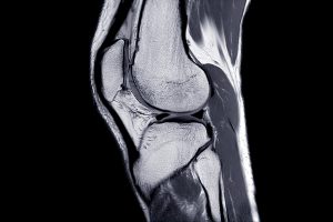
A New Patch Could Fix Broken Hearts

Heart disease is by far one of the most common medical conditions in the world, and has a high risk of morbidity. While some efforts in tissue engineering have sought to resolve cardiac tissue damage, they often require the use of existing heart cells, which can introduce a variety of complications to its integration into the human body. So, a group of bioengineers at Trinity College in Dublin sought to eliminate the need for cells by creating a patch that mimics both the mechanical and electrical properties of cardiac tissue.

Using thermoelastic polymers, the engineers, led by Ussher Assistant Professor in Biomedical Engineering Michael Monaghan, Ph.D., created a patch that could withstand multiple rounds of stretching and exhibited elasticity: two of the biggest challenges in designing synthetic cardiac tissues. With the desired mechanical properties working, the team then coated the patches with an electroconductive polymer that would allow for the necessary electrical signaling of cardiac tissue without decreasing cell compatibility in the patch. So far, the patch has demonstrated success in both mechanical and electrical behaviors in ex vivo models, suggesting promise that it might be able to work in the human body, too.

3-D Printing a New Tissue Engineering Scaffold

While successful tissue engineering innovations often hold tremendous promise for advances in personalized medicine and regeneration, creating the right scaffold for cells to grow on either before or after implantation into the body can be tricky. One common approach is to use 3-D printers to extrude scaffolds into customizable shapes. But the problem is that not all scaffold materials that are best for the body will hold up their structure in the 3-D printing process.

A team of biomedical engineers at Rutgers University led by Chair of Biomedical Engineering David I. Schreiber, Ph.D., hopes to apply the use of hyaluronic acid — a common natural molecule throughout the human body — in conjunction with polyethylene glycol to create a gel-like scaffold. The hope is that the polyethylene glycol will improve the scaffold’s durability, as using hyaluronic acid alone creates a substance that is often too weak for tissue engineering use. Envisioning this gel-like scaffold as a sort of ink cartridge, the engineers hope that they can create a platform that’s customizable for a variety of different cells that require different mechanical properties to survive. Notably, this new approach can specifically control both the stiffness and the ligands of the scaffold, tailoring it to a number of tissue engineering applications.

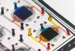
A New Portable Chip Can Track Wide Ranges of Brain Activity

Understanding the workings of the human brain is no small feat, and neuroscience still has a long way to go. While recent technology in brain probes and imaging allows for better understanding of the organ than ever before, that technology often requires immense amounts of wires and stationary attachments, limiting the scope of brain activity that can be studied. The answer to this problem? Figure out a way to implant a portable probe into the brain to monitor its everyday signaling pathways.

That’s exactly what researchers from the University of Arizona, George Washington University, and Northwestern University set out to do. Together, they created a small, wireless, and battery-free device that can monitor brain activity by using light. The light-sensing works by first tinting some neurons with a dye that can change its brightness according to neuronal activity levels. Instead of using a battery, the device relies on energy from oscillating magnetic fields that it can pick up with a miniature antenna. Led in part by the University of Arizona’s Gutruf Lab, the new device holds promise for better understanding how complex brain conditions like Alzheimer’s and Parkinson’s might work, as well as what the mechanisms of some mental health conditions look like, too.

People & Places

Each year, the National Academy of Engineering (NAE) elects new members in what is considered one of the highest professional honors in engineering. This year, NAE elected 87 new members and 18 international members, including a former Penn faculty member and alumna Susan S. Margulies, Ph.D. Now a professor of Biomedical Engineering at Georgia Tech and Emory University, Margulies was recognized by the NAE for her contributions to “elaborating the traumatic injury thresholds of brain and lung in terms of structure-function mechanisms.” Congratulations, Dr. Margulies!

Nimmi Ramanujam, Ph.D., a Distinguished Professor of Bioengineering at Duke University, was recently announced as having one of the highest-scoring proposals for the MacArthur Foundation’s 100&Change competition for her proposal “Women-Inspired Strategies for Health (WISH): A Revolution Against Cervical Cancer.” Dr. Ramanujam’s proposal, which will enter the next round of competition for the grant, focuses on closing the cervical cancer inequity gap by creating a new model of women-centered healthcare.

Dr. Danielle Bassett and Dr. Jason Burdick Named to Highly Cited Researchers List

by Sophie Burkholder

One way to measure the success or influence of a researcher is to consider how many times they’re cited by other researchers. Every published paper requires a reference section listing relevant earlier papers, and the Web of Science Group keeps track of how many times different authors are cited over the course of a year.

Danielle Bassett, Ph.D.

In 2019, two members of the Penn Bioengineering department, Jason Burdick, Ph.D., and Danielle Bassett, Ph.D., were named Highly Cited Researchers, indicating that each of them placed within the top 1% of citations in their field based on the Web of Science’s index. For the past year, only 6,300 researchers were recognized with this honor, a number that makes up a mere 0.1% of researchers worldwide. Bassett’s lab looks at the use of knowledge, brain, and dynamic networks to understand bioengineering problems at a systems-level analysis, while Burdick’s lab focuses on advancements in tissue engineering through polymer design and development.

Robert D. Bent Chair
Jason Burdick, PhD

Burdick’s and Bassett’s naming to the list of Highly Cited Researchers demonstrates that their research had an outsized influence over current work in the field of bioengineering in the last year, and that new innovations continue to be developed from foundations these two Penn researchers created. To be included among such a small percentage of researchers worldwide indicates that Bassett and Burdick are sources of great impact and influence in bioengineering advancements today.

BE Seminar Series: February 27th with Michael Yaszemski, M.D., Ph.D.

Our next Penn Bioengineering seminar will be held this Thursday. We hope to see you there!

Michael Yaszemski, M.D., Ph.D.

Speaker: Michael Yaszemski, M.D., Ph.D.
The Krehbiel Endowed Professor of Orthopedic Surgery and Biomedical Engineering
Mayo Clinic

Date: Thursday, February 27, 2020
Time: 12:00-1:00 pm
Location: Room 337, Towne Building

Title: “Musculoskeletal Tissue Engineering”


Abstract:

The field of Tissue Engineering/Regenerative Medicine is replete with advances that have been translated to human use. However, our job is not done when a treatment for a specific disease or traumatic event has been invented and translated to humans. In order to be available to the population nationwide (or globally), our novel treatment must be manufactured, transported to the user, and administered by a physician to that user. In addition, novel treatments for rare diseases may not be amenable to manufacture by a company, and perhaps would be best manufactured by an academic medical center. I will discuss these issues that occur after successful translation of a novel treatment to human use, as well as potential strategies to address them.

Bio:

Dr. Michael Yaszemski is the Krehbiel Family Endowed Professor of Orthopedic Surgery and Biomedical Engineering at Mayo Clinic and director of its Polymeric Biomaterials and Tissue Engineering Laboratory. He is a retired USAF Brigadier General. He has served as the president of the Mayo medical staff. He received both bachelor’s and master’s degrees in chemical engineering from Lehigh University in 1977 and 1978, an M.D. from Georgetown University in 1983 and a Ph.D. in chemical engineering from Massachusetts Institute of Technology in 1995.  He served as a member of the Lehigh University Board of Trustees.

Alex Hughes Receives the First MIRA Award of Penn SEAS

by Sophie Burkholder

Alex Hughes, Ph.D.

We would like to congratulate Assistant Professor in Bioengineering Alex Hughes, Ph.D., on receiving the Maximizing Investigators’ Research Award (MIRA) from the National Institutes of Health (NIH), which funds investigators to create flexible and forward-thinking research programs. Hughes is the first recipient of this award in Penn’s School of Engineering and Applied Science, marking a major accomplishment for him and his lab.

The award recognizes Hughes’ efforts to create new  tools used for tissue engineering, in particular by fusing concepts from developmental biology into tissue construction efforts. Hughes believes this approach will have impacts on fundamental understanding human disease, leading to new strategies to combat them. Hughes and his lab specifically focus on kidney disease. As Hughes says, “defects in the kidney and urinary tract account for up to a third of all birth defects.” Furthermore, because kidney development involves many different kinds of cell interactions, there’s a gap in understanding exactly how these defects occur.

Unlike other grants that focus on funding projects, the MIRA prioritizes the people behind the research, giving them funding as a sign of faith in the future work they’ll choose to do. “The MIRA has allowed us significant leeway to integrate several complementary approaches here,” Hughes says. Because of this flexibility, Hughes and his lab thinks it will allow them to reach for more innovative and risky approaches in their research, in the hopes that this will lead to a better understanding of kidney defects and modes of treatment for them.

Jason Burdick Named National Academy of Inventors Fellow

Robert D. Bent Chair
Jason Burdick, PhD

Jason Burdick, Robert D. Bent Professor in the Department of Bioengineering, has been named a Fellow of the National Academy of Inventors (NAI), an award of high professional distinction accorded to academic inventors. Elected Fellows have demonstrated a prolific spirit of innovation in creating or facilitating outstanding inventions that have made a tangible impact on quality of life, economic development and the welfare of society.

Burdick’s research interests include developing degradable polymeric biomaterials that can be used for tissue engineering, drug delivery, and fundamental polymer studies. His lab focuses on developing polymeric materials for biomedical applications with specific emphasis on tissue regeneration and drug delivery. Burdick believes that advances in synthetic chemistry and materials processing could be the answer to organ and tissue shortages in medicine. The specific targets of his research include: scaffolding for cartilage regeneration, controlling stem cell differentiation through material signals, electrospinning and 3D printing for scaffold fabrication, and injectable hydrogels for therapies after a heart attack.

Read the full story on the Penn Engineering blog.

BE Grace Hopper Lecture: Powering Tumor Cell Migration Through Hetergeneous Microenvironments

We hope you will join us for the 2019 Bioengineering Grace Hopper lecture by Dr. Cynthia Reinhart-King.

Date: Thursday, April 4, 2019
Time: 3:30-4:30 PM
Location: Glandt Forum, Singh Center, 3205 Walnut Street

Dr. Cynthia Reinhart-King, Engineering, BME, Photo by Joe Howell

Speaker: Cynthia Reinhart-King, Ph.D.
Cornelius Vanderbilt Professor of Engineering, Director of Graduate Studies, Biomedical Engienering
Vanderbilt University

Title: “Powering Tumor Cell Migration Through Heterogeneous Microenvironments”

Abstract:
To move through tissues, cancer cells must navigate a complex, heterogeneous network of fibers in the extracellular matrix. This network of fibers also provides chemical, structural and mechanical cues to the resident cells. In this talk, I will describe my lab’s efforts to understand the forces driving cell movements in the tumor microenvironment. Combining tissue engineering approaches, mouse models, and patient samples, we create and validate in vitro systems to understand how cells navigate the tumor stroma environment. Microfabrication and native biomaterials are used to build mimics of the paths created and taken by cells during metastasis. Using these platforms, we have described a role for a balance between cellular energetics, cell and matrix stiffness, and confinement in determining migration behavior. Moreover, we have extended this work into investigating the role of the mechanical microenvironment in tumor angiogenesis to show that mechanics guides vessel growth and integrity. I will discuss the mechanical influences at play during tumor progression and the underlying biological mechanisms driving angiogenesis and metastatic cell migration as a function of the ECM with an eye towards potential therapeutic avenues.

Bio:
Cynthia Reinhart-King is the Cornelius Vanderbilt Professor of Engineering and the Director of Graduate Studies in Biomedical Engineering at Vanderbilt University.  Prior to joining the Vanderbilt faculty in 2017, she was on the faculty of Cornell University where she received tenure in the Department of Biomedical Engineering. She obtained undergraduate degrees in chemical engineering and biology at MIT and her PhD at the University of Pennsylvania in the Department of Bioengineering as a Whitaker Fellow working with Daniel Hammer. She then completed postdoctoral training as an Individual NIH NRSA postdoctoral fellow at the University of Rochester.  Her lab’s research interests are in the areas of cell mechanics and cell migration specifically in the context of cancer and atherosclerosis. Her lab has received funding from the American Heart Association, the National Institutes of Health, the National Science Foundation and the American Federation of Aging Research.  She has been awarded the Rita Schaffer Young Investigator Award in 2010 and the Mid-Career Award in 2018 from the Biomedical Engineering Society, an NSF CAREER Award, the 2010 Sonny Yau ‘72 Excellence in Teaching Award, a Cook Award for “contributions towards improving the climate for women at Cornell,” and the Zellman Warhaft Commitment to Diversity Award from the Cornell College of Engineering. She is a fellow of the Biomedical Engineering Society and the American Institute for Medical and Biological Engineering, and she is a New Voices Fellow of the National Academies of Science, Engineering and Medicine. She is currently a standing member of the NIH CMT study section panel and Secretary of the Biomedical Engineering Society.

Information on the Grace Hopper Lecture:
In support of its educational mission of promoting the role of all engineers in society, the School of Engineering and Applied Science presents the Grace Hopper Lecture Series. This series is intended to serve the dual purpose of recognizing successful women in engineering and of inspiring students to achieve at the highest level.
Rear Admiral Grace Hopper was a mathematician, computer scientist, systems designer and the inventor of the compiler. Her outstanding contributions to computer science benefited academia, industry and the military. In 1928 she graduated from Vassar College with a B.A. in mathematics and physics and joined the Vassar faculty. While an instructor, she continued her studies in mathematics at Yale University where she earned an M.A. in 1930 and a Ph.D. in 1934. Grace Hopper is known worldwide for her work with the first large-scale digital computer, the Navy’s Mark I. In 1949 she joined Philadelphia’s Eckert-Mauchly, founded by the builders of ENIAC, which was building UNIVAC I. Her work on compilers and on making machines understand ordinary language instructions lead ultimately to the development of the business language, COBOL. Grace Hopper served on the faculty of the Moore School for 15 years, and in 1974 received an honorary degree from the University. In support of the accomplishments of women in engineering, each department within the School invites a prominent speaker to campus for a one or two-day visit that incorporates a public lecture, various mini-talks and opportunities to interact with undergraduate and graduate students and faculty. The lecture is open to everyone!

Week in BioE (March 22, 2019)

by Sophie Burkholder

A New Microscopy Technique Could Reduce the Risk of LASIK Surgery

Though over ten million Americans have undergone LASIK vision corrective surgery since the option became available about 20 years ago, the procedure still poses some risk to patients. In addition to the usual risks of any surgery however, LASIK has even more due to the lack of a precise way to measure the refractive properties of the eye, which forces surgeons to make approximations in their measurements during the procedure. In an effort to eliminate this risk, a University of Maryland team of researchers in the Optics Biotech Laboratory led by Giuliano Scarcelli, Ph. D., designed a microscopy technique that would allow for precise measurements of these properties.

Using a form of light-scattering technology called Brillouin spectroscopy, Scarcelli and his lab found a way to directly determine a patient’s refractive index – the quantity surgeons need to know to be able to measure and adjust the way light travels through the eye. Often used as a way to sense mechanical properties of tissues and cells, this technology holds promise for taking three-dimensional spatial observations of these structures around the eye. Scarcelli hopes to keep improving the resolution of the new technique, to further understanding of the eye, and reduce even more of the risks involved with LASIK surgery.

Taking Tissue Models to the Final Frontier

Space flight is likely to cause deleterious changes to the composition of bacterial flora, leading to an increased risk of infection. The environment may also affect the susceptibility of microorganisms within the spacecraft to antibiotics, key components of flown medical kits, and may modify the virulence of bacteria and other microorganisms that contaminate the fabric of the International Space Station and other flight platforms.

“It has been known since the early days of human space flight that astronauts are more prone to infection,” says Dongeun (Dan) Huh, Wilf Family Term Assistant Professor in Bioengineering at Penn Engineering. “Infections can potentially be a serious threat to astronauts, but we don’t have a good fundamental understanding of how the microgravity environment changes the way our immune system reacts to pathogens.”

In collaboration with G. Scott Worthen, a physician-scientist in neonatology at the Children’s Hospital of Philadelphia, Huh will attempt to answer this question by sending tissues-on-chips to space. Last June, the Center for the Advancement of Science in Space (CASIS) and the National Center for Advancing Translational Sciences (NCATS), part of the National Institutes of Health (NIH), announced that the duo had received funding to study lung host defense in microgravity at the International Space Station.

Huh and Worthen aim to model respiratory infection, which accounts for more than 30 percent of all infections reported in astronauts. The project’s goals are to test engineered systems that model the airway and bone marrow, a critical organ in the immune system responsible for generating white blood cells, and to combine the models to emulate and understand the integrated immune responses of the human respiratory system in microgravity.

Read the rest of the article on Penn Engineering’s Medium Blog. Media contacts Evan Lerner and Janelle Weaver.

Sappi Limited Teams Up with the University of Maine to Develop Paper Microfluidics

At the Westbrook Technology Center of Sappi, a global pulp and paper company, researchers found ways to apply innovations in paper texture for medical use. So far, these include endeavors in medical test devices and patches for patient diagnostics. In collaboration with the Caitlin Howell, Ph.D., Assistant Professor of Chemical and Biomedical Engineering at the University of Maine, Sappi hopes to continue advances in these unconventional uses of their paper, especially as the business in paper for publishing purposes declines.

Sappi’s projects with the university focus on the development of paper microfluidics devices as what’s now becoming a widespread solution for obstacles in point-of-care diagnostics. One project in particular, called Sharklet, uses a paper that mimics shark skin as a way to impede unwanted microbial growth on the device – a key characteristic needed for its transition into commercial use. Beyond this example, Sappi’s work in developing paper microfluidics underscores the benefits of these devices in their mass producibility and adaptability.

New Observations of the WNT Pathway Deepen the Understanding of Protein Signaling in Cellular Development

Scientists at Rice University recently found that a protein signaling pathway called WNT, typically associated with its role in early organism development, can both listen for signals from a large amount of triggers and influence cell types throughout embryonic development. These new findings, published in PNAS, add to the already known functions of WNT, deepening our understanding of it and opening the doors to new potential applications of it in stem cell research.

Led by Aryeh Warmflash, Ph. D., researchers discovered that the WNT pathway is different between stem cells and differentiated cells, contrary to prior belief that it was the same for both. Using CRISPR-Cas9 gene editing technology, the Warmflash lab observed that the WNT signaling pathway is actually context-dependent throughout the process of cellular development. This research brings a whole new understanding to the way the WNT pathway operates, and could open the doors to new forms of gene therapy and treatments for diseases like cancers that involve genetic pathway mutations.

People and Places

In a recent article from Technical.ly Philly, named Group K Diagnostics on a list of ten promising startups in Philadelphia. Group K Diagnostics founder Brianna Wronko graduated with a B.S.E. from Penn’s Department of Bioengineering in 2017, and her point-of-care diagnostics company raised over $2 million in funding last year. Congratulations Brianna!

We would also like to congratulate Pamela K. Woodward, M.D., on her being named as the inaugural Hugh Monroe Wilson Professor of Radiology at the Washington University School of Medicine in St. Louis. Also a Professor of Biomedical Engineering at the university, Dr. Woodward leads a research lab with a focus on cardiovascular imaging, including work on new standards for diagnosis of pulmonary blood clots and on an atherosclerosis imaging agent.

Lastly, we would like to congratulate all of the following researchers on their election to the National Academy of Engineering:

  • David Bishop, Ph. D., a professor at the College of Engineering at Boston University whose current research involves the development of personalized heart tissue as an all-encompassing treatment for patients with heart disease.
  • Joanna Aizenberg, Ph. D., a professor of chemistry and chemical biology at Harvard University who leads research in the synthesis of biomimetic inorganic materials
  • Gilda Barabino, Ph. D., the dean of the City College of New York’s Grove School for Engineering whose lab focuses on cartilage tissue engineering and treatments for sickle cell disease.
  • Karl Deisseroth, M.D., Ph. D., a professor of bioengineering at Stanford University whose research involves the re-engineering of brain circuits through novel electromagnetic brain stimulation techniques.
  • Rosalind Picard, Ph.D., the founder and director of the Affective Computing Research Group at the Massachusetts Institute of Technology’s Media Lab whose research focuses on the development of technology that can measure and understand human emotion.
  • And finally, Molly Stevens, Ph. D., the Research Director for Biomedical Material Sciences at the Imperial College of London with research in understanding biomaterial interfaces for biosensing and regenerative medicine.

Week in BioE (July 31, 2018)

New Data Analysis Methods

Like many other fields, biomedical research is experiencing a data explosion. Some estimates suggest that the amount of data generated from the health sciences is now doubling every eighteen months, and experts expect it to double every seventy-three days by 2020.  One challenge that researchers face is how to meaningfully analyze this data tsunami.

The challenge of interpreting data occurs at all scales, and a recent collaboration shows how new approaches can allow us to handle the volumes of data emerging at the level of individual cells to infer more about how biology “works” at this level.  Wharton Statistics Department researchers Mo Huang and Jingshu Wang (PhD Student and Postdoctoral Researcher, respectively) collaborated with Arjun Raj’s lab in Bioengineering and published their findings in recent issues of Nature Methods and Proceedings of the National Academy of Sciences.  One study focused on a de-noising technique called SAVER to provide more precise data from single cell experiments and significantly improves the ability to detect trends in a dataset, similar to how increasing sample size helps improve the ability to determine differences between experimental groups.  The second method, termed DESCEND, creates more accurate characterization of gene expression that occur in individual cells. Together these two methods will improve data collection for biologists and medical professionals working  to diagnose, monitor, and treat diseased cells.

Dr. Raj’s team contributed data to the cause and acted as consultants on the biological aspects of this research. Further collaboration involved Mingyao Li, PhD, Professor of Biostatistics at the Perelman School of Medicine, and Nancy Zhang, PD, Professor Statistics at the Wharton School. “We are so happy to have had the chance to work with Nancy and Mingyao on analyzing single cell data,” said Dr. Raj. “The things they were able to do with our data are pretty amazing and important for the field.”

Advancements in Biomaterials

This blog features many new biomaterials techniques and substances, and there are several exciting new developments to report this week. First, the journal of Nature Biomedical Engineering published a study announcing a new therapy to treat or even eliminate lung infections, such as those acquired while in hospital or as the result of cystic fibrosis, which are highly common and dangerous. Researchers identified and designed viruses to target and kill the bacteria which causes these infections, but the difficulty of administering them to patients has proven prohibitive. This new therapy, developed by researchers at the Georgia Institute of Technology, is administered as a dry powder directly to the lungs and bypasses many of the delivery problems appearing in past treatments. Further research on the safety of this method is required before clinical trials can begin.

A team at Harvard University published another recent study in Nature Biomedical Engineering announcing their creation of a tissue-engineered scale model of the left human heart ventricle. This model is made from degradable fibers that simulate the natural fibers of heart tissue. Lead investigator Professor Kevin Kit Parker, PhD, and his team eventually hope to build specific models culled from patient stem cells to replicate the features of that patient’s heart, complete with the patient’s unique DNA and any heart defects or diseases. This replica would allow researchers and clinicians to study and test various treatments before applying them to a specific patient.

Lastly, researchers at the Tufts University School of Engineering published in the Proceedings of the National Academy of Sciences on their creation of flexible magnetic composites that respond to light. This material is capable of macroscale motion and is extremely flexible, allowing its adaptation into a variety of substances such as sponges, film, and hydrogels. Author and graduate student Meg Li explained that this material differs from similar substances in its complexity; for example, in the ability for engineers to dictate specific movements, such as toward or away from the light source. Co-author Fiorenzo Omenetto, PhD, suggests that with further research, these movements could be controlled at even more specific and detailed levels.

CFPS: Getting Closer to “On Demand” Medicine

A recent and growing trend in medicine is the move towards personalized or “on demand” medicine, allowing for treatment customized to specific patients’ needs and situations. One leading method is Cell-Free Protein Synthesis (CFPS), a way of engineering cellular biology without using actual cells. CFPS is used to make substances such as medicine, vaccines, and chemicals in a sustainable and portable way. One instance if the rapid manufacture of insulin to treat diabetic patients. Given that many clinics most in need of such substances are found in remote and under-served locations far away from well-equipped hospitals and urban infrastructure, the ability to safely and quickly create and transport these vital substances to patients is vitally important.

The biggest limiting factor to CFPS is difficulty of replicating Glycosylation, a complex modification that most proteins undergo. Glycosylation is important for proteins to exert their biological function, and is very difficult to synthetically duplicate. Previously, achieving successful Glycosylation was a key barrier in CFPS. Fortunately, Matthew DeLisa, PhD, the Williams L. Lewis Professor of Engineering at Cornell University and Michael Jewett, PD, Associate Professor of Chemical and Biological Engineering at Northwestern University, have created a “single-pot” glycoprotein biosynthesis that allows them to make these critical molecules very quickly. The full study was recently published in Nature Communications. With this new method, medicine is one step closer to being fully “on demand.”

People and Places

The Institute of Electrical and Electronics Engineers (IEEE) interviewed our own Penn faculty member Danielle Bassett, PhD, the Edwardo D. Glandt Faculty Fellow and Associate Professor in Bioengineering, for their website. Dr. Bassett, who shares a joint appointment with Electrical Systems Engineering (ESE) at Penn, has published groundbreaking research in Network Neuroscience, Complex Systems, and more. In the video interview (below), Dr. Bassett discusses current research trends in neuroscience and their applications in medicine.

Finally, a new partnership between Case Western Reserve University and Cleveland Clinic seeks to promote education and research in biomedical engineering in the Cleveland area. Cleveland Clinic Lerner Research Institute‘s Chair of Biomedical Engineering, Geoff Vince, PhD, sees this as an opportunity to capitalize on the renown of both institutions, building on the region’s already stellar reputation in the field of BME. Dozens of researchers from both institutions will have the opportunity to collaborate in a variety of disciplines and projects. In addition to professional academics and medical doctors, the leaders of this new partnership hope to create an atmosphere that can benefit all levels of education, all the way down to high school students.