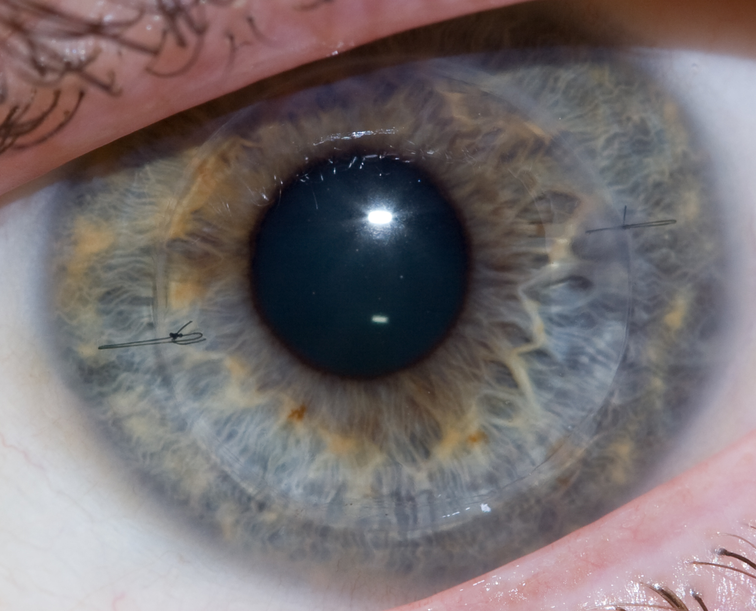
A human eye that received a cornea transplant one year postoperatively.
Disorders of or damage to the cornea — the clear covering over the lens of the eye — can be threatening to vision, and for the last century, corneal transplantation has been a cornerstone of treatment for these conditions. However, corneal transplants are complicated by two key facts: first, as with virtually all transplant procedures, donor organs are in short supply; and second, rejection is common, and recipients of transplants face repeated procedures or a lifetime of steroid eyedrops to prevent rejection.
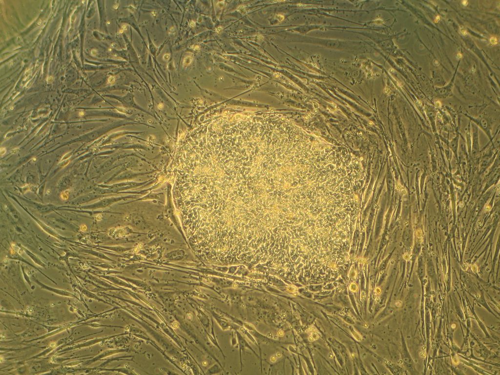
One way of obviating these issues is the use of synthetic materials, which can now be manufactured with three-dimensional printing. In a new study from scientists at the Institute of Genetic Medicine at Newcastle University in the UK, to be published this summer in Experimental Eye Research, synthetic corneal tissue was 3D printed using a bioink loaded with encapsulated keratocytes (corneal cells), in combination with computer modeling based on actual corneas. The study is only proof to show that printing a biological replicate of the cornea is possible, but it lays the groundwork for future studies in animals.
Engineering Brain Recovery
One of the reasons why stroke is such a damaging event is the inability of damaged brain tissue to regenerate. Angiogenesis, the growth of new blood vessels, can help to regenerate brain tissue but properly guiding the process of angiogenesis is rather difficult.
However, a new report in Nature Materials indicates success using an injectable biogel for this purpose. In the report, a team led by Tatiana Segura, PhD, Professor of Biomedical Engineering at Duke with colleagues at UCLA, details its engineering of an injectable gel using nanoparticles consisting of heparin (a blood-thinning agent to prevent unwanted blood clotting) and vascular endothelial growth factor (VEGF) to stimulate brain regeneration. After injecting the gel in a mouse model of stroke, the mice showed a significant improvement in recovery compared to animals not receiving the engineered nanomaterial.
Here at Penn, D. Kacy Cullen, PhD, Research Associate Professor of Neurosurgery in the Perelman School of Medicine, has been investigating the use of implantable tissue-engineered brain pathways to treat and perhaps reverse the effects of neurodegnerative diseases like Parkinson’s disease. Penn Today has the story, with video of Dr. Cullen and photos and quotes from several of our own Bioengineering students.
Streamlining Environmental Bioengineering
Outside of the health sciences, bioengineering has applications in diverse fields, including energy development and environmental protection. Biofuels are one application for bioengineering that received a major boost recently. In an article published in NPJ Systems Biology and Applications, engineers from the US Department of Energy’s Lawrence Berkeley National Laboratory describe how they used machine learning to better predict the ability of engineered microbes to produce biofuel. With this information, they can then better adjust fuel-producing microbial pathways to maximize production. The machine learning model is a significant improvement over earlier, traditionally algorithmic approaches requiring complex differential equations. The time saved could, over generations of adjustments, result in a significant increase in output.
More on Pilots
Last week, we discussed how the cognitive load borne by airline pilots differs between simulated and real flight. Other scientists, it turns out, are looking at ways that pilots — in particular, fighter pilots — can overcome fatigue. With more than $1 million in grants from the US Department of Defense, Merhavan Singh, PhD, Dean of the Graduate School of Biomedical Sciences at the University of North Texas Health Science Center, and Kai Shen, PhD, Associate Professor in the Department of Chemistry and Forensic Science at Savannah State University in Georgia, are investigating compounds targeting the sigma 1 receptor, which the scientists believe could combat fatigue and also have neuroprotective effects if activated. This is particularly important among fighter pilots serving in conflict, who are often sleep deprived but must remain alert during missions.
People and Places
Having achieved success in its mission, the University of Alabama at Birmingham’s PREP Scholars Program, which supports underrepresented minority students in pursuing graduate study in bioengineering and biomedical engineering, has received an additional $1.8 million in support from the National Institutes of Health. The money will enable the funding of 40 students over the next five years.
Jeffrey Collins Wolchok, PhD, and Kartik Balachandran, PhD, both associate professors in the Department of Biomedical Engineering at the University of Arkansas, have received a $375,000 grant from the National Science Foundation to study the long-term effects of multiple concussions on the brain. With the increased emphasis in the scientific community and media on traumatic brain injury and chronic traumatic encephalopathy, including among former athletes, the two scientists will develop brain on a chip technology to examine the issue.
Finally, this week, the Best College Reviews website published its Top 10 list of online Master’s programs in biomedical engineering. Purdue University’s program finished in first place, with appearances on the list by Colorado State, UC Riverside, Stevens Tech, and Worcester Tech.
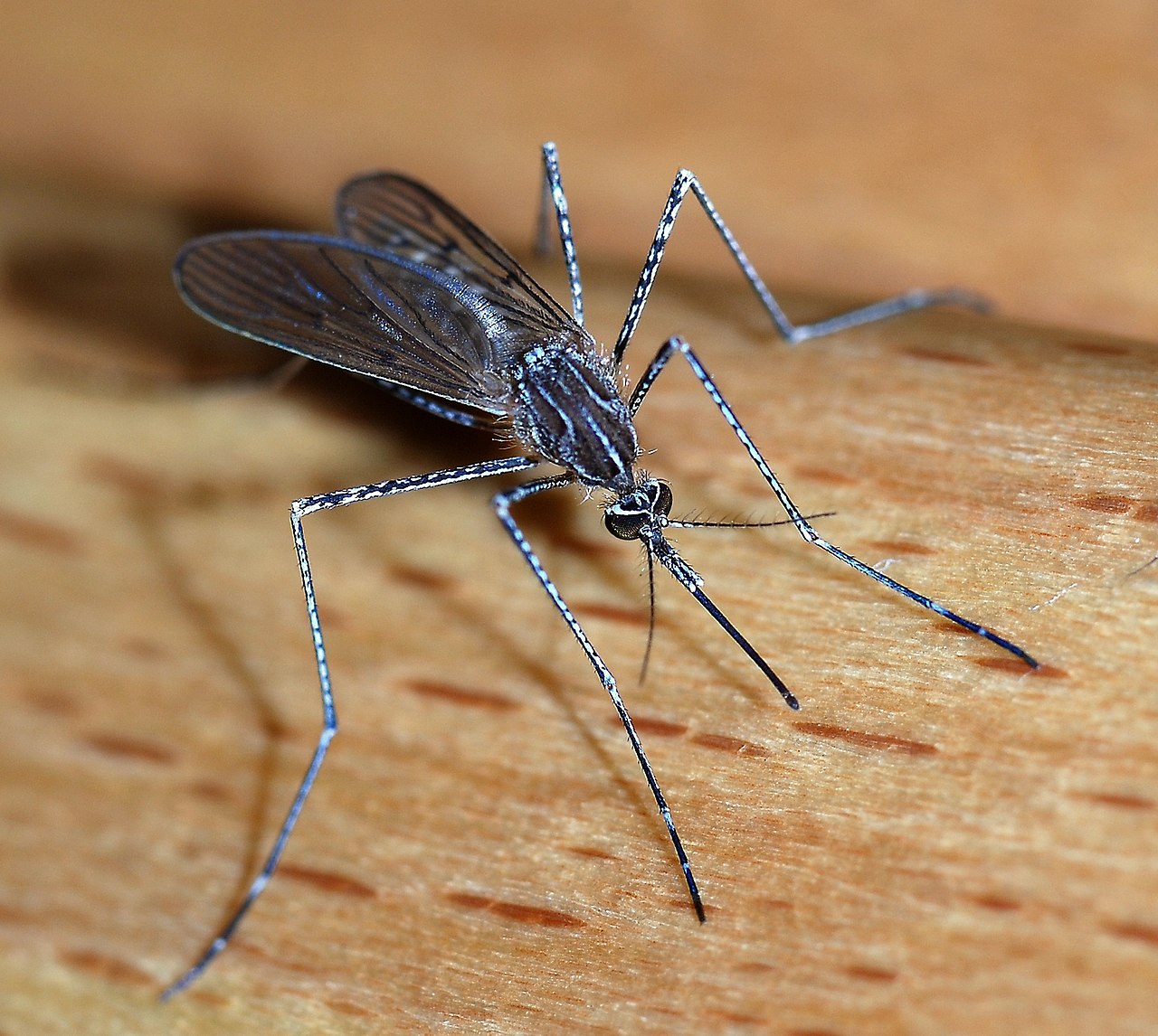
We’ve talked before at this site about the difficulty involved in implanting devices in the brain. One chief problem is that any implant to record brain signals causes small amounts of damage that causes signal quality to deteriorate over time. One approach to overcoming this problem uses flexible materials that can move with brain tissue movement, rather than resisting the movement to cause damage.
One of the more recent designs was inspired by an NPR report on mosquitoes. Dr. Andrew Shoffstall, a postdoc in the lab of Jeffrey Capadona, PhD, Associate Professor of Biomedical Engineering at Case Western Reserve University (CWRU), saw the report and used the mechanism that mosquitoes use when biting people to design a new device, which the CWRU team describes in an article in Scientific Reports.
The authors studied the buckling force when mosquitoes puncture the skin, using this design to invent new microneedles for brain implant recordings. The group fashioned a 3D-printed plastic device to mimic the process used by the mosquito. They tested the device, first mechanically and then in rat brains, finding that the device could successfully implant a microelectrode in 8 out of 8 trials. Certainly the device will require much more rigorous testing, but if successful, it could change the way implants are inserted into human patients.
Big News About Small Things
Speaking of implants, they continue to decrease in size. Scientists at Stanford University created a wireless device that is the size of a rice grain. Reporting in IEEE Transactions on Biomedical Circuits and Systems, the scientists, led by Amin Arbabian, PhD, Assistant Professor of Electrical Engineering at Stanford, and including Dr. Felicity Gore, a postdoc in the Department of Bioengineering, describe the design and fabrication of this implant. The implant was designed to stimulate peripheral nerves using either platinum electrodes connected directly to the nerve or light from a blue LED to stimulate optogenetic channels expressed in the neurons. The group conducted an in vivo experiment, using the device to stimulate the sciatic nerve of a frog, and they showed the device’s feasibility. Powered by ultrasound transmitted through the skin, the device has no external wire connections. The size of the implant, combined with its ability to target single nerves, could revolutionize how pain is treated, among other applications.
Meanwhile, here at Penn, the creation of very small things is getting a very big boost. In a new collaboration among schools and centers, the university’s Center for Targeted Therapeutics and Translational Nanomedicine has established the Chemical and Nanoparticle Synthesis Core (CNSC). The director, Andrew Tsourkas, PhD, is a Professor in the Department of Bioengineering and the Undergraduate Chair. The mission of the CNSC is to provide a concierge level service for Penn faculty interested in synthesizing new molecules for therapy development, as well as new nanoparticles for advanced diagnostics.
A Leap Forward With Stem Cells
Over the last decade, stem cell research has resulted in significant contributions to medical science. One application is the modeling of organs and organ systems for studies before in vivo investigations. However, stem cell projects involving the heart have been limited by the inability to get these cells to a mature state.
However, in a letter published in Nature, researchers at Columbia University and the University of Minho in Portugal describe how they used electrical and mechanical stimulation of human induced pluripotent stem cells to create more mature cells. The authors, led by Gordana Vunjak-Novakovic, PhD, University Professor and Mikati Foundation Professor of Biomedical Engineering and Medical Sciences at Columbia, describe how, after four weeks of culturing under the described conditions, the cells displayed multiple characteristics of maturity, although some electromechanical properties of mature cells remained lacking. These findings show that engineering the physical environment that surrounds cells during development is a key factor for the engineering design of replacement tissue.
Individualizing First Aid
Personalized medicine has begun to affect the way that doctors treat several diseases with genetic bases, notably cancer. However, first aid has lagged a bit behind in personalization, in part because the urgency of first aid care emphasizes fast, practical solutions that work for everyone. However, in a presentation at Philadelphia’s Franklin Institute last month, Jonathan Gerstenhaber, PhD, Assistant Professor of Instruction in the Department of Bioengineering at Temple University, demonstrated a prototype device that uses 3D printing technology to produce personalized bandages when they are needed.
Dr. Gersternhaber created a 3D printer that will print bandages directly onto the skin of the patient. Customizing the fit of the bandage with the printing technology would make them last longer, and the ‘on demand’ production of the bandage provides a chance to individualize the bandage design even in the urgent care setting. The device uses electrospinning technology to create bandages from soy protein, which, as a natural substance, can actually speed healing. Having completed the prototype, Dr. Gerstenhaber has moved onto portable models, as well as a larger device that can make bandages across a larger surface area.
Solving Two Problems in Glaucoma Care
Glaucoma is one of the earliest medical uses for cannabis, commonly known as marijuana. The cannabinoids in the cannabis plan have the effect of lowering intraocular pressure, which is the primary mechanism underlying glaucoma. However, the intoxicating effects of cannabis pose a problem for many patients. Thus, most patients still rely on eyedrops containing other drugs. Getting the dosage correct with eyedrops is tricky, however, because of the continual blinking and tearing of the eye.
Now, in a new article published in Drug Delivery and Translational Research, a team of researchers led by Vikramaditya G. Yadav, PhD, Assistant Professor of Chemical and Biological Engineering at the University of British Columbia, describes how they developed a nanoparticle hydrogel medication to deliver a cannabinoid. The authors tested the gel in situ, with good results. The authors imagine that such a gel could be used by patients at bedtime, and during the night, the drug would be dispensed by the gel and be gone by morning.
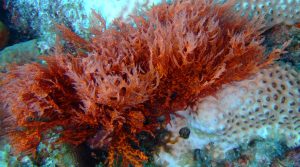
Chondrus crispus, a common red algae from which carrageenan is extracted.
Medicine has made tremendous strides since the 1960s, as evidenced by the increased survival rates of combat soldiers since Vietnam. Nevertheless, blood loss remains the most common cause of death of soldiers on the battlefield. Finding a way for medics or soldiers to stop bleeding can significantly cut down on these deaths, but current approaches are either very expensive or not easy to use in combat.
According to a new paper published in Acta Biomaterialia, a solution to this problem could come from seaweed — or more precisely, from kappa-carrageenan, a type of polymeric carbohydrate produced by certain types of edible seaweed. Akhilesh K. Gaharwar, PhD, Assistant Professor of Biomedical Engineering at Texas A&M, led a study team who developed and tested an injectable hydrogel nanoengineered from kappa-carrageenan.
The authors combined kappa-carrageenan with clay-based nanoparticles to yield a hydrogel that can be injected into wounds. When the gel solidifies, it both stanches the flow of blood and helps to generate new tissue. The gel performed well in in vitro experiments. The next step will be to test the gel in animal models of wounds.
A New Understanding of Anatomy
A group of scientists collaborating among Mount Sinai Medical Center, NYU, Weill Cornell Medical Center, and the University of Pennsylvania, including Penn Bioengineering secondary faculty member Rebecca Wells, MD, published a paper in Scientific Reports detailing the heretofore unknown extent of the human interstitium and providing a new understanding of these fluid-filled compartments beneath the skin surface. The study used confocal laser endomicroscopy, which can examine structures at depths of 60-70 µm, to look at human hepatobiliary tissue. They found a reticular pattern of fluid-filled sinuses not detected before, which is connected to the lymph nodes and similar to structures found in other organs and organ systems.
On the basis of their findings, the authors suggest that our current understanding of the anatomy might be revised. Much more research is necessary, but they also believe that the fluid-filled spaces might play important roles in cancer metastasis and a number of other disease processes.
Bringing Bioprinting to the Masses
Three-dimensional printing is one of the great innovations of the last decade, and it has transformed numerous fields inside and outside of science. In the health sciences, the ability to manufacture 3D biomaterials holds enormous promise. Unfortunately, the costs of 3D printing remain prohibitive; the available models range between $10,000 and $200,000 in cost, not including the raw materials, software, etc. However, engineers at Carnegie Mellon University (CMU) might have devised a solution. In a paper published in HardwareX, Adam Feinberg, PhD, Associate Professor of Biomedical Engineering at CMU, and his coauthors describe their development of a syringe-pump large volume extruder (LVE).
Syringe pump extruders, which inject raw material into 3D printers, are already used to print biomaterials. However, achieving cheap, fast, and precise printing of 3D materials is a major technical challenge. The LVE, which is based on open-source hardware and software, significantly increases the size of the extruder without compromising speed, and it can print at sizes as small as 100 µm. The authors estimate that the materials necessary to build their bioprinter would cost less than $500 — orders of magnitude less than current models that are slower and unable to print using large volumes. Their source materials are online here.
People and Places
Missouri dominates this week’s news, with a new program at one institution and a symposium at another. At the University of Missouri, the College of Engineering has announced that it will begin offering an undergraduate program in biomedical engineering in the fall. Ninety miles away at Missouri University of Science and Technology, a symposium will be held this week — the first to be convened on the topic of biomedical humanities. The event is a collaboration between Missouri S&T’s Center for Science, Technology, and Society and the Center for Biomedical Research.
Colorado State University’s Department of Biomedical Engineering is celebrating its 10th anniversary. In that time, the department has added more than 20 faculty members to its original cohort of 29.
Finally, we offer our congratulations to Jelena Kovačević, PhD, who has been named the new dean at NYU’s Tandon School of Engineering. A graduate of Columbia and the University of Belgrade, Dr. Kovačević, who is an electrical engineer with broad interest in biomedical applications, moves to NYU from CMU and is the first-ever female dean of Tandon. Congratulations Jelena!
One of the key processes in embryonic development and growth through childhood and adolescence is that of how tissue folds into the specific shapes required for them to function in the body. For instance, mesenchymal stem cells, which form a variety of tissues including bones, muscles, and fat, are required to “know” what shapes to take on as they form organ systems and other structures. Therefore, a big concern in tissue engineering is determining how to control these processes of tissue folding.
Just in time for his arrival at Penn Bioengineering, Dr. Alex Hughes, a new assistant professor in the department, is the lead author on a new paper in Developmental Cell that explores this concern. The study was coauthored with Dr. Zev Gartner of the University of California, Berkeley, where Dr. Hughes just finished a postdoctoral fellowship. In the paper, the authors used three-dimensional cell-patterning techniques, embryonic tissue explants, and finite element modeling to determine that the folding process involves the interaction of a protein called myosin II with the extracellular matrix, itself the molecular material that provides a structural framework for developing tissues. With the knowledge gained in the initial experiments, the authors were then able to reproduce the tissue folding process in the lab.
“Bioengineers are currently thinking about building tissues,” Dr. Hughes says, “not just at the level of organoids, but at the level of organs in the body. One of my interests at Penn is to harness developmental principles that link these length scales, allowing us to design medically relevant scaffolds and machines.”
Equipped with the knowledge gleaned from this research, future studies could contribute further to the ability to generate tissues and even organ systems in laboratories. Ultimately, this knowledge could revolutionize transplant medicine, as well as variety of other fields.
Repairing heart tissue after a heart attack is a major focus of tissue engineering. A key challenge here is keeping grafted cardiomyocytes in place within the tissue to promote repair. As we reported a couple of weeks ago, using tissue spheroids and nanowires is one approach to overcome this challenge. Another approach involves manipulating the cell cycle — the process by which normal cells reproduce, grow, and eventually die.
In the latest advance in cellular engineering for this purpose, Jianyi Zhang, M.D., Ph.D., chair of the Department of Biomedical Engineering at the University of Alabama, Birmingham (UAB) and T. Michael and Gillian Goodrich Endowed Chair of Engineering Leadership, published an article in Circulation Research showing how to control key cell-cycle activators to improve the success rate of cardiomyocyte transplants. Dr. Zhang and his coauthors, using a mouse model of myocardial infarction, engineered the transplanted cells so that they expressed much higher levels of cyclin d2, a protein that plays a key role in cell division. Cardiac function improved significantly, and infarct size decreased in mice receiving these engineered the cells. The authors plan to test their discovery next in larger animal models.
Use of stem cells in tissue regeneration isn’t limited to the heart, of course. Stephanie Willerth, Ph.D., Canada Research Chair in Biomedical Engineering at the University of Victoria in Canada, is one of two recipients from that school of an Ignite Award from the British Columbia Innovation Council. Dr. Willerth will use her award to create “bioink” for three-dimensional printers. The bioink will convert skin cells into pluripotent stem cells using technology developed by Aspect Biosystems, a biotech company in Vancouver. Once induced, the pluripotent stem cells can be converted again into a number of different cell types. Dr. Willerth’s specific focus is building brain tissue with this technology.
Making Music
Prosthetic limbs have been a standard of care for amputees and people with underdeveloped arms or legs. Many current prostheses are designed to resemble actual limbs and use myoelectrical interfaces to re-create normal movements. Alternatively, other prostheses designed for specific purposes, such as the Flex-Foot Cheetah prosthetic foot for running, do not resemble the human limb but are optimized for a specific prosthetic function.
Now, a group of undergraduate bioengineering students at George Mason University (GMU) produced a prosthetic arm to play the violin. The students, who were instructed by Laurence Bray, Ph.D., associate chair of the Department of Bioengineering at GMU, were connected with a local fifth grader from nearby Alexandria, Va., named Isabella Nicola. Nicola was born without a left hand and only part of her left arm, and she had been learning violin using a prosthesis designed for her by her music teacher. The teacher, a GMU alumnus, reached out the department for help.
The design team used a three-dimensional printer to create a prosthetic arm for Isabella. The prosthesis is made of durable, lightweight plastic and includes a built-in bow, which Isabella can use to play her instrument. The prosthesis is hot pink — the color of Isabella’s choosing. She can now play the violin much more easily than before. Whether a symphony chair is in her future is up to her.
People and Places
The University of New Hampshire will use a five-year Center of Biomedical Research Excellence grant by the National Institutes of Health to create the Center of Integrated Biomedical and Bioengineering Research. The center will unite several colleges under the rubric of bioengineering and biomedical engineering. Similarly, the University of Iowa will use a $1.4 million grant from the Roy J. Carver Charitable Trust, an Iowa-based charity, to add a biomedical engineering laboratory for its College of Engineering.
Finally, congratulations to University of Minnesota Ph.D. BME student Lizzy Crist, who has been named the NCAA’s Woman of the Year, for her undergraduate record as a scholar-athlete (soccer) at Washington University in St. Louis. She joins last year’s winner, MIT biological engineering student Margaret Guo, a swimmer who is now an M.D./Ph.D. student at Stanford.
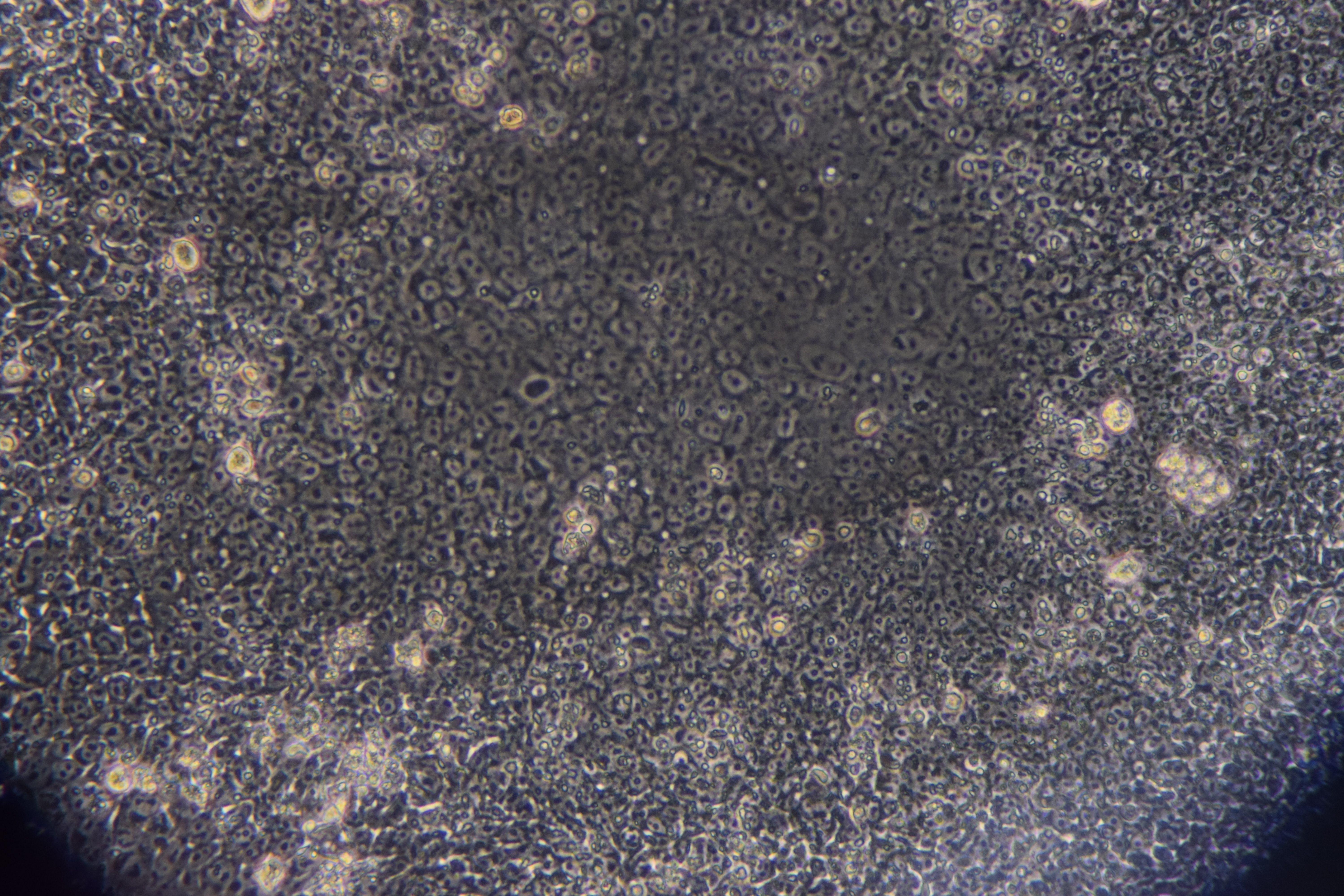
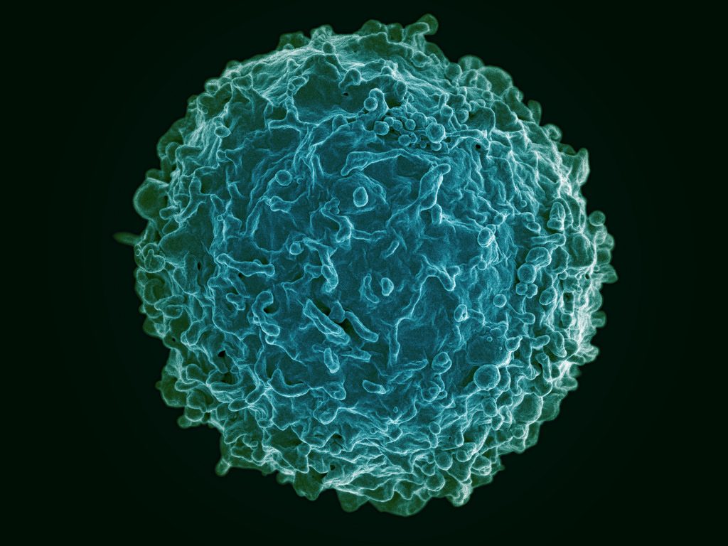
The human immune system deploys a variety of cells to counteract pathogens when they enter the body. B cells are a type of white blood cell specific to particular pathogens, and they form part of the adaptive immune system. As these cells develop, the cells with the strongest reactions to antigens are favored over others. This process is called clonal selection. Given the sheer number of pathogens out there, the number of different clonal lineages for B cells is estimated to be around 100 billion. A landscape like that can be difficult to navigate without a map.
Luckily, an atlas was recently published in Nature Biotechnology. It is the work of scientists collaborating between Penn’s own Perelman School of Medicine and faculty from the School of Biomedical Engineering, Science and Health Systems at our next-door neighbor, Drexel University. Using tissue samples from an organ donor network, the authors, led by Nina Luning Prak, MD, PhD, of Penn and Uri Heshberg, Ph.D., of Drexel, submitted the samples to a process called deep immune repertoire profiling to identify unique clones and clonal lineages. In total, they identified nearly a million lineages and mapped them to two networks: one in the gastointestinal tract and one that connects the blood, bone marrow, spleen, and lungs. This discovery suggests that the networks might be less complicated than initially thought. Also, it confirms a key role for the immune system in the gut.
Not only does this B cell atlas provide valuable information to the scientific community, but it also could serve as the basis for immune-based therapies for diseases. If we can identify these lineages and how clonal selection occurs, we could identify the most effective immunological cells and perhaps engineer them in the lab. At the very least, the extent to which scientists understand how B cells are formed and develop has received an enormous push with this research.
Understanding Muscle Movement
Natural movements of limbs require the coordinated activation of several muscle groups. Although the molecular composition of muscle is known, there remains a poor understanding of how these molecules coordinate their actions to confer power, strength, and endurance to muscle tissue. New fields of synthetic biology require this new knowledge to efficiently produce naturally inspired muscle substitutes.
Responding to this challenge, scientists at Carnegie Mellon University, including Philip R. LeDuc, Ph.D., William J. Brown Professor of Mechanical Engineering and Professor of Biomedical Engineering, have developed a computational system to better understand how mixtures of specific myosins affect muscle properties. Their method, published in PNAS, uses a computer model to show that mixtures of myosins will unexpectedly produce properties that are not the average of myosin molecular properties. Instead, the myosin mixtures coordinate and complement each other at the molecular level to create emergent behaviors, which lead to a robustness in how the muscle functions across a broad range. Dr. LeDuc and his colleagues then confirmed their model in lab experiments using muscle tissue from chickens. In the future, this new computational method could be used for other types of tissue, and it could prove useful in developing treatments for a variety of disorders.
Determining Brain Connectivity
How the brain forms and keeps memories is one of the greatest challenges in neuroscience. The hippocampus is a brain region considered critical for remembering sequences and events. The connections made by the hippocampus to other brain regions is considered critical for the hippocampus to integrate and remember experiences. However, this broad connectivity of the hippocampus to other brain areas raises a critical question: What connections are essential for rewiring the brain for new memories?
To offer an explanation for this question, a team of scientists in Hong Kong published a paper in PNAS in which they report on a study conducted in rats using resting-state function MRI. The study team, led by Ed X. Wu, Ph.D., of the University of Hong Kong, found that stimulation of a region deep in the hippocampus would propagate more broadly out into many areas of the cortex. The stimulation frequency affected how far this signal propagated from the hippocampus and pointed out the ability for frequency-based information signals to selectively connect the hippocampus to the rest of the brain. Altering the frequency of stimulation could affect visual function, indicating that targeted stimulation of the brain could have widespread functional effects throughout the brain.
Although human and rodent brains are obviously different, these findings from rats offer insights into how brain connectivity emerges in general. Similar studies in humans will be needed to corroborate these findings.
Seeing Inside a Tumor
Years of research have yielded the knowledge that the most effective treatments for cancer are often individualized. Knowing the genetic mutation involved in oncogenesis, for instance, can provide important information about the right drug to treat the tumor. Another important factor to know is the tumor’s chemical makeup, but far less is known about this factor due to the limitations of imaging.
However, a new study published in Nature Communications is offering some hope in this regard. In the study, scientists led by Xueding Wang, Ph.D., associate professor of biomedical engineering and radiology at the University of Michigan, used pH-sensing nanoprobes and multiwavelength photoacoustic imaging to determine tumor types in phantoms and animals. This new technology is based on the principle that cancerous cells frequently lower the pH levels in tissue, and designing probes with properties that are pH sensitive provides a method to find tumors with imaging methods and also treat these tumors.
With this technology, Dr. Wang and his colleagues were able to obtain three-dimensional images of pH levels inside of tumors. Importantly, it allowed them to noninvasively view the changes in a dye injected inside the tumor. Although a clinical application is years away, the information obtained using the Michigan team’s techniques could add significantly to our knowledge about tumorigenesis and tumor growth.
The Role of Bacteria in MS
The growing awareness of how bacteria interact with humans to affect health has led to the emergence of new scientific areas (e.g., human microbiome). Research findings from scientists collaborating between Caltech and UCSF suggest bacteria can play a role in the onset of multiple sclerosis. These investigators include Sarkis K. Mazmanian, Ph.D., Luis B. and Nelly Soux Professor of Microbiology and a faculty member in the Division of Biology and Biological Engineering at Caltech. Reporting their research results in PNAS, the researchers found several bacteria elevated in the MS microbiome. Study results showed that these bacteria regulated adaptive immune responses and helped to create a proinflammatory milieu. The identification of the bacteria interacting with immunity in MS patients could result in better diagnosis and treatment of this disabling disease.
People and Places
Faculty members at the University of California, Irvine, including biomedical engineer Zoran Nenadic, Ph.D., have received an $8 million grant to develop a brain-computer interface. The research using this grant aims to restore function in people with spinal cord injuries. Also, at the University of Texas, Austin, the lab of Amy Brock, Ph.D., an assistant professor in the Department of Biomedical Engineering, has received a three-year $180,000 R21 grant from the National Cancer Institute to develop a barcoding platform to isolate cancer cell lineages and to identify genetic targets for treatment.
Bone injuries and bone loss can constitute major challenges for patients and the people who treat them. Beyond the need for bone grafts or artificial implants in cases such as severe fractures, cancers metastasizing to the bones can be disabling and disfiguring. Doctors are able to use autologous bone grafts, in which patients are their own bone donors and provides grafts from other bones in their bodies. However, the grafting process compromises the bone from the donor site. In addition, there are specific problems in cases of long bones, such as those in the arms and legs. With these bones, no site of the body can provide sufficient material without becoming severely compromised itself due to bone loss.
Stem cells have been intensively investigated as a source of bone grafts. With their ability to produce a variety of cell lines from the same source, these cells have the potential to be used in a variety of clinical situations. The mechanisms underlying the determination of the type of cell that an individual stem cell will become are known. However, the ability to produce living bone cells in the laboratory had remained elusive – until now. In an article published online last week by Nature Biomedical Engineering, a group of scientists led in part by Professor Matthew Dalby, a cellular engineer with the Institute of Molecular, Cell and Systems Biology at the University of Glasgow, United Kingdom, reported its success.
Professor Dalby’s tissue engineering team used a nanoscale bioreactor to stimulate mesenchymal stem cells into osteogenesis (bone creation). The bioreactor applied vibrations on a microscopic scale of 1,000 hertz with 15 nanometers of vertical displacement. In their previous work, Professor Dalby and his colleagues could generate only one bone cell sample at a time. In the current paper, they showed the ability to generate multiple cells for three-dimensional tissue. In addition, they showed that the cells could be generated in environments with less rigidity than that in which osteogenesis normally occurs. This is an important advance because the body provides optimal conditions of stiffness for this process, but the lab does not. Should the techniques in the paper prove viable on a greater scale, they could revolutionize the field of bone grafting.
Microfluidics in the News
Since their introduction, organs on a chip (OOCs) have proliferated in the field of bioengineering. These chips use microfluidics technology to create a model of an organ system in the body. However, until now, OOCs have not been used to model the human placenta – the tissue that connects the embryonic sac to the uterine wall during pregnancy.
Responding to the lack of a OOC model of the placenta, two professors at Florida International University (FAU) have developed a placenta OOC. Sarah E. Du, Ph.D., assistant professor of ocean and mechanical engineering, and Andrew Oleinkov, Ph.D., associate professor of biomedical science, have collaborated to create this chip, which they to intend to use to determine the effects of malaria on the placental microenvironment. A $400,000 grant from the NIH will certainly help.
With malaria causing more than 200,000 perinatal deaths annually, beyond the burden we cited last week, there is an urgent need to determine the exact effects of this parasitic infection on the placenta. Without this knowledge, the development of technologies to mitigate or even prevent these effects will be much more difficult. In addition, because of the obvious ethical constraints on prospective testing in natural history studies, the placenta OOC offers an ideal model.
Elsewhere in the field of microfluidics, an NIH grant to scientists at the University of Illinois, Urbana-Champaign, has gone toward the development of a new test chip to detect sepsis, a condition in which the body’s reaction to infection results in inflammation of the blood vessels and which can cause lethal shock unless detected and treated promptly. The UIUC team developing this more rapid diagnostic technology is led by Rashid Bashir, Ph.D., professor of bioengineering and associate dean of UIUC’s Carle Illinois College of Medicine. Dr. Bashir was lead author on a paper published over the summer in Nature Communications.
Among the more remarkable aspects of the chip developed by Professor Bashir and his colleagues is that it can diagnose sepsis with a single drop of blood. Therefore, in addition to the device’s portability and size, which allows it to be used at the point of care, it is only necessary to use 10 microliters of blood to complete the test. Other available lab tests for sepsis can require as much as 300 times as much blood. Testing its device against the gold standard of flow cytometry, the UIUC team found that the findings obtained with its biochip were strongly correlated with those from flow cytometry. Unlike the new chip, flow cytometry cannot be performed outside the lab.
Since a large proportion of sepsis patients are treated in intensive care units, the ICU is a likely setting in which the biochip could be used, particularly because some ICUs might be in hospitals where the staff does not have 24-hour lab access. The ability to use this chip at the bedside immediately, rather than waiting until the next morning or longer, could make a key difference in detecting and treating sepsis.
Brains on the Internet
For years, Ray Kurzweil, the computer scientist turned author and inventor, has been discussing a future in which, he claims, the distinction between human and artificial intelligence will disappear. For example, Kurweil imagines brains being uploaded to computers. While what Kurzeil imagines has yet to materialize, scientists in South Africa have created the “Brainternet,” which streams brain waves onto the Internet in real time.
As a student project at the School of Electrical and Information Engineering of the University of Witerstand in Johannesburg led by Adam Pantanowitz, a lecturer in the school, the Brainternet was developed from pre-existing technology. The project starts with portable electroencephalography (EEG), which is worn by the subject and which transmits its signal by telemetry to a Raspberry Pi computer. Then, using open source software, the computer live streams the data to an application programming interface, which in turn allows the data to be published at a website accessible to others.
Beyond being an innovative use of these technologies, the Brainternet could be used in telemedicine applications. For instance, it could be helpful in situations where a specialist neurologist is not in the immediate geographic vicinity. Moreover, for research projects involving EEG measurement during tasks or under certain types of external stimulation, the Brainternet could allow for a much larger sample size to be enrolled, owing to its portability and use of the Internet.
People and Places
Dawn Elliott, Ph.D., chair of the Department of Biomedical Engineering at the University of Delaware, has been elected president of the Biomedical Engineering Society (BMES), for which she had served as treasurer. Dr. Elliott’s term as president will begin in October 2018 and last for two years. As president, she plans to take a closer look at education in the field to determine how bioengineering and biomedical engineering departments can graduate the most successful students. We wish her the best of luck and hearty congratulations.
At Columbia, a new way of treating lung disease is under development. As reported recently in Science Advances, a Columbia research group, headed by Gordana Vunjak-Novakovic, Ph.D., from the Department of Biomedical Engineering, developed a way to prepare grafted lung tissue for transplantation that could make the process easier. The challenge has been removing the epithelial cells, which ultimately make up the surface of the organ, from potential grafts without damaging the blood vessels. Applying a detergent solution to lung tissue from rats, Dr. Vunjak-Novakovic’s team was able to obtain grafts that could subsequently be used as scaffolds for human pulmonary cells and stem cell-derived lung epithelial cells. Although this approach remains in a very early state, the results here indicate promise for this technology for end-stage lung diseases such as emphysema.
Eliminating Obesity and Diabetes With Injections
You’ve probably heard that there’s an epidemic of obesity in the United States. Obesity carries an enormous health cost because it is linked to a variety of major health complications, including diabetes and heart disease. At a cell level, white fat cells require more energy to work off than brown fat cells. Approaches to fight obesity now include efforts to increase the number of brown fat cells. Scientists at Purdue University might have found a significant shortcut to creating more brown fat cells. By inhibiting the Notch signaling pathway, Meng Deng, Ph.D., of the Weldon School of Biomedical Engineering and his colleagues were able to cause white fat cells to convert into brown cells. Reporting their results in Molecular Therapy, the team used nanoparticles loaded with dibenazapine, a chemical used widely in pharmacology, to treat obese mice with targeted injections of the drug-laden nanoparticles. Results showed that the reduction of white fat in the mice was correlated with improved glucose metabolism and reduced body weight. While it’s not yet time to cancel the gym membership, an easier way to combat obesity could be on the horizon.
Diabetes is a chronic health condition with treatments that include diet management and/or insulin injections. In a new twist on diabetes treatments, scientists at the University of Toronto have shown, in a recent PNAS study, that pancreatic islets cells, which produce insulin, could be injected subcutaneously to reverse diabetes in mice. While the idea of transplanting islets into the pancreas has been investigated for some time, this is the first time that transplants were placed under the skin, far away from the pancreas. Impressively, the modules could be retrieved and reused. If future investigations are successful, these modules could form the basis of a treatment for type 1 (so-called juvenile) diabetes, which is caused by autoimmune destruction of the pancreatic islets.
News from New England
Feng Zhang, Ph.D., associate professor in the Departments of Brain and Cognitive Sciences and of Biological Engineering at MIT, is one of five scientists to receive the Albany Medical Prize in Medicine and Biomedical Research for his work on CRISPR-Cas9 gene editing technology. We offer Dr. Zhang our heartfelt congratulations.
Across the river from Cambridge in Medford, Tufts University has announced that its newly completed Science and Engineering Complex (SEC) will open this semester and will combine classrooms and laboratories — specifically what the developers are calling “lab neighborhoods,” or spaces for collaboration among laboratories working on related research questions. Bruce Panilaitis, Ph.D., a research assistant professor in the Department of Biomedical Engineering, is the director of the SEC, and his department will also have offices there.
Continuing with our series of interviews with new faculty members, we feature this interview with Dr. Joel Boerckel, who has a dual appointment in the Department of Bioengineering at Penn and the Perelman School of Medicine’s Department of Orthopaedic Surgery. Dr. Boerckel’s research concerns the mechanobiology of development and regeneration. Here, he speaks with Andrew Mathis about his career to this point and where he sees the fields of tissue engineering and regenerative medicine heading over the future. Enjoy!
Synthetic biology (SynBio) is an important field within bioengineering. Now, SynBio and its relationships with nanotechnology and microbiology will get a big boost with a $6 million grant from the National Science Foundation awarded to the lab of Jason Gleghorn, Ph.D., assistant professor of biomedical engineering at the University of Delaware. The grant, which comes from the NSF’s Established Program to Stimulate Competitive Research, will fund research to determine the interactions between a single virus and single microbe, using microfluidics technology so that the lab staff can examine the interactions in tiny droplets of fluid, rather than using pipettes and test tubes. They believe their research could impact healthcare broadly, as well as perhaps help agriculture by increasing crop yields.
While must SynBio research is medical, the technology is now also being used in making commercial products that will compete with other natural or chemically synthesized products. Antony Evans’s company Taxa Biotechnologies has developed a fragrant moss that he hopes can compete against the sprays and other chemicals you see on the store shelves. Using SynBio principles, Taxa isolates the gene in plants causing odor and transplants these genes to a simple moss in a glass terrarium that, with sufficient sunlight, water, carbon dioxide, will provide one of three scents completely naturally. Technically, the mosses are genetically modified organisms (GMOs), but since people aren’t eating them, they aren’t likely to generate the controversy raised by GMO foods. Taxa has also been working on transplanting bioluminescence genes to plants to provide light without requiring electricity, all as a part of a larger green campaign.
A Few Good Brains
A division of the U.S. Department of Defense, the Targeted Neuroplasticity Training (TNT) program of the Defense Advanced Research Projects Agency (DARPA) will fund the research of Stephen Helms Tillery, Ph.D., of the School of Biological & Health Systems Engineering at Arizona State University, who is investigating methods of enhancing cognitive performance using external stimulation. The ASU project is using transdermal electrical neuromodulation to apply electrical stimulation via electrodes placed on the scalp to determine the effects on awareness and concentration. DARPA hopes to obtain insight into how to improve decision making among troops who are actively deployed. The high-stress environment of a military deployment, combined with the fact that soldiers tend to get suboptimal amounts of sleep, leaves them with fatigue that can cloud judgment in moments of life or death. If the DARPA can find a way to alleviate that fatigue and clarify decision-making processes, it would likely save lives.
Circulatory Science
End-stage organ failure can be treated by transplantation, but waiting lists are long and the number of donors still insufficient, so alternatives are continually sought. In the field of regenerative medicine, which is partly dedicated to finding alternatives, scientists at Ohio State have developed a technology called tissue nanotransfection, which can generate any cell type within a patient’s own body. In a paper published in Nature Nanotechnology, professors Chandan Sen and James Lee and their research team describe how they used nanochip technology to reprogram skin cells into vascular cells. After injecting these cells into the injured legs and brains of mice and pigs, they found the cells could help to restore blood flow. The applications to organ systems is potentially limitless.
For cardiac patients whose conditions can be treated without need for a transplant, who make up the vast majority of this cohort, stents and valve prostheses are crucial tools. However, these devices and the procedures to implant them have high complication rates. Currently, patients receiving prosthetic valves made in part of metal must take blood thinners to prevent clots, and these drugs can greatly diminish quality of life and limit activity, particularly in younger patients. At Cornell, Jonathan Butcher, Ph.D., associate professor of biomedical engineering, is developing a prosthetic heart valve with small niches in the material loaded with biomaterials to maintain normal heart function and prevent clotting. While it has been possible for some time to coat the surface of an implant with a drug or chemical to facilitate its integration and function, these niches allow for a larger depot of such a material to be distributed over a longer period of time, increasing the durability of the positive effects of these procedures.
Smartphone Spectrometry
A number of medical diagnoses are accomplished by testing of bodily fluids, and spectrometry is a key technology in this process. However, spectrometers are expensive and usually not very portable, posing a challenge for health professionals working outside of traditional care settings. Now, a team led by Brian Cunningham, Ph.D., from the University of Illinois, Urbana-Champaign, has published in Lab on a Chipa paper detailing their creation of a smartphone-integrated spectroscope. Called the spectral Transmission-Reflectance-Intensity (TRI)-Analyzer, it uses microfluidics technology to provide point-of-care analysis to facilitate treatment decisions. The authors liken it to a Swiss army knife in terms of versatility and stress that the TRI Analyzer is less a specialized device than a mobile laboratory. The device costs $550, which is several times less than common lab-based instruments.
New Chair at Stanford
Stanford’s Department of Bioengineering has announced that Jennifer Cochran, Ph.D., will begin a five-year term as department chair beginning on September 1. Dr. Cochran arrived at Stanford in 2005 after earning degrees at the University of Delaware and MIT. Cochran has two connections to Penn – she is currently serving as a member of our department advisory board and completed her postdoctoral training in Penn Medicine. Our heartiest congratulations to her!