Week in BioE (September 29, 2017)

An Immune Cell Atlas

atlas
A human B cell

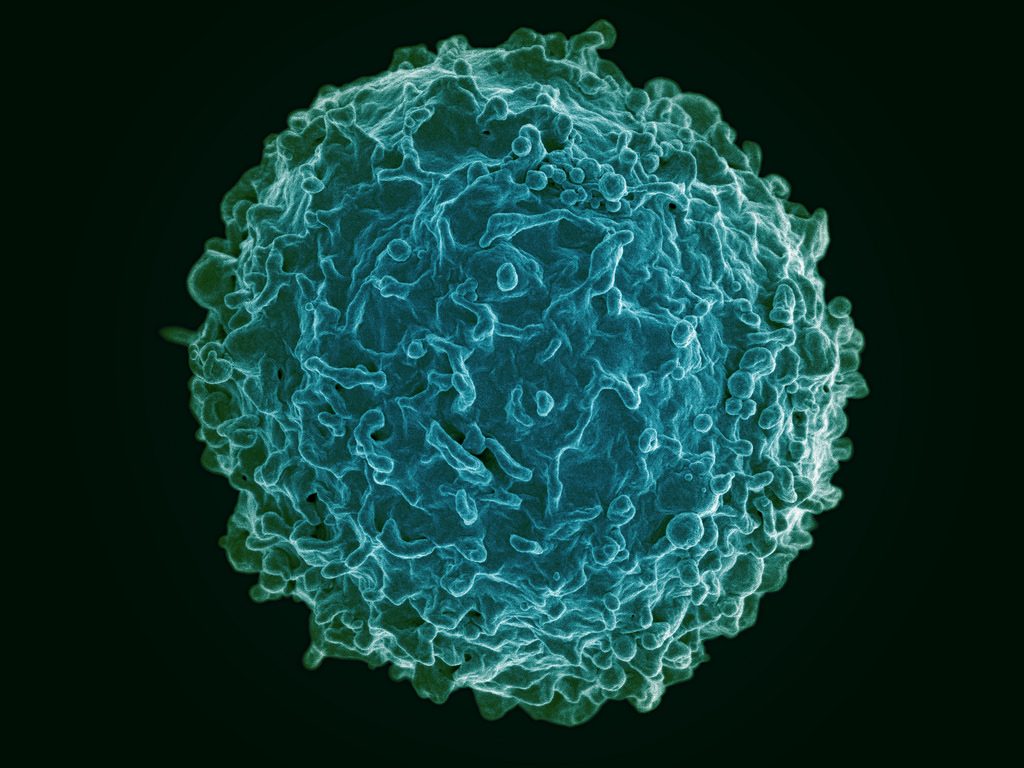
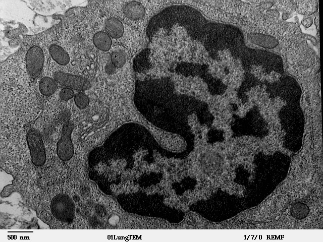
The human immune system deploys a variety of cells to counteract pathogens when they enter the body. B cells are a type of white blood cell specific to particular pathogens, and they form part of the adaptive immune system. As these cells develop, the cells with the strongest reactions to antigens are favored over others. This process is called clonal selection. Given the sheer number of pathogens out there, the number of different clonal lineages for B cells is estimated to be around 100 billion. A landscape like that can be difficult to navigate without a map.

Luckily, an atlas was recently published in Nature Biotechnology.  It is the work of scientists collaborating between Penn’s own Perelman School of Medicine and faculty from the School of Biomedical Engineering, Science and Health Systems at our next-door neighbor, Drexel University. Using tissue samples from an organ donor network, the authors, led by Nina Luning Prak, MD, PhD, of Penn and Uri Heshberg, Ph.D., of Drexel, submitted the samples to a process called deep immune repertoire profiling to identify unique clones and clonal lineages. In total, they identified nearly a million lineages and mapped them to two networks: one in the gastointestinal tract and one that connects the blood, bone marrow, spleen, and lungs. This discovery suggests that the networks might be less complicated than initially thought. Also, it confirms a key role for the immune system in the gut.

Not only does this B cell atlas provide valuable information to the scientific community, but it also could serve as the basis for immune-based therapies for diseases. If we can identify these lineages and how clonal selection occurs, we could identify the most effective immunological cells and perhaps engineer them in the lab. At the very least, the extent to which scientists understand how B cells are formed and develop has received an enormous push with this research.

Understanding Muscle Movement

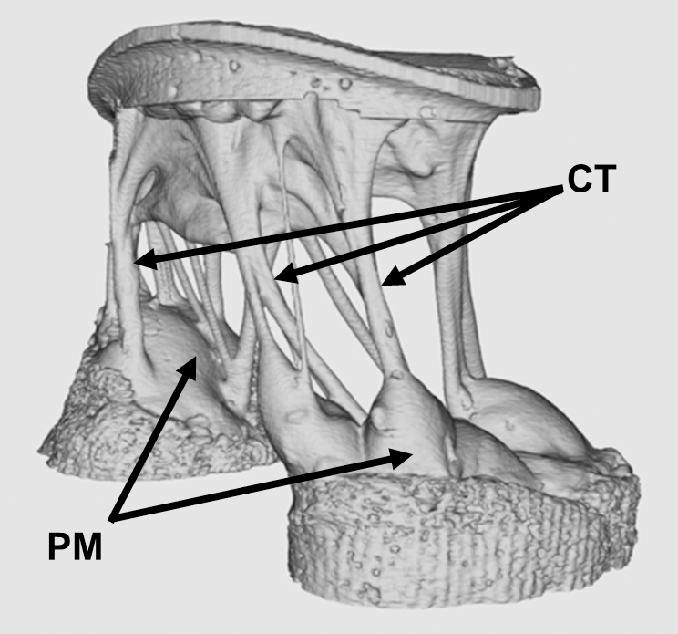
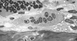
Natural movements of limbs require the coordinated activation of several muscle groups. Although the molecular composition of muscle is known, there remains a poor understanding of how these molecules coordinate their actions to confer power, strength, and endurance to muscle tissue. New fields of synthetic biology require this new knowledge to efficiently produce naturally inspired muscle substitutes.

Responding to this challenge, scientists at Carnegie Mellon University, including Philip R. LeDuc, Ph.D.,  William J. Brown Professor of Mechanical Engineering and Professor of Biomedical Engineering, have developed a computational system to better understand how mixtures of specific myosins affect muscle properties. Their method, published in PNAS, uses a computer model to show that mixtures of myosins will unexpectedly produce properties that are not the average of myosin molecular properties. Instead, the myosin mixtures coordinate and complement each other at the molecular level to create emergent behaviors, which lead to a robustness in how the muscle functions across a broad range. Dr. LeDuc and his colleagues then confirmed their model in lab experiments using muscle tissue from chickens. In the future, this new computational method could be used for other types of tissue, and it could prove useful in developing treatments for a variety of disorders.

Determining Brain Connectivity

How the brain forms and keeps memories is one of the greatest challenges in neuroscience. The hippocampus is a brain region considered critical for remembering sequences and events. The connections made by the hippocampus to other brain regions is considered critical for the hippocampus to integrate and remember experiences. However, this broad connectivity of the hippocampus to other brain areas raises a critical question: What connections are essential for rewiring the brain for new memories?

To offer an explanation for this question, a team of scientists in Hong Kong published a paper in PNAS in which they report on a study conducted in rats using resting-state function MRI. The study team, led by Ed X. Wu, Ph.D., of the University of Hong Kong, found that stimulation of a region deep in the hippocampus would propagate more broadly out into many areas of the cortex. The stimulation frequency affected how far this signal propagated from the hippocampus and pointed out the ability for frequency-based information signals to selectively connect the hippocampus to the rest of the brain. Altering the frequency of stimulation could affect visual function, indicating that targeted stimulation of the brain could have widespread functional effects throughout the brain.

Although human and rodent brains are obviously different, these findings from rats offer insights into how brain connectivity emerges in general. Similar studies in humans will be needed to corroborate these findings.

Seeing Inside a Tumor

Years of research have yielded the knowledge that the most effective treatments for cancer are often individualized. Knowing the genetic mutation involved in oncogenesis, for instance, can provide important information about the right drug to treat the tumor. Another important factor to know is the tumor’s chemical makeup, but far less is known about this factor due to the limitations of imaging.

However, a new study published in Nature Communications is offering some hope in this regard. In the study, scientists led by Xueding Wang, Ph.D., associate professor of biomedical engineering and radiology at the University of Michigan, used pH-sensing nanoprobes and multiwavelength photoacoustic imaging to determine tumor types in phantoms and animals. This new technology is based on the principle that cancerous cells frequently lower the pH levels in tissue, and designing probes with properties that are pH sensitive provides a method to find tumors with imaging methods and also treat these tumors.

With this technology, Dr. Wang and his colleagues were able to obtain three-dimensional images of pH levels inside of tumors. Importantly, it allowed them to noninvasively view the changes in a dye injected inside the tumor. Although a clinical application is years away, the information obtained using the Michigan team’s techniques could add significantly to our knowledge about tumorigenesis and tumor growth.

The Role of Bacteria in MS

The growing awareness of how bacteria interact with humans to affect health has led to the emergence of new scientific areas (e.g., human microbiome). Research findings from scientists collaborating between Caltech and UCSF suggest bacteria can play a role in the onset of multiple sclerosis. These investigators include Sarkis K. Mazmanian, Ph.D., Luis B. and Nelly Soux Professor of Microbiology and a faculty member in the Division of Biology and Biological Engineering at Caltech. Reporting their research results in PNAS, the researchers found several bacteria elevated in the MS microbiome. Study results showed that these bacteria regulated adaptive immune responses and helped to create a proinflammatory milieu. The identification of the bacteria interacting with immunity in MS patients could result in better diagnosis and treatment of this disabling disease.

People and Places

Faculty members at the University of California, Irvine, including biomedical engineer Zoran Nenadic, Ph.D., have received an $8 million grant to develop a brain-computer interface. The research using this grant aims to restore function in people with spinal cord injuries. Also, at the University of Texas, Austin, the lab of Amy Brock, Ph.D., an assistant professor in the Department of Biomedical Engineering, has received a three-year $180,000 R21 grant from the National Cancer Institute to develop a barcoding platform to isolate cancer cell lineages and to identify genetic targets for treatment.

Week in BioE (September 22, 2017)

Live Bone Cells Grown in Lab

osteogenesis
An osteoclast, one type of bone cell.

Bone injuries and bone loss can constitute major challenges for patients and the people who treat them. Beyond the need for bone grafts or artificial implants in cases such as severe fractures, cancers metastasizing to the bones can be disabling and disfiguring. Doctors are able to use autologous bone grafts, in which patients are their own bone donors and provides grafts from other bones in their bodies. However, the grafting process compromises the bone from the donor site. In addition, there are specific problems in cases of long bones, such as those in the arms and legs. With these bones, no site of the body can provide sufficient material without becoming severely compromised itself due to bone loss.

Stem cells have been intensively investigated as a source of bone grafts. With their ability to produce a variety of cell lines from the same source, these cells have the potential to be used in a variety of clinical situations. The mechanisms underlying the determination of the type of cell that an individual stem cell will become are known. However, the ability to produce living bone cells in the laboratory had remained elusive – until now.  In an article published online last week by Nature Biomedical Engineering, a group of scientists led in part by Professor Matthew Dalby, a cellular engineer with the Institute of Molecular, Cell and Systems Biology at the University of Glasgow, United Kingdom, reported its success.

Professor Dalby’s tissue engineering team used a nanoscale bioreactor to stimulate mesenchymal stem cells into osteogenesis (bone creation). The bioreactor applied vibrations on a microscopic scale of 1,000 hertz with 15 nanometers of vertical displacement. In their previous work, Professor Dalby and his colleagues could generate only one bone cell sample at a time. In the current paper, they showed the ability to generate multiple cells for three-dimensional tissue. In addition, they showed that the cells could be generated in environments with less rigidity than that in which osteogenesis normally occurs. This is an important advance because the body provides optimal conditions of stiffness for this process, but the lab does not. Should the techniques in the paper prove viable on a greater scale, they could revolutionize the field of bone grafting.

Microfluidics in the News

Since their introduction, organs on a chip (OOCs) have proliferated in the field of bioengineering. These chips use microfluidics technology to create a model of an organ system in the body. However, until now, OOCs have not been used to model the human placenta – the tissue that connects the embryonic sac to the uterine wall during pregnancy.

Responding to the lack of a OOC model of the placenta, two professors at Florida International University (FAU) have developed a placenta OOC. Sarah E. Du, Ph.D., assistant professor of ocean and mechanical engineering, and Andrew Oleinkov, Ph.D., associate professor of biomedical science, have collaborated to create this chip, which they to intend to use to determine the effects of malaria on the placental microenvironment. A $400,000 grant from the NIH will certainly help.

With malaria causing more than 200,000 perinatal deaths annually, beyond the burden we cited last week, there is an urgent need to determine the exact effects of this parasitic infection on the placenta. Without this knowledge, the development of technologies to mitigate or even prevent these effects will be much more difficult. In addition, because of the obvious ethical constraints on prospective testing in natural history studies, the placenta OOC offers an ideal model.

Elsewhere in the field of microfluidics, an NIH grant to scientists at the University of Illinois, Urbana-Champaign, has gone toward the development of a new test chip to detect sepsis, a condition in which the body’s reaction to infection results in inflammation of the blood vessels and which can cause lethal shock unless detected and treated promptly. The UIUC team developing this more rapid diagnostic technology is led by Rashid Bashir, Ph.D., professor of bioengineering and associate dean of UIUC’s Carle Illinois College of Medicine. Dr. Bashir was lead author on a paper published over the summer in Nature Communications.

Among the more remarkable aspects of the chip developed by Professor Bashir and his colleagues is that it can diagnose sepsis with a single drop of blood. Therefore, in addition to the device’s portability and size, which allows it to be used at the point of care, it is only necessary to use 10 microliters of blood to complete the test. Other available lab tests for sepsis can require as much as 300 times as much blood. Testing its device against the gold standard of flow cytometry, the UIUC team found that the findings obtained with its biochip were strongly correlated with those from flow cytometry. Unlike the new chip, flow cytometry cannot be performed outside the lab.

Since a large proportion of sepsis patients are treated in intensive care units, the ICU is a likely setting in which the biochip could be used, particularly because some ICUs might be in hospitals where the staff does not have 24-hour lab access. The ability to use this chip at the bedside immediately, rather than waiting until the next morning or longer, could make a key difference in detecting and treating sepsis.

Brains on the Internet

For years, Ray Kurzweil, the computer scientist turned author and inventor, has been discussing a future in which, he claims, the distinction between human and artificial intelligence will disappear. For example, Kurweil imagines brains being uploaded to computers. While what Kurzeil imagines has yet to materialize, scientists in South Africa have created the “Brainternet,” which streams brain waves onto the Internet in real time.

As a student project at the School of Electrical and Information Engineering of the University of Witerstand in Johannesburg led by Adam Pantanowitz, a lecturer in the school, the Brainternet was developed from pre-existing technology. The project starts with portable electroencephalography (EEG), which is worn by the subject and which transmits its signal by telemetry to a Raspberry Pi computer. Then, using open source software, the computer live streams the data to an application programming interface, which in turn allows the data to be published at a website accessible to others.

Beyond being an innovative use of these technologies, the Brainternet could be used in telemedicine applications. For instance, it could be helpful in situations where a specialist neurologist is not in the immediate geographic vicinity. Moreover, for research projects involving EEG measurement during tasks or under certain types of external stimulation, the Brainternet could allow for a much larger sample size to be enrolled, owing to its portability and use of the Internet.

People and Places

Dawn Elliott, Ph.D., chair of the Department of Biomedical Engineering at the University of Delaware, has been elected president of the Biomedical Engineering Society (BMES), for which she had served as treasurer. Dr. Elliott’s term as president will begin in October 2018 and last for two years. As president, she plans to take a closer look at education in the field to determine how bioengineering and biomedical engineering departments can graduate the most successful students. We wish her the best of luck and hearty congratulations.

InnoWorks Academy Engages Local Teens

InnoWorks

The week of August 21-25, 20 students from area middle schools visited Penn to participate in InnoWorks, an educational initiative of the United InnoWorks Academy (UIA), a non-profit organization founded in 2003 by William Hwang, M.D., Ph.D., when he was an undergraduate engineering major at Duke.  Today, InnoWorks offers programs at 20 colleges and universities in the U.S. and Caribbean. In the program, undergraduate student volunteers host middle schoolers from disadvantaged backgrounds to foster the teenagers’ interest in science and engineering.

InnoWorks
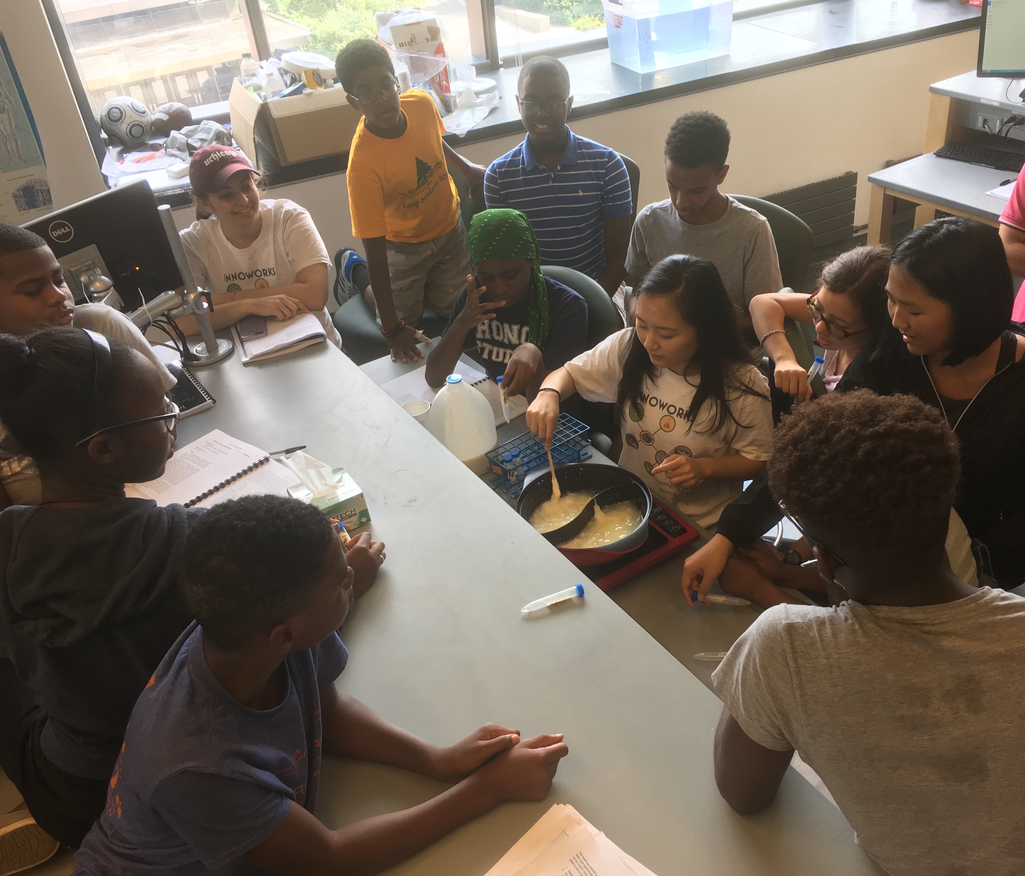
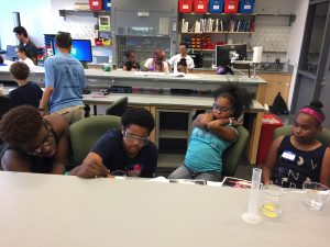
Each day of the week, from 9:15 a.m. to 5:00 p.m., the group of teens attended presentations, conducted experiments, and competed against one another in activities designed to have them apply the principles they learned about. Each day was dedicated to different topics: materials science on Monday; biology on Tuesday; chemistry and earth science on Wednesday; physics and computer science on Thursday; and a wrap-up day on Friday. In addition, over the course of the week, the students were scored for their activities, with a winner announced at the end of the week.

For instance, on Friday, students participating in InnoWorks competed in the Egg Drop Challenge. Using materials provided for them, the students designed their own parachutes for eggs, which they used in dropping the eggs from the second floor of Skirkanich Hall to the ground floor. The students did so well that the competition had to be extended to the third floor.

On another day, Dr. Kushol Gupta, a research assistant professor at Penn conducting research on HIV and assistant director of the Penn Band, talked to the students about the physics of music. Sarah Tang and Monroe Kennedy of Dean Vijay Kumar’s GRASP lab discussed the current state of robotics and drones and showed video clips of some of their work. It was among the most popular popular parts of the week.

InnoWorks
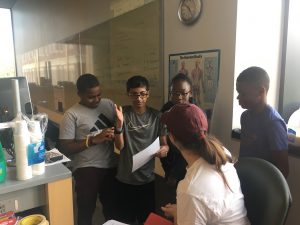
Here at Penn, the codirectors of the chapter are Jacqueline Valeri, a senior bioengineering major, and Monica Shen, a senior biology major. Asked about InnoWorks this year, Jackie said, “The InnoWorks program is a great camp to be involved with because the hands-on, interactive engagement really gets our students excited about science. We try to do really minimal lecturing time and maximize the opportunity for the students to actually do experiments, demonstrations, and design challenges. As codirector of this year’s program, it was really awesome to see what a great group of students we had this year and how they flourished throughout the week. This is my third year participating in Penn’s InnoWorks chapter and it has been one of the most rewarding, fun experiences that I’ve had at Penn!”

Week in BioE (September 15, 2017)

A Closer Look

7T-MRI
Seven-tesla MRI of the porcine mitral valve showing the papillary muscle (PM) and chordae tendinae (CT).

Since its invention in the early 1970s, magnetic resonance imaging (MRI) has played an increasingly important role in the diagnosis of illness. In addition, over time, the technology of MRI has evolved enormously, with the ability to render more detailed three-dimensional images using stronger magnetic fields . However, imaging tissues under mechanical loads (e.g., beating heart, lung breathing) are still difficult to image precisely with MRI.

A new study in PLOS One, led by Morten Jensen, Ph.D., of the University of Arkansas, breaks an important technical barrier in high resolution imaging for tissues under mechanical load. Using 3D-printed mounting hardware and 7-tesla MRI, this group produced some of the highest quality images yet produced of the mitral valve exposed to physiological pressures (see above). In the longer term, this method could point to new corrective surgical procedures that would greatly improve the repair procedures for mitral valves.

Oncology Breakthroughs

Among the most significant challenges faced by surgical oncologists is developing a ‘clear margin,’ meaning that the tissue remaining after tumor excision is free of any tumor cells. If the margins are not free of tumor cells, the cancer is more likely to recur. However, until now, it has been impossible to determine if cancer cells were still present in tissue margins before finishing surgery because of the time required to test specimens.

At the University of Texas, Austin, however, scientists are getting closer to overcoming this obstacle. In a recent study published in Science Translational Medicine, this team of scientists presents the MasSpecPen — short for mass spectrometry pen. This device is capable of injecting a tiny drop of water into tissue, extracting the water after it mixes with the tissue, and quickly analyzing the sample’s molecular components. The authors, who included biomedical engineering faculty member Thomas E. Milner, Ph.D., tested the device using ex vivo samples from 253 patients with different varieties of cancer, including breast, lung, and thyroid cancers. The MasSpecPen provided sensitivity, specificity, and accuracy exceeding 96% in all cases. Although it has yet to be tested intraoperatively, if effective under those conditions, the device could become an essential part of the surgeon’s arsenal.

If the MasSpecPen could render surgical treatment of cancer more effective, a device developed at SUNY Buffalo could help doctors diagnose lung cancer earlier. Lung cancer is a particularly deadly variety of cancer because patients don’t feel any discomfort until the cancer has spread to other areas of the body. In collaboration with Buffalo’s Roswell Park Center Institute, microchip manufacturer Intel, and local startup Garwood Medical Devices, a team of scientists, including Professor Edward P. Furlani, Ph.D., from Buffalo’s Department of Chemical and Biomedical Engineering, was awarded a grant from the National Science Foundation to develop a subcutaneous implant incorporating a nanoplasmonic biochip to detect biomarkers of lung cancer. A wearable smart band would receive data from the biochip and would act as an early warning system for lung cancer. The biomarkers selected for the biochip would optimally predict lung cancer risk much earlier than the metastasis stage. If the system that the team develops is successful in diagnosing lung cancer before it spreads, it could greatly improve survival and cure rates.

Feverish Growth

Worldwide but particularly in the Global South, malaria remains a major public health concern. According to a Global Burden of Disease study in 2015, there were nearly 300 million cases of the disease in a single year, with 731,000 fatalities. One of the earliest treatments to combat malaria was invented by British colonialists, who added quinine to the tonic used in the gin and tonic cocktail. More recently the drug artemisinin was developed for fighting malaria. However, this drug and its derivatives are very expensive. The primary reason for this cost is that the drug is extracted from the sweet wormwood plant, which is in short supply. In hopes of producing a greater supply of artemisinin, scientists collaborating among Denmark, Malaysia, and the Netherlands report in Frontiers in Bioengineering and Biotechnology that transplanting the genes responsible for producing atremisinin into Physcomitrella patens, a common moss, led to a much faster production rate of the drug than what is possible with the wormwood plant. The process proved simpler and less expensive than earlier attempts to transplant genes into tobacco plants. If this potential is harnessed correctly, it could make an enormous difference in lessening the global burden of malaria. 

Understanding Fear

We’ve known for years about the flight-or-fight response — the adrenergic response of our bodies to danger, which we share in common with a number of other animals. Once the decision to flee is made, however, we know far less about what determines the escape strategy used. According to Malcolm A. MacIver, professor of biomedical engineering and mechanical engineering in Northwestern University’s McCormick School of Engineering, part of the escape strategy depends on how far away the attacker is. In a paper he coauthored that was published in Current Biology, Dr. MacIver studied threat responses in larval zebrafish and found that a fast-looming stimulus produced either freezing or escape at a shorter interval following the threat perception; when the perceived threat was slow looming, longer latency following the perception of the threat was seen, resulting in a greater variety of types of escape behaviors. While it might seem a giant leap between observing behaviors in fish and higher life forms, the basic mechanism in the “oldest” parts of the brain, from an evolutionary standpoint, are less different than we might think.

People and Places

The University of Maryland has announced that construction on a new building to serve as the home of its Department of Bioengineering will be finished by the end of September. The building is to be named A. James Clark Hall, after a builder, philanthropist, and alumnus of Maryland’s School of Engineering. Further south, George Mason  University in Fairfax, Va., has announced that the new chair of its Department of Engineering there will be Michael Buschmann, Ph.D., an alumnus of MIT and faculty member since the 1990s at École Polytechnique in Montreal. Congratulations, Dr. Buschmann!


Undergraduates Converge at Penn for REU

REU
This year’s summer students

This past summer, 10 undergraduate from 10 colleges came to Penn for 10 weeks (May 30 to August 4) for the Summer Undergraduate Research Experience (SURE), also known as the Research Experience for Undergraduates (REU). During the program, the students were hosted in the laboratories of faculty in Penn’s Schools of Engineering and Applied Science (including Penn Bioengineering faculty Beth Winkelstein, Dan Huh, and Jason Burdick) and Arts and Sciences and the Perelman School of Medicine. These students were hosted under the aegis of the Center for Engineering MechanoBiology (CEMB), a National Science Foundation-funded collaboration among Penn, Washington University (WashU) in St. Louis, New Jersey Institute of Technology (NJIT), Alabama State University, Bryn Mawr College, Boston University, and the University of Texas at Austin.

The students all worked on individual research projects. At the end of the 10-week term, three abstracts from this research were chosen for presentation at the forthcoming annual meeting of the Biomedical Engineering Society (BMES), which will be held October 11-14 in Phoenix. The three students are Kimberly DeLuca (NJIT), John Durel (Univ. of Virginia), and Olivia Leavitt (Worcester Polytech).

The CEMB Web site at WashU has a nice page up featuring the program and this summer’s students.

Penn’s 2017 Summer Undergraduate Research Experience At-a-Glance

Bioengineers Get Support to Study Chronic Pain

chronic pain
Zhiliang Cheng, Ph.D.

Zhiliang Cheng, Ph.D., a research assistant professor in the Department of Bioengineering at the University of Pennsylvania, has received an R01 grant from the National Institute of Neurological Disorders and Stroke to study chronic pain. The grant, which provides nearly $1.7 million over the next five years, will support the work of Dr. Cheng, Bioengineering Professor Andrew Tsourkas, and Vice Provost for Education and Professor Beth Winkelstein, in developing a novel nanotechnology platform for greater effectiveness in radiculopathy treatment.

Based on the idea that phospholipase-A2 (PLA2) enzymes, which modulate inflammation, play an important role in pain due to nerve damage, the group’s research seeks to develop PLA2-responsive multifunctional nanoparticles (PRMNs) that could both deliver anti-inflammatory drugs and magnetic resonance contrast agents to sites of pain so that the molecular mechanisms at work in producing chronic pain can be imaged, as well as allowing for the closer monitoring of treatment.

This research builds on previous findings by Drs. Cheng, Tsourkas, and Winkelstein. In a 2011 paper, Drs. Tsourkas and Winkelstein used superparamagnetic iron oxide nanoparticles to enhance magnetic resonance imaging of neurological injury in a rat model. Based on the theory of reactive oxygen species playing a role in pain following neural trauma, a subsequent paper published in July with Sonia Kartha as first author and Dr. Cheng as a coauthor found that a type of nanoparticle called polymersomes could be used to deploy superoxide dismutase, an antioxidant, to sites of neuropathic pain. The current grant-supported study combines the technologies developed in the previous studies.

“To the best of our knowledge, no studies have sought to combine and/or leverage this aspect of the inflammatory and PLA2 response for developing effective pain treatment. We hypothesize that this theranostic agent, which integrates both diagnostic and therapeutic functions into a single system, offers a unique opportunity and tremendous potential for monitoring and treating patients with direct, clinically translational impact,” Dr. Cheng said.

Noordergraaf Fellows Conduct Summer Research

Each year, the Penn Department of Bioengineering chooses undergraduate students to receive fellowships for summer research. These fellowships, which provide a $3,500 stipend for use over 10 weeks, were endowed by the Abraham Noordergraaf Student Summer Bioengineering Research Fund. Dr. Noordergraaf, who died in 2014, was a founding member and first chair of the Penn BE Department. In keeping with Dr. Noordergraaf’s research focus on the cardiovascular system, fellows with a focus on this system are favored but not exclusively awarded.

Noordergraaf
Brianna Karpowicz

The fellows for the summer of 2017 were Brianna Karpowicz, Jacqueline Valeri, and Alejandro Villasmil. Brianna is a junior bioengineering major working in the lab of Professor Yale Cohen. In her research, Brianna worked with Dr. Cohen in the Auditory Research Laboratory, examining the modeling of multisensory perceptual decision making and specifically seeking to better understand the mechanisms underlying the relationship between sensory information and perception.

Noordergraaf
Alejandro Villasmil

Alejandro Villasmil, who is a senior bioengineering major working in Professor Beth Winkelstein’s lab, used his Noordergraaf’s grant to study chronic pain in neck injury. To better understand this problem, Alejandro helped to model injury to the facet capsular ligament — one of the structures in the neck — by examining how painful and nonpainful stimuli affected the axonal structure. He found using fluorescence technology that uniaxial tension resulted in axonal changes resulting in pain.

Noordergraaf
Jacqueline Valeri

Finally, Jacqueline Valeri is a senior bioengineering major doing research in the lab of Professor Jennifer Phillips-Cremins. In Professor Cremins’s lab, Jackie undertook research on stem cells, specifically examining the question of whether light could be used to control and modulate the fate of these cells — a field called optogenetics. She helped to design two light boxes to stimulate the interaction between two proteins as a first step toward ultimately attempting to control pluripotent stem cells using light, specifically determining what cell lines these stem cells ultimately produce.

We congratulate our Noordergraaf award winners!

Week in BioE (September 8, 2017)

A Breath of Fresh Air

lung grafts
A macrophage in the alveolus of a lung.

At Columbia, a new way of treating lung disease is under development. As reported recently in Science Advances, a Columbia research group, headed by Gordana Vunjak-Novakovic, Ph.D., from the Department of Biomedical Engineering, developed a way to prepare grafted lung tissue for transplantation that could make the process easier. The challenge has been removing the epithelial cells, which ultimately make up the surface of the organ, from potential grafts without damaging the blood vessels. Applying a detergent solution to lung tissue from rats, Dr. Vunjak-Novakovic’s team was able to obtain grafts that could subsequently be used as scaffolds for human pulmonary cells and stem cell-derived lung epithelial cells.  Although this approach remains in a very early state, the results here indicate promise for this technology for end-stage lung diseases such as emphysema.

Eliminating Obesity and Diabetes With Injections

You’ve probably heard that there’s an epidemic of obesity in the United States. Obesity carries an enormous health cost because it is linked to a variety of major health complications, including diabetes and heart disease. At a cell level, white fat cells require more energy to work off than brown fat cells. Approaches to fight obesity now include efforts to increase the number of brown fat cells. Scientists at Purdue University might have found a significant shortcut to creating more brown fat cells. By inhibiting the Notch signaling pathway, Meng Deng, Ph.D., of the Weldon School of Biomedical Engineering and his colleagues were able to cause white fat cells to convert into brown cells. Reporting their results in Molecular Therapy, the team used nanoparticles loaded with dibenazapine, a chemical used widely in pharmacology, to treat obese mice with targeted injections of the drug-laden nanoparticles. Results showed that the reduction of white fat in the mice was correlated with improved glucose metabolism and reduced body weight. While it’s not yet time to cancel the gym membership, an easier way to combat obesity could be on the horizon.

Diabetes is a chronic health condition with treatments that include diet management and/or insulin injections. In a new twist on diabetes treatments, scientists at the University of Toronto have shown, in a recent PNAS study, that pancreatic islets cells, which produce insulin, could be injected subcutaneously to reverse diabetes in mice. While the idea of transplanting islets into the pancreas has been investigated for some time, this is the first time that transplants were placed under the skin, far away from the pancreas. Impressively, the modules could be retrieved and reused. If future investigations are successful, these modules could form the basis of a treatment for type 1 (so-called juvenile) diabetes, which is caused by autoimmune destruction of the pancreatic islets.

News from New England

Feng Zhang, Ph.D., associate professor in the Departments of Brain and Cognitive Sciences and of Biological Engineering at MIT, is one of five scientists to receive the Albany Medical Prize in Medicine and Biomedical Research for his work on CRISPR-Cas9 gene editing technology. We offer Dr. Zhang our heartfelt congratulations.

Across the river from Cambridge in Medford, Tufts University has announced that its newly completed Science and Engineering Complex (SEC) will open this semester and will combine classrooms and laboratories — specifically what the developers are calling “lab neighborhoods,” or spaces for collaboration among laboratories working on related research questions. Bruce Panilaitis, Ph.D., a research assistant professor in the Department of Biomedical Engineering, is the director of the SEC, and his department will also have offices there.

Week in BioE (September 1, 2017)

Overcoming CP With Robotics

robotic exoskeletonCerebral palsy (CP) remains one of the most common congenital birth defects, affecting 500,000 American newborns per year. Gait disorders from CP are common, and crouch gait — characterized by misdirection and improper bending of the feet, causing excessive knee bending and the appearance of crouching — is among the most difficult to correct.

Researchers at Northern Arizona University recently developed a new exoskeleton to treat crouch gait. In an article published in Science Translational Medicine, Zach Lerner, Ph.D., assistant professor of mechanical engineering and a faculty member with NAU’s Center for Bioengineering Innovation, tested a robotic, motorized exoskeleton in seven patients with crouch gait. Six of the seven participants using the exoskeleton show improvements on par with surgical procedures to correct crouch gait. Although commercial availability of the exoskeleton will require testing in much larger patient groups, the device is an encouraging development in the treatment of a difficult disorder.

Brain Science News

A couple of weeks ago, we discussed here how the Department of Defense supports research using electrical stimulation of the scalp to direct brain activity. At the University of Texas at Arlington (UTA), bioengineering professor Hanli Liu, Ph.D., received a NIH grant to test how infrared light, rather than electrical stimulation, can achieve similar effects on the brain. In collaboration with two other UTA professors, Professor Liu uses Transcranial Infrafred Brain Stimulation (TIBS) to project infrared light onto the forehead to enhance blood flow and oxygen supply to the underlying area of the brain. With the grant, she and her colleagues intend to develop imaging tools that will provide greater insight into how both TIBS and the brain itself work.

Even as we learn more about the brain, the devastating effects of neurodegenerative diseases show us how much we still don’t know. Certain drugs can slow the inevitable advance of the disease, but beginning treatment early is important to maintaining a sense of normalcy. At Case Western Reserve University, Anant Madabhushi, Ph.D., professor of biomedical engineering, is developing computer technology to distinguish Alzheimer’s from other disorders and to predict onset earlier and more accurately.  Reporting their outcomes in Scientific Reports from testing in nearly 150 patients, Dr. Madabhushi and his colleagues used a variety of clinical measures (blood biomarkers, imaging data, neuropsychological testing) instead of a single test and developed a much more accurate test for detecting Alzheimer’s disease. Their approach, called cascaded multiview canonical correlation (CaMCCo), used the ordered analysis of different tests to stratify different patient groups at each stage, rather than developing a single combined measure all at once. More work will be needed to determine how this approach can lead to earlier detection of Alzheimer’s, but its accuracy is very encouraging for future studies.

Causes for Congratulations

Rose-Hulman Institute of Technology has announced that Kay C. Dee, Ph.D., is among the recipients of this year’s Inspiring Leaders in STEM Award from Insight Into Diversity magazine.  Professor Dee, Associate Dean of Learning and Technology and Professor of Biology and Biomedical Engineering at Rose-Hulman, is the former head of her department. As a dean, she has focused on several issues, including easier access for students with disabilities. Congratulations to Dr. Dee!

Also, several bioengineering and biomedical engineering departments across the country are celebrating birthdays. The departments at both the University of Virginia (biomedical engineering) and the University of Michigan (bioengineering) are 50 years old, with Michigan also celebrating the 20th birthday of their biomedical engineering department. The comparative baby of the group, the Department of Biomedical Engineering at Tulane University, turns 40. Happy birthday all!