Week in BioE: March 29, 2019

by Sophie Burkholder

New Studies in Mechanobiology Could Open Doors for Cellular Disease Treatment

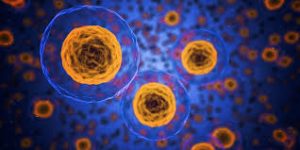
When we think of treatments at the cellular level, we most often think of biochemical applications. But what if we began to consider more biomechanical-oriented approaches in the regulation of cellular life and death? Under a grant from the National Science Foundation (NSF),Worcester Polytechnic Institute’s (WPI) Head of the Department of Biomedical Engineering Kristen Billiar, Ph.D., performs research that looks at the way mechanical stimuli can affect and trigger programmed cell death.

Billiar, who received his M.S.E. and Ph.D. from Penn, began his research by first noticing the way that cells typically respond to the mechanical stimuli in their everyday environment, such as pressure or stretching, with behaviors like migration, proliferation, or contraction. He and his research team hope to find a way to eventually predict and control cellular responses to their environment, which they hope could open doors to more forms of treatment for disorders like heart disease or cancer, where cellular behavior is directly linked to the cause of the disease.

Self-Learning Algorithm Could Help Improve Robotic Leg Functionality

Obviously, one of the biggest challenges in the field of prosthetics is the extreme difficulty in creating a device that perfectly mimics whatever the device replaces for its user. Particularly with more complex designs that involve user-controlled motion for joints in the limbs or hands, the electrical circuits implemented are by no means a perfect replacement of the neural connections in the human body from brain to muscle. But recently at the University of Southern California Viterbi School of Engineering, a team of researchers led by Francisco J. Valero-Cuevas, Ph. D.,  developed an algorithm with the ability to learn new walking tasks and adapt to others without any additional programming.

The algorithm will hopefully help to speed the progress of robotic interactions with the world, and thus allow for more adaptive technology in prosthetics, that responds to and learns with their users. The algorithm Valero-Cuevas and his team created takes inspiration from the cognition involved with babies and toddlers as they slowly learn how to walk, first through random free play and then from pulling on relevant prior experience. In a prosthetic leg, the algorithm could help the device adjust to its user’s habits and gait preferences, more closely mimicking the behavior of an actual human leg.

Neurofeedback Can Improve Behavioral Performance in High-Stress Situations

We’re all familiar with the concept of being “in the zone,” or the feeling of extraordinary focus that we can sometimes have in situations of high-stress. But how can we understand this shift in mindset on a neuroengineering level? Using the principal of the Yerkes-Dodson law, which says that there is a state of brain arousal that is optimal for behavioral performance, a team of biomedical engineering researchers at Columbia University hope to find ways of applying neurofeedback to improving this performance in demanding high-stress tasks.

Led by Paul Sajda, Ph.D., who received his doctoral degree from Penn, the researchers used a brain-computer interface to collect electroencephalography (EEG) signals from users immersed in virtual reality aerial navigation tasks of varying difficulty levels. In doing so, they were able to make connections between stressful situations and brain activity as transmitted through the EEG data, adding to the understanding of how the Yerkes-Dodson law actually operates in the human body and eventually demonstrating that the use of neurofeedback reduced the neural state of arousal in patients. The hope is that neurofeedback may be used in the future to help treat emotional conditions like post-traumatic stress disorder (PTSD).

Ultrasound Stimulation Could Lead to New Treatments for Inflammatory Arthritis

Arthritis, an autoimmune disease that causes painful inflammation in the joints, is one of the more common diseases among older patients, with more than 3 million diagnosed cases in the United States every year. Though extreme measures like joint replacement surgery are one solution, most patients simply treat the pain with nonsteroidal anti-inflammatory drugs or the adoption of gentle exercise routines like yoga. Recently however, researchers at the University of Minnesota led by Daniel Zachs, M.S.E., in the Sensory Optimization and Neural Implant Coding Lab used ultrasound stimulation treatment as a way to reduce arthritic pain in mice. In collaboration with Medtronic, Zachs and his team found that this noninvasive ultrasound stimulation greatly decreased joint swelling in mice who received the treatment as opposed to those that did not. They hope that in the future, similar methods of noninvasive treatment will be able to be used for arthritic patients, who otherwise have to rely on surgical remedies for serious pain.

People and Places

Leadership and Inspiration: EDAB’s Blueprint for Engineering Student Life

To undergraduates at a large university, the administration can seem like a mysterious, all-powerful entity, creating policy that affects their lives but doesn’t always take into account the reality of their day-to-day experience. The Engineering Deans’ Advisory Board (EDAB) was designed to bridge that gap and give students a platform to communicate with key decision makers.

The 13-member board meets once per week for 60 to 90 minutes. The executive board, comprised of four members, also meets weekly to plan out action items and brainstorm. Throughout his interactions with the group, board president Jonathan Chen, (ENG ‘19, W ‘19), has found a real kinship with his fellow board members, who he says work hard and enjoy one another’s company in equal measure.

Bioengineering major Daphne Cheung (ENG’19) joined the board as a first-year student because she saw an opportunity to develop professional skills outside of the classroom. “For me, it was about trying to build a different kind of aptitude in areas such as project management, and learning how to work with different kinds of people, including students and faculty, and of course, the deans,” she says.

Read the full story on Penn Engineering’s Medium Blog. Media contact Evan Lerner.

Purdue University College of Engineering and Indiana University School of Medicine Team Up in New Engineering-Medicine Partnership

The Purdue University College of Engineering and the Indiana University School of Medicine recently announced a new Engineering-Medicine partnership, that seeks to formalize ongoing and future collaborations in research between the two schools. One highlight of the partnership is the establishment of a new M.D./M.S. degree program in biomedical engineering that will allow medical students at Indiana University to receive M.S.-level training in engineering technologies as they apply to clinical practice. The goal of this new level of collaboration is to further involve Purdue’s engineering program in the medical field, and to exhibit the benefits that developing an engineering mindset can have for medical students. The leadership of this new partnership includes

BE Grace Hopper Lecture: Powering Tumor Cell Migration Through Hetergeneous Microenvironments

We hope you will join us for the 2019 Bioengineering Grace Hopper lecture by Dr. Cynthia Reinhart-King.

Date: Thursday, April 4, 2019
Time: 3:30-4:30 PM
Location: Glandt Forum, Singh Center, 3205 Walnut Street

Dr. Cynthia Reinhart-King, Engineering, BME, Photo by Joe Howell

Speaker: Cynthia Reinhart-King, Ph.D.
Cornelius Vanderbilt Professor of Engineering, Director of Graduate Studies, Biomedical Engienering
Vanderbilt University

Title: “Powering Tumor Cell Migration Through Heterogeneous Microenvironments”

Abstract:
To move through tissues, cancer cells must navigate a complex, heterogeneous network of fibers in the extracellular matrix. This network of fibers also provides chemical, structural and mechanical cues to the resident cells. In this talk, I will describe my lab’s efforts to understand the forces driving cell movements in the tumor microenvironment. Combining tissue engineering approaches, mouse models, and patient samples, we create and validate in vitro systems to understand how cells navigate the tumor stroma environment. Microfabrication and native biomaterials are used to build mimics of the paths created and taken by cells during metastasis. Using these platforms, we have described a role for a balance between cellular energetics, cell and matrix stiffness, and confinement in determining migration behavior. Moreover, we have extended this work into investigating the role of the mechanical microenvironment in tumor angiogenesis to show that mechanics guides vessel growth and integrity. I will discuss the mechanical influences at play during tumor progression and the underlying biological mechanisms driving angiogenesis and metastatic cell migration as a function of the ECM with an eye towards potential therapeutic avenues.

Bio:
Cynthia Reinhart-King is the Cornelius Vanderbilt Professor of Engineering and the Director of Graduate Studies in Biomedical Engineering at Vanderbilt University.  Prior to joining the Vanderbilt faculty in 2017, she was on the faculty of Cornell University where she received tenure in the Department of Biomedical Engineering. She obtained undergraduate degrees in chemical engineering and biology at MIT and her PhD at the University of Pennsylvania in the Department of Bioengineering as a Whitaker Fellow working with Daniel Hammer. She then completed postdoctoral training as an Individual NIH NRSA postdoctoral fellow at the University of Rochester.  Her lab’s research interests are in the areas of cell mechanics and cell migration specifically in the context of cancer and atherosclerosis. Her lab has received funding from the American Heart Association, the National Institutes of Health, the National Science Foundation and the American Federation of Aging Research.  She has been awarded the Rita Schaffer Young Investigator Award in 2010 and the Mid-Career Award in 2018 from the Biomedical Engineering Society, an NSF CAREER Award, the 2010 Sonny Yau ‘72 Excellence in Teaching Award, a Cook Award for “contributions towards improving the climate for women at Cornell,” and the Zellman Warhaft Commitment to Diversity Award from the Cornell College of Engineering. She is a fellow of the Biomedical Engineering Society and the American Institute for Medical and Biological Engineering, and she is a New Voices Fellow of the National Academies of Science, Engineering and Medicine. She is currently a standing member of the NIH CMT study section panel and Secretary of the Biomedical Engineering Society.

Information on the Grace Hopper Lecture:
In support of its educational mission of promoting the role of all engineers in society, the School of Engineering and Applied Science presents the Grace Hopper Lecture Series. This series is intended to serve the dual purpose of recognizing successful women in engineering and of inspiring students to achieve at the highest level.
Rear Admiral Grace Hopper was a mathematician, computer scientist, systems designer and the inventor of the compiler. Her outstanding contributions to computer science benefited academia, industry and the military. In 1928 she graduated from Vassar College with a B.A. in mathematics and physics and joined the Vassar faculty. While an instructor, she continued her studies in mathematics at Yale University where she earned an M.A. in 1930 and a Ph.D. in 1934. Grace Hopper is known worldwide for her work with the first large-scale digital computer, the Navy’s Mark I. In 1949 she joined Philadelphia’s Eckert-Mauchly, founded by the builders of ENIAC, which was building UNIVAC I. Her work on compilers and on making machines understand ordinary language instructions lead ultimately to the development of the business language, COBOL. Grace Hopper served on the faculty of the Moore School for 15 years, and in 1974 received an honorary degree from the University. In support of the accomplishments of women in engineering, each department within the School invites a prominent speaker to campus for a one or two-day visit that incorporates a public lecture, various mini-talks and opportunities to interact with undergraduate and graduate students and faculty. The lecture is open to everyone!

Penn Bioengineers: Cells Control Their Own Fate by Manipulating Their Environment

by Lauren Salig

In these images, the researchers labeled new proteins white, and antibodies against other proteins in different colors. The co-localization of new proteins and antibodies show how cells can impact their local environments.

As different as muscle, blood, brain and skin cells are from one another, they all share the same DNA. Stem cells’ transformation into these specialized cells — a process called cell fate determination — is controlled through various signals from their surroundings.

A recent Penn Engineering study suggests that cells may have more control over their fate than previously thought.

Jason Burdick, Robert D. Bent Professor of Bioengineering, and Claudia Loebel, a postdoctoral researcher in his lab, led the study. Robert Mauck, Mary Black Ralston Professor for Education and Research in Orthopaedic Surgery at Penn’s Perelman School of Medicine, also contributed to the research.

Their study was published in Nature Materials.

Read the full story in the Penn Engineering Medium Blog. Media contact Evan Lerner.

Week in BioE (March 22, 2019)

by Sophie Burkholder

A New Microscopy Technique Could Reduce the Risk of LASIK Surgery

Though over ten million Americans have undergone LASIK vision corrective surgery since the option became available about 20 years ago, the procedure still poses some risk to patients. In addition to the usual risks of any surgery however, LASIK has even more due to the lack of a precise way to measure the refractive properties of the eye, which forces surgeons to make approximations in their measurements during the procedure. In an effort to eliminate this risk, a University of Maryland team of researchers in the Optics Biotech Laboratory led by Giuliano Scarcelli, Ph. D., designed a microscopy technique that would allow for precise measurements of these properties.

Using a form of light-scattering technology called Brillouin spectroscopy, Scarcelli and his lab found a way to directly determine a patient’s refractive index – the quantity surgeons need to know to be able to measure and adjust the way light travels through the eye. Often used as a way to sense mechanical properties of tissues and cells, this technology holds promise for taking three-dimensional spatial observations of these structures around the eye. Scarcelli hopes to keep improving the resolution of the new technique, to further understanding of the eye, and reduce even more of the risks involved with LASIK surgery.

Taking Tissue Models to the Final Frontier

Space flight is likely to cause deleterious changes to the composition of bacterial flora, leading to an increased risk of infection. The environment may also affect the susceptibility of microorganisms within the spacecraft to antibiotics, key components of flown medical kits, and may modify the virulence of bacteria and other microorganisms that contaminate the fabric of the International Space Station and other flight platforms.

“It has been known since the early days of human space flight that astronauts are more prone to infection,” says Dongeun (Dan) Huh, Wilf Family Term Assistant Professor in Bioengineering at Penn Engineering. “Infections can potentially be a serious threat to astronauts, but we don’t have a good fundamental understanding of how the microgravity environment changes the way our immune system reacts to pathogens.”

In collaboration with G. Scott Worthen, a physician-scientist in neonatology at the Children’s Hospital of Philadelphia, Huh will attempt to answer this question by sending tissues-on-chips to space. Last June, the Center for the Advancement of Science in Space (CASIS) and the National Center for Advancing Translational Sciences (NCATS), part of the National Institutes of Health (NIH), announced that the duo had received funding to study lung host defense in microgravity at the International Space Station.

Huh and Worthen aim to model respiratory infection, which accounts for more than 30 percent of all infections reported in astronauts. The project’s goals are to test engineered systems that model the airway and bone marrow, a critical organ in the immune system responsible for generating white blood cells, and to combine the models to emulate and understand the integrated immune responses of the human respiratory system in microgravity.

Read the rest of the article on Penn Engineering’s Medium Blog. Media contacts Evan Lerner and Janelle Weaver.

Sappi Limited Teams Up with the University of Maine to Develop Paper Microfluidics

At the Westbrook Technology Center of Sappi, a global pulp and paper company, researchers found ways to apply innovations in paper texture for medical use. So far, these include endeavors in medical test devices and patches for patient diagnostics. In collaboration with the Caitlin Howell, Ph.D., Assistant Professor of Chemical and Biomedical Engineering at the University of Maine, Sappi hopes to continue advances in these unconventional uses of their paper, especially as the business in paper for publishing purposes declines.

Sappi’s projects with the university focus on the development of paper microfluidics devices as what’s now becoming a widespread solution for obstacles in point-of-care diagnostics. One project in particular, called Sharklet, uses a paper that mimics shark skin as a way to impede unwanted microbial growth on the device – a key characteristic needed for its transition into commercial use. Beyond this example, Sappi’s work in developing paper microfluidics underscores the benefits of these devices in their mass producibility and adaptability.

New Observations of the WNT Pathway Deepen the Understanding of Protein Signaling in Cellular Development

Scientists at Rice University recently found that a protein signaling pathway called WNT, typically associated with its role in early organism development, can both listen for signals from a large amount of triggers and influence cell types throughout embryonic development. These new findings, published in PNAS, add to the already known functions of WNT, deepening our understanding of it and opening the doors to new potential applications of it in stem cell research.

Led by Aryeh Warmflash, Ph. D., researchers discovered that the WNT pathway is different between stem cells and differentiated cells, contrary to prior belief that it was the same for both. Using CRISPR-Cas9 gene editing technology, the Warmflash lab observed that the WNT signaling pathway is actually context-dependent throughout the process of cellular development. This research brings a whole new understanding to the way the WNT pathway operates, and could open the doors to new forms of gene therapy and treatments for diseases like cancers that involve genetic pathway mutations.

People and Places

In a recent article from Technical.ly Philly, named Group K Diagnostics on a list of ten promising startups in Philadelphia. Group K Diagnostics founder Brianna Wronko graduated with a B.S.E. from Penn’s Department of Bioengineering in 2017, and her point-of-care diagnostics company raised over $2 million in funding last year. Congratulations Brianna!

We would also like to congratulate Pamela K. Woodward, M.D., on her being named as the inaugural Hugh Monroe Wilson Professor of Radiology at the Washington University School of Medicine in St. Louis. Also a Professor of Biomedical Engineering at the university, Dr. Woodward leads a research lab with a focus on cardiovascular imaging, including work on new standards for diagnosis of pulmonary blood clots and on an atherosclerosis imaging agent.

Lastly, we would like to congratulate all of the following researchers on their election to the National Academy of Engineering:

  • David Bishop, Ph. D., a professor at the College of Engineering at Boston University whose current research involves the development of personalized heart tissue as an all-encompassing treatment for patients with heart disease.
  • Joanna Aizenberg, Ph. D., a professor of chemistry and chemical biology at Harvard University who leads research in the synthesis of biomimetic inorganic materials
  • Gilda Barabino, Ph. D., the dean of the City College of New York’s Grove School for Engineering whose lab focuses on cartilage tissue engineering and treatments for sickle cell disease.
  • Karl Deisseroth, M.D., Ph. D., a professor of bioengineering at Stanford University whose research involves the re-engineering of brain circuits through novel electromagnetic brain stimulation techniques.
  • Rosalind Picard, Ph.D., the founder and director of the Affective Computing Research Group at the Massachusetts Institute of Technology’s Media Lab whose research focuses on the development of technology that can measure and understand human emotion.
  • And finally, Molly Stevens, Ph. D., the Research Director for Biomedical Material Sciences at the Imperial College of London with research in understanding biomaterial interfaces for biosensing and regenerative medicine.

BE’s Michael Mitchell Wins Controlled Release Society T. Nagai Award

Michael Mitchell, Ph.D.

Michael Mitchell, Skirkanich Assistant Professor of Innovation in the Department of Bioengineering, is the recipient of the Controlled Release Society (CRS) 2019 T. Nagai Postdoctoral Research Achievement Award, which comes with a $3,000 honorarium.

According to the CRS website, “The Controlled Release Society T. Nagai Postdoctoral Research Achievement Award has been established to recognize an individual postdoctoral candidate who has recently completed outstanding postdoctoral research in controlled release science and technology, and the postdoc’s advisor who played an integral role in those achievements.”

Mitchell and his postdoctoral advisor at MIT, Robert Langer, will receive the award at the 2019 CRS annual meeting this July in Valencia, Spain.

The sole recipient of this award, Mitchell was recognized for his work on engineering controlled release technologies for cancer gene therapy and immunotherapy. Mitchell focuses on improving the way drugs are delivered within the body by combining approaches from engineering, biology, machine learning, and data science to better target diseased cells. Mitchell’s work helps to lay the foundation for a new class of therapeutic strategies against hematologic cancers such as multiple myeloma and leukemia.

For this research, Mitchell also received the Burroughs Wellcome Fund Career Award at the Scientific Interface in 2016, the NIH Director’s New Innovator Award in 2018, and a Rising Star Award in Cellular and Molecular Bioengineering from the Biomedical Engineering Society in 2019. He joined the Penn faculty in January 2018 after completing an NIH NCI postdoctoral fellowship at the Koch Institute for Integrative Cancer Research at MIT.

Originally posted on the Penn Engineering Medium blog. Media contacts Evan Lerner. 

Penn BE Undergrads Make Biology More Accessible with Open-Source Plate Reader

The annual International Genetically Engineered Machine (iGEM) competition challenges students to expand the field of synthetic biology to solve tangible problems. While most iGEM projects involve imbuing microorganisms with useful new traits and adding them to a global toolkit, Penn Engineering students took a unique approach to the iGEM challenge by creating an open-source blueprint for a mechanical instrument that could make biological research more accessible.

Penn Bioengineering undergraduate Andrew Clark and recent graduates Karol Szymula, now a research assistant in Penn’s Complex Systems Lab, and Michael Patterson, now the lab engineer for Penn Bioengineering’s Instructional Laboratories, contributed to the project that originated through the 2017 iGEM challenge. Graduate student Michael Magaraci, who started Penn’s iGEM program as an undergraduate, and Sevile Mannickarottu, director of Instructional Laboratories, also participated. Brian Chow, Assistant Professor in Bioengineering at Penn, who helped create the iGEM competition when he was an MIT graduate student, oversaw the project.

Read the full story at Penn Engineering’s Medium Blog. Media contacts Evan Lerner and Lauren Salig.

Week in BioE (March 15, 2019)

by Sophie Burkholder

Synthetic Spinal Discs from a Penn Research Team Might Be the Solution to Chronic Back Pain

Spinal discs, the concentric circles of collagen fiber found between each vertebra of the spine, can be the source of immense back pain when ruptured. Especially for truck and bus drivers, veterans, and cigarette smokers, there is an increased risk in spinal disc rupture due to overuse or deterioration over time. But these patients aren’t alone. In fact, spinal discs erode over time for almost everyone, and are one of the sources of back pain in older patients, especially when the discs erode so much that they allow direct bone-to-bone contact between two or more vertebrae.

Robert Mauck, Ph.D.

Robert Mauck, Ph.D., who is the director of the McKay Orthopaedic Research Laboratory here at Penn and a member of the Bioengineering Graduate Group Faculty, led a research team in creating artificial spinal discs, with an outer layer made from biodegradable polymer and an inner layer made with a sugar-like gel. Their findings appear in Science Translational Medicine. These synthetic discs are also seeded with stem cells that produce collagen over time, meant to replace the polymer as it degrades in vivo over time. Though Mauck and his time are still far from human clinical trials for the discs, they’ve shown some success in goat models so far. If successful, these biodegradable discs could lead to a solution for back pain that integrates itself into the human body over time, potentially eliminating the need of multiple invasive procedures that current solutions require. Mauck’s work was recently featured in Philly.com.

An Untethered, Light-Activated Electrode for Innovations in Neurostimulation

Neurostimulation, a process by which nervous system activity can be purposefully modulated, is a common treatment for patients with some form of paralysis or neurological disorders like Parkinson’s disease. This procedure is typically invasive, and because of the brain’s extreme sensitivity, even the slightest involuntary movement of the cables, electrodes, and other components involved can lead to further brain damage through inflammation and scarring. In an effort to solve this common problem, researchers from the B.I.O.N.I.C. Lab run by Takashi D.Y. Kozai, Ph. D., at the University of Pittsburgh replaced long cables with long wavelength light and a formerly tethered electrode with a smaller, untethered one.

The research team, which includes Pitt senior bioengineering and computer engineering student Kaylene Stocking, centered the device on the principle of the photoelectric effect – a concept first described in a publication by Einstein as the local change in electric potential for an object when hit with a photon. Their design, which includes a 7-8 micron diameter carbon fiber implant, is now patent pending, and Kozai hopes that it will lead to safer and more precise advancements in neurostimulation for patients in the future.

A New Microfluidic Chip Can Detect Cancer in a Drop of Blood

Many forms of cancer cannot be detected until the disease has progressed past the point of optimum treatment time, increasing the risk for patients who receive late diagnoses of these kinds of cancer. But what if the diagnostic process could be simplified and made more efficient so that even a single drop of blood could be enough input to detect the presence of cancer in a patient? Yong Zeng, Ph.D., and his team of researchers at the University of Kansas in Lawrence sought to answer that question.

They designed a self-assembled 3D-nanopatterned microfluidic chip to increase typical microfluidic chip sensitivity so that it can now detect lower levels of tumor-associated exosomes in patient blood plasma. This is in large part due to the nanopatterns in the structure of the chip, which promote mass transfer and increase surface area, which in turn promotes surface-particle interactions in the device. The team applied the device to their studies of ovarian cancer, one of the notoriously more difficult kinds of cancer to detect early on in patients.

A Wearable Respiration Monitor Made from Shrinky Dinks

Michelle Khine, Ph. D., a professor of biomedical engineering at the University of California, Irvine incorporates Shrinky Dinks into her research. After using them once before in a medical device involving microfluidics, her lab recently worked to incorporate them into a wearable respiration monitor – a device that would be useful for patients with asthma, cystic fibrosis, and other chronic pulmonary diseases. The device has the capability to track the rate and volume of its user’s respiration based on measurements of the strain at the locations where the device makes contact with the user’s abdomen.

Paired with Bluetooth technology, this sensor can feed live readings to a smartphone app, giving constant updates to users and doctors, as opposed to the typical pulmonary function test, which only provides information from the time at which the test takes a reading. Though Khine and her team have only tested the device on healthy patients so far, they look forward to testing with patients who have pulmonary disorders, in hopes that the device will provide more comprehensive and accessible data on their respiration.

People and Places

Ashley Kimbel, a high school senior from Grissom High School in Huntsville, Alabama, designed a lightweight prosthetic leg for local Marine, Kendall Bane, after an attack in Afghanistan led him to amputate one of his legs below the knee. Bane, who likes to keep as active as possible, said the new lighter design is more ideal for activities like hiking and mountain biking, especially as any added weight makes balance during these activities more difficult. Kimbel used a CAD-modeling software produced by Siemens called Solid Edge, which the company hopes to continue improving in accessibility so that more students can start projects like Kimbel’s.

This week, we would like to congratulate Angela Belcher, Ph.D., on being named the new head of the Department of Biological Engineering at the Massachusetts Institute of Technology (MIT). With her appointment to this role, now half of the MIT engineering department heads are women. Belcher’s research is in the overlap of materials science and biological engineering, with a particular focus on creating nanostructures based on the evolution of ancient organisms for applications in medical diagnostics, batteries, solar cells, and more.

We would also like to congratulate Eva Dyer, Ph.D., and Chethan Pandarinath, Ph.D., both of whom are faculty members at the Walter H. Coulter Department of Biomedical Engineering at Georgia Tech and Emory University, on receiving research fellowships from the Alfred P. Sloan Foundation. Dr. Dyer, who formerly worked with Penn bioengineering faculty member Dr. Konrad Kording while he was at Northwestern University, leads research in the field of using data analysis methods to quantify neuroanatomy. Dr. Pandarinth leads the Emory and Georgia Tech Systems Neural Engineering Lab, where he works with a team of researchers to use properties of artificial intelligence and machine learning to better understand large neural networks in the brain.


BE Seminar Series: March 14th

The BE Seminar Series continues this week. We hope to see you there!

Shuichi Takayama, Ph.D.

Speaker: Shuichi Takayama, Ph.D.
Professor, GRA Eminent Scholar, Price Gilbert, Jr. Chair in Regenerative Engineering and Medicine
Wallace H. Coulter Department of Biomedical Engineering
Georgia Institute of Technology and Emory University

Date: Thursday, March 14th, 2019
Time: 12:00 pm
Location: Room 337, Towne Building

“Microfluidics and Immuno-Materials for Organs-on-a-Chip”

This presentation will describe microfluidic technologies to conveniently produce life-like pulsatile flows along with applications to study of lung injury, enhancement of in vitro fertilization, and analysis of frequency-dependent cellular responses. The microfluidic technologies range from adaptation of piezo-electric actuator arrays from Braille displays to design of microfluidic circuits that can be designed to switch fluid flow on and off periodically on their own. The presentation will also describe engineered materials to mimic an aspect of the innate immune system to combat bacterial infection. More specifically, reconstituted chromatin microwebs inspired by neutrophil extracellular traps. Using a defined composition reconstituted chromatin microweb, we reveal impact of microweb DNA-histone ratio on bacteria capture. Additionally, we found that E. coli, including clinical isolates and resistant strains, are killed more efficiently by the last-resort antibiotic, colistin, when bound to microwebs. Recent efforts towards incorporation of these materials into human cell systems will also be described. Time permitting, topics on organoids, fibrosis, liquid-liquid phase separation, and scaling may be incorporated.

Dr. LeAnn Dourte Appointed Practice Associate Professor

LeAnn Dourte, Ph.D.

The Department of Bioengineering at the University of Pennsylvania would like to congratulate LeAnn Dourte, Ph.D. on her recent promotion to Practice Associate Professor. Formerly a Senior Lecturer, this promotion reflects Dr. Dourte’s innovative approaches to pedagogy since arriving at Penn in 2011. The title of Practice Associate Professor reflects exceptional accomplishment in teaching, leadership, and educational programs. As an official member of the faculty, this formalizes Dourte’s role as a leader in pedagogy and educational scholarship, furthering empowering her to think creatively and progressively about higher education.

As a key member of the BE teaching faculty, Dourte has regularly taught core undergraduate subjects such as Intro to Biomechanics (BE 200) and Biomaterials (BE 220) as well as popular graduate electives Biomechanics and Biotransport (BE 510) and Biomechatronics (BE 570). In particular, she spearheaded the department’s initiative to improve classroom and learning experiences through experimentation with the Structured, Active, In-Class Learning (or SAIL) model of education which emphasizes teamwork and dynamic problem-solving. Dourte worked with the Center for Teaching and Learning to understand this model and assess how it would best be applicable for BE students, and then presented her findings at national conferences and to the BE faculty, helping to introduce them to these techniques and advise them on best practices. Other BE faculty such as Chris Fang-Yen, Ph.D., Jennifer Philips-Cremins, Ph.D., and Danielle Bassett, Ph.D., have experimented with and incorporated these ideas into their courses. Thanks to Dourte’s efforts, BE has been integral in demonstrating the success of SAIL classes to the School of Engineering and then spreading this philosophy to other schools at Penn. In addition to her pedagogical interests, Dourte is also highly involved in the Department’s student wellness initiative, serving on the Department’s Climate Committee and as the Wellness Ambassador.

Now that she has achieved this latest milestone, Dourte has set her sights on goals for the future. She intends to work with Associate Dean for Undergraduate Education Dr. Russ Composto to strengthen initiatives to assist SEAS’s First-Generation, Low-Income (FGLI) students. She will be working with Dr. Bassett, who specializes in network science, to learn more about how students learn, and what tools can be developed to assess students in addition to traditional exams and homework; to tell more easily when students are missing key concepts; and to intervene sooner in moments of crisis. And finally, Dourte will also be one of three representatives from Penn at an upcoming national education summit this May to discuss the future of Bioengineering curricula.

“Our best educators are teachers for the rest of the faculty, as well as the students,” says Department Chair Dr. David Meaney. “We are enormously proud of the prestige and expertise that LeAnn shares with all of us. I was fortunate to teach biomechanics with LeAnn for many years, and saw her outstanding ability as an educator in person.”

Once again, we would like to extend hearty congratulations to Dr. Dourte on this well-deserved recognition of her leadership and both in and out of the classroom.