Collaboration in Research by Bioengineering Faculty

Jennifer Phillips-Cremins
Danielle Bassett

In faculty matters, specialization is the name of game. The areas in which individual professors conduct their research and teach are highly specific, with often no overlap between the areas of expertise of people in the same departments. Given the broad range of topics covered by the term, bioengineering is particularly complex in the array of subjects researched by faculty.

Now and then, however, these paths converge. Most recently, Jennifer Phillips-Cremins, Ph.D., Assistant Professor of Bioengineering, and Danielle Bassett, Ph.D., Eduardo D. Glandt Faculty Fellow and Associate Professor of Bioengineering, collaborated on a paper published in Nature Methods. Dr. Cremins’s research has focused on genome folding, an intricate process by which DNA in the nuclei of cells creates loops that result in  specific forms of gene regulation. Dr. Bassett’s area is network science and systems theory. Both professors apply their research in the area of central nervous development.

In the new paper, Drs. Cremins and Bassett, along with members of both their labs and colleagues from the Department of Genetics, developed a a graph theory-based method for detecting genome folding, called 3DNetMod, which outperformed earlier models used for the same purpose. In addition, Dr. Cremins is profiled in the same issue of Nature Methods, where she discusses how her past education and experience have resulted in her career achievements thus far.

Week in BioE (February 27, 2018)

Pain Relief Using Spearmint Aromatherapy

spearmintPain is the body’s way of telling you there’s something wrong. For most of us, the pain goes away after the body fixes itself. However, more than 10% of Americans suffer from chronic pain after the healing period. Many chronic pain patients need drugs to reduce their symptoms.  Given the pervasive use of opioid drugs to treat chronic pain, opioid addiction is common among chronic pain patients.

However, a remarkably clever and elegant cellular engineering technology may provide a new approach for treating chronic pain. Martin Fussenegger, Ph.D., a professor in the Department of Biosystems Science and Bioengineering at the Swiss Federal Institute of Technology, is the lead author of a new study published in Nature Biomedical Engineering combining cellular and genetic engineering to alleviate pain using cells as factories to produce spearmint. The strategy employed by the authors used engineered human cells to express huwentoxin IV, a blocker of sodium channels regulating pain signals in neurons, upon exposure to carvone, a terpenoid found in spearmint.

Testing their concept in a mouse model of pain, the authors found that mice exposed to spearmint both orally and via aromatherapy showed fewer signs of pain. Looking forward, Dr. Fussenegger and his colleagues believe that their technology, called AromaCell, should be tested next in human cell lines to alleviate concerns about immunological responses to the cells when implanted into patients.

Press Button to Bleed

Another recent article in Nature Biomedical Engineering details the work of the Boston-area biotech firm Seventh Sense Biosystems on their push-button blood collection device, called TAP. As we have discussed here before, currently used blood-drawing procedures are often uncomfortable to patients because of the sharp needle prick used to collect blood. TAP was designed to collect 100 microliters of whole blood using a device the size of a stethoscope bell in a “virtually painless” manner.

The scientists from Seventh Sense designed the patch using microneedle technology. With this approach, they designed TAP with multiple microneedles deployed at high velocity to collect blood from capillaries — the tiniest vessels that connect veins and arteries and that lie closest to the surface of the skin — rather than from a vein tied off with a tourniquet. Testing the device in 144 volunteers, the study authors found that the device was as accurate as current methods for obtaining blood to measure hemoglobin (important for diabetics) and was significantly less painful.

Seventh Sense predicts this disposable device will cost only $5 per use, but this is still almost double the materials cost for standard blood draws. However, the company believes that the pain-free nature of and time saved with TAP will offset the higher cost of the device.

Advances in Global Health

The positively epidemic nature of human papilloma virus (HPV), affecting nearly one quarter of all Americans, has drawn particular attention over the last decade or so. The clear association between HPV and cervical cancer (as well as head and neck cancers) has led to the development and deployment of vaccines (controversial due to the sexually transmitted nature of HPV) and to increased calls for more regular and accurate screening. In developing nations, implementing either effective vaccination or early screening programs remains an uphill struggle.

Responding to the need for more accessible screening technologies, Jessica Ramella-Roman, Ph.D., Associate Professor of Biomedical Engineering at Florida International University (FIU), and Purnima Madhivanan, Ph.D., an epidemiology professor at FIU, traveled to Mysore, India, to install a device developed by Dr. Ramella-Roman at the Public Health Research Institute of India. The device is a hand-held imaging tool that uses a technology called Mueller matrix imaging to provide high-resolution digital images of the cervix in about 5 seconds. The resolution of the images eliminates the need to use dyes or stains to detect malignant cells. The testing of the device is currently ongoing.

Elsewhere in global health, researchers at Google have teamed with medical faculty from Stanford to produce a machine learning algorithm that could examine the human retina and determine whether the person in question is at risk for cardiovascular disease. They report their findings in Nature Biomedical Engineering.

The technology is not ready for actual patients yet — the study authors concede that the algorithm does not outperform the currently available technologies. However, if improved with additional research and testing, the algorithm could be deployed virtually anywhere, including in patients’ homes.

People and Places

Yale University has launched a new Center for Biomedical Data Science, dedicated to collecting, studying, and managing big data. The interim directors are Mark Gerstein, Ph.D., Albert L Williams Professor of Biomedical Informatics, Molecular Biophysics, and Biochemistry, and Hongyu Zhao, Ph.D., Ira V. Hiscock Professor of Biostatistics and Professor of Genetics and Professor of Statistics and Data Science.

The University of Virginia has announced a partnership with Smithfield Bioscience, a subsidiary of Smithfield Foods, Inc. The goal of the partnership is to advance a variety of tissue engineering applications using tissue samples from pigs. George J. Christ, Ph.D., Professor of Biomedical Engineering and Orthopaedic Surgery, heads UVA’s $3 million Center for Advanced Biomanufacturing, which is involved in the partnership.

Finally, we offer our congratulations to Guoqiang Yu, Ph.D., Professor of Biomedical Engineering at the University of Kentucky and a former research faculty member in physics here at Penn, for being awarded a two-year $420,000 R21 research grant from the NIH’s Eunice Kennedy Shriver National Institute of Child Health and Human Development. Dr. Yu will use the money to develop a device to measure cerebral hemodynamics in neonatal ICU patients.

Week in BioE (February 20, 2018)

Modeling Hemostasis With Microfluidics

hemostasis
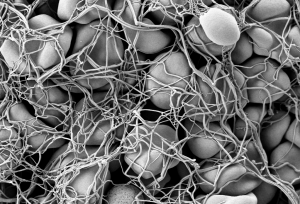
Electronic scanning microscopic image of red blood cells forming a clot

Hemostasis is the process by which blood stops flowing from damaged blood vessels. It is a complex process involving multiple molecules and forces, and our current understanding is limited by our inability to test these factors simultaneously in the laboratory. Some tests, for instance, can tell us much about clotting — a part of hemostasis — but little about the other elements at play. In particular, the role in hemostasis of the endothelium, which is the cell layer that lines the blood vessels, has generally been omitted from previous studies.

However, a new article in Nature Communications details the use of microfluidics technology, which is often used to model organ systems outside the body, to engineer a more complete model of hemostasis. Led by Wilbur A. Lam, M.D., Ph.D., Assistant Professor in the Wallace H. Coulter Department of Biomedical Engineering at Georgia Tech, the study authors fabricated microfluidics devices and then seeded vascular channels in the devices with human aortic and umbilical vein cells to simulate the endothelium.

Using the device, the authors were able to visualize hemostatic plug formation with whole blood and with blood from subjects with hemophilia. Although the authors concede that their model best represents capillary bleeding, rather than bleeding from larger vessels, they are confident that their model reliably represents the interaction of the endothelium with multiple varieties of blood cells.

Shedding Light on Cancer Response and Resistance

Penn’s founder, Benjamin Franklin, has a famous axiom: “an ounce of prevention is worth a pound of cure.” If Franklin were alive today, he would likely agree with two common axioms in cancer treatment: 1) if you can’t see it, you can’t treat it; and 2) if you treat it, treat all of it. Recent publications from investigators at Columbia and the University of Maryland reveal how imaging technologies can help predict to  outcomes and how nanotechnology is offering a new therapeutic tools for fighting cancer.

Using diffuse optical tomography (DOT), which employs near-infrared spectroscopy to obtain three-dimensional images, scientists at Columbia have shown in an article in Radiology that treatment response could be predicted as early as two weeks after the start of therapy. The authors, led by Andreas H. Hielscher, PhD, Professor of Biomedical Engineering, Electrical Engineering, and Radiology at Columbia, applied DOT in 40 women with breast cancer undergoing chemotherapy. They found that DOT imaging features were associated, some very strongly, with treatment outcomes at 5 months.  Given their positive findings, the authors intend to continue testing DOT in a larger cohort prospective study.

Another major issue in cancer chemotherapy is multidrug resistance (MDR),  a highly frustrating complication resulting from lengthy treatment. In MDR, cancer types can find ways to overcome the effects of chemotherapy, resulting in relapse, often with deadly consequences. Therefore, among the challenges that oncologists face is the need to predict MDR, preferably before treatment even begins.

Based on the knowledge that adenosine triphosphate (ATP), a common organic molecule in energy generation, is involved in MDR, scientists at the University of Maryland engineered nanoparticles that could target cancer cells and, when exposed to near-infrared laser irradiation, reduce the amount of ATP in the cells . The scientists, led by Xiaoming Shawn He, Ph.D., Professor of Bioengineering at Maryland, published their findings in Nature Communications.

Dr. He’s team tested their nanoparticles in vitro and subsequently in mice and found decreased tumor sizes in mice treated with the particles, as well as more deaths of cancer cells. In addition, two of seven mice treated with the nanoparticles plus light experienced complete tumor eradication. The findings offer hope that MDR could be overcome with direct delivery of targeted treatment to resistant tumors.

Preserving the Tooth

A frustrating problem often encountered by dentists is the growth of new cavities around existing fillings. Microbes are often critical catalysts for these new cavities. Using antimicrobial agents at cavity-repair sites could make a real difference. However, mesoporous silica has proved suboptimal for this purpose.

However, help might be on the way. A study in a recent issue of Scientific Reports, written by a trio of authors led by Benjamin D. Hatton, Ph.D., Assistant Professor at the Institute of Biomaterials & Biomedical Engineering of the University of Toronto, reports the successful synthesis of 500-nm nanocomposite spheres combining silica with octenidine dihydrochloride, a common antiseptic. The newly synthesized nanospheres outperformed earlier attempts with mesoporous silica. The authors will continue to develop these nanoparticles to deliver other drugs for longer periods of time.

New Review Blazes the TRAIL

TRAIL
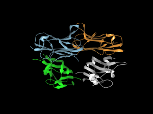
Drawing of tumor necrosis factor alpha

Cell signaling and the proteins involved in it participate in virtually every process in the body, whether normal or pathological. Much of this signaling involves proteins called cytokines, and of particular interest among them are tumor necrosis factors (TNFs), whose job it is to carry out apoptosis — the process by which cells die at predetermined time points as part of their normal life cycle. Among this family of cytokines, TNF-related apoptosis-inducing ligand (TRAIL) has been of particular interest to oncologists.

The process by which TRAIL combines with or binds to other molecules that modulate the life cycle of cancer cells can interfere with the ability of these molecules to facilitate the growth of cancer cells into tumors. However, attempts to deploy the cytokine to interfere in the process that produces cancer have been unsuccessful because of issues regarding inefficient delivery of TRAIL to the relevant sites, poor circulation of the cytokine in the blood, and the development of resistance to TRAIL. Bioengineers have been hard at work attempting to overcome these barriers.

In a new article published in ACS Nano coauthored by Michael J. Mitchell, Ph.D., Skirkanich Assistant Professor of Innovation at Penn Bioengineering, and Robert Langer, Ph.D., David H. Koch Institute Professor at MIT, these engineered solutions are reviewed and assessed. The review covers nanoparticle technologies with potential to solve the problems encountered thus far, including a range of materials (polymers, lipids, inorganic), cell-nanoparticle hybrids, and therapeutic cells genetically engineered using nanoparticles.

“The TRAIL protein is a essential component of our immune system,” Dr. Mitchell says, “and it kills tumor cells without harming normal ones. However, it remains challenging to deliver the protein into tumors, and tumors can also be resistant to the protein. We and others are now exploiting nanotechnology, genetic engineering, and immune cell-biomaterial hybrids to overcome these key biological barriers to cancer therapy.”

Week in BioE (February 12, 2018)

Engineering Recovery?

opioidsThe introduction of morphine in the 19th century to alleviate pain revolutionized medicine in a way few innovations do, but it brought with it a grave unintended consequence: addiction. In today’s society, opioid addiction is creating the biggest health crisis of the last half century. Affecting nearly 1 in 100 people, opioid addiction occurs more than type 1 diabetes, multiple sclerosis, or a number of other diseases. The addiction crisis also appears in global affairs and impacts our national security: heroin production in Afghanistan over the last 40 years has been critical to funding military actions by insurgent groups against both the US and, in the past, the Soviet Union.

However, bioengineers at Stanford have begun to tackle the issue of production and might have begun to tackle the issue of addiction. In the lab of Christine Smolke, Ph.D., Professor of Bioengineering at Stanford, they have been genetically engineering yeast to produce opioids. They described the process in a 2015 article from Science. Now, in a recent interview in Fast Company, Dr. Smolke discusses the possibility of using the yeast producing method she pioneered to produce opioids without addiction potential. These alternative drugs are very expensive to produce, and Dr. Smolke’s process could provide safer, less addictive compounds to people in need.

Breaking the Barrier

Among the challenges faced by bioengineers working on therapies for brain disease is the blood-brain barrier (BBB), a tightly regulated boundary between the circulatory system and the brain that prevents all but the tiniest molecules from getting into the brain. The poor permeability of the BBB to many molecules means that one needs to use higher drug dosages to reach the brain, which is one of the reasons why most psychiatric medications have a broad array of side effects.
One way of circumventing this issue is to deliver the drugs directly to the brain, rather than using oral or intravenous delivery methods that need to cross the BB. Here, the challenge is one of size — unless a needle used to administer such a drug is very small, it will invariably damage brain tissue, which can have devastating consequences. Answering this call has been Robert Langer, Ph.D., David H. Koch Institute Professor at MIT, whose lab has successfully microfabricated delivery cannulas as small as 30 microns, about one-third the diameter of human hair. As they report in Science Translational Medicine, the new cannula can target brain areas as small as 1 cubic millimeter.
Dr. Langer and his colleagues used the new cannula to create an implantable device, called the miniaturized neural drug delivery system (MiNDS), that they subsequently tested in rats and rhesus monkeys. They found that the device could modulate neuronal activity in both animals. In addition, MiNDS could also record and transmit information from the treatment site to enable feedback control. Going forward, the study authors envision the use of non-metallic materials to fashion cannulas and hydrogel coatings to facilitate MR imaging and increase biocompatibility.

Unlocking the Mystery of IPF

Idiopathic pulmonary fibrosis (IPF) is a lung disease that causes permanent scarring of the lung tissue. The disease affects around five million people worldwide, mainly people 50 or older, and the five-year mortality rate is very high. Although risk factors, such as cigarette smoking, have been identified, as the word “idiopathic” implies, the cause is unknown, making it difficult to create effective therapies other than ones that merely slow the progression of the disease.
However, thanks to a new discovery, we might be closer to effective treatments. In an article in the Journal of Clinical Investigation Insight, a team of scientists from Yale University report that the tissue lesions that constitute IPF are made up of roughly one-fifth pericytes — a type of contractile cell that plays an important role in the proper function of capillaries, including those in the lungs.
The study authors, led by Anjelica Gonzalez, Ph.D., Donna L. Dubinsky Associate Professor of Biomedical Engineering at Yale, found that IPF caused  pericytes to take on the properties of myofibroblasts, a cell type that is important to the wound-healing process. They found further that treatment of these myofibroblast-like pericytes with nintedanib, a drug approved for IPF treatment, reversed this effect. Armed with this knowledge, we come a step closer to designing and producing more effective therapies for IPF, as well as for diseases with similar effects.

People and Places

Washington University in St. Louis has announced it will launch a Ph.D. program in imaging science, to enroll its first cohort this fall. The program will be headed by Mark Anastasio, Ph.D., Professor of Biomedical Engineering and a 1993 recipient of an MSE from Penn.  WashU’s program is only the second such program in the country, following the program at the Rochester Institute of Technology.
Closer to home, Johns Hopkins is the recipient of a $50 million donation from the United Arab Emirates. The money will be used to create the Sheikh Khalifa Stroke Institute, which will unite faculty members from biomedical engineering, neurology, and rehabilitation medicine to advance research into stroke.

Week in BioE (February 2, 2018)

Broccoli + Yogurt = Cancer Prevention?

broccoliGeorge H.W. Bush refused to eat it, but maybe he should start. It turns out that broccoli, combined with bioengineered yogurt, could provide effect cancer prevention. We’ve known for some time that compounds in certain fresh vegetables can increase chemoprevention, but the levels are usually too low to be effective, or they can’t be assimilated optimally by the body.  However, scientists in Singapore found that engineered bacteria, when ingested by mice with colorectal cancer, had anticancer effects. The bacteria caused the secretion of an enzyme by the cancer cells that transformed glucosinolates — compounds found in vegetables — into molecules with anticancer efficacy. The scientists report their findings in Nature Biomedical Engineering.

The authors programmed an E. coli cell line to bind to heparan sulfate proteoglycan, a cell surface protein that occurs in colorectal cancer cells. Once the engineered bacteria bound to the cancer cells, the bacteria secreted myrosinase, an enzyme that commonly occurs in many plants to defend them against aphids. In the cell model employed by the authors, myrosinase caused the conversion of glucosinolates into sulforaphane, which in turn could inhibit cancer cell growth.

The scientists then applied their system in a mouse model of colorectal cancer, feeding the mice yogurt infused with the engineered bacteria. They found that the mice fed broccoli plus the yogurt developed fewer and smaller tumors than mice fed broccoli alone. Additional testing is necessary, of course, but the study authors believe that their engineered bacteria could be used both as a preventive tool in high-risk patients and as a supplement for cancer patients after surgery to remove their tumors.

The Gates of CRISPR

About two years ago, software giant Microsoft unveiled Azimuth, a gene-editing tool for CRISPR/Casa9 that it had developed in collaboration with scientists at the Broad Institute. Now, in response to concerns that CRIPR may edit more of the genome than a bioengineer wants, the team has introduced a tool called Elevation. A new article in Nature Biomedical Engineering discusses the new tool.

In the article, the team, co-led by John C. Doench, Ph.D., Institute Scientist at the Broad Institute, describes how it developed Azimuth and Elevation, both of which are machine learning models, and deployed the tools to compare their ability to predict off-target editing with the ability of other approaches. The Elevation model outperformed the other methods. In addition, the team has implemented a cloud-based service for end-to-end RNA design, which should alleviate some of the time and resource handicaps that scientists face in using CRISPR.

Reducing Infant Mortality With an App

Among the challenges still faced in the developing world with regard to health care is high infant mortality, with the most common cause being perinatal asphyxia, or lack of oxygen reaching the infant during delivery. In response, Nigerian graduate student Charles C. Onu, a Master’s student in the computer science lab of Doina Precup, Ph.D., at McGill University in Montreal, founded a company called Ubenwa, an Igbo word that means “baby’s cry.”

With Ubenwa and scientists from McGill, Onu developed a smartphone app and a wearable that apply machine learning to instantly diagnose birth asphyxia based on the sound of a baby’s cry. In initial testing, the device performed well, with sensitivity of more than 86% and specificity of more than 89%. You can read more about the development and testing of Ubenwa at Arxiv.

People and Places

Several universities have announced that they are introducing new centers for research in bioengineering. Purdue University secured $27 million in funding from Semiconductor Research Corp. for its Center for Brain-inspired Computing Enabling Autonomous Intelligence, or C-BRIC, which opened last month. The center will develop, among other technologies, robotics that can operate without human intervention.

In Atlanta, Emory University received a $400 million pledge from the Robert W. Woodruff Foundation for two new centers — the Winship Cancer Institute Tower and a new Health Sciences Research Building. The latter will host five research teams, including one specializing in biomedical engineering. Further north in Richmond, Virginia Commonwealth University announced that it will begin construction on a new $92 million Engineering Research Building in the fall.  The uppermost floors of the new building will include labs for the college’s Department of Biomedical Engineering.

Finally, North Carolina’s Elon College will introduce a bachelor’s degree program in engineering in the fall. The program will offer concentrations in biomedical engineering and computer engineering. Sirena Hargrove-Leak, Ph.D., is director of the program.