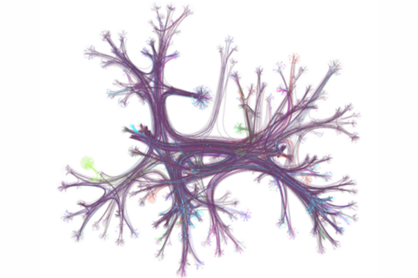
A hyperlink network from English Wikipedia, with only 0.1% of articles (nodes) and their connections (edges) visualized. Seven different reader journeys through this network are highlighted in various colors. The network is organized by topic and displayed using a layout that groups related articles together. (Image: Dale Zhou)
At one point or another, you may have gone online looking for a specific bit of information and found yourself “going down the Wiki rabbit hole” as you discover wholly new, ever-more fascinating related topics — some trivial, some relevant — and you may have gone so far down the hole it’s difficult to piece together what brought you there to begin with.
According to the University of Pennsylvania’s Dani Bassett, who recently worked with a collaborative team of researcher to examine the browsing habits of 482,760 Wikipedia readers from 50 different countries, this style of information acquisition is called the “busybody.” This is someone who goes from one idea or piece of information to another, and the two pieces may not relate to each other much.
“The busybody loves any and all kinds of newness, they’re happy to jump from here to there, with seemingly no rhyme or reason, and this is contrasted by the ‘hunter,’ which is a more goal-oriented, focused person who seeks to solve a problem, find a missing factor, or fill out a model of the world,” says Bassett.
In the research, published in the journal Science Advances, Bassett and colleagues discovered stark differences in browsing habits between countries with more education and gender equality versus less equality, raising key questions about the impact of culture on curiosity and learning.
Dani S. Bassett is the J. Peter Skirkanich Professor at the University of Pennsylvania with a primary appointment in the School of Engineering and Applied Science’s Department of Bioengineering and secondary appointments in the School of Arts & Sciences’ Department of Physics & Astronomy, Penn Engineering’s Department of Electrical and Systems Engineering, and the Perelman School of Medicine’s Departments of Neurology and Psychiatry.
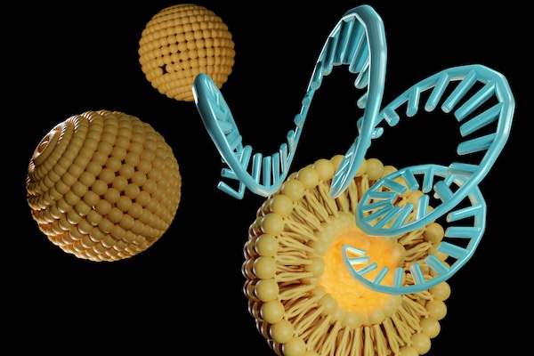
By adjusting the chemical structure of lipid nanoparticles (LNPs), Penn Engineers have discovered how to target specific organs, a major breakthrough in precision medicine. (Love Employee via Getty Images)
Penn Engineers have discovered a novel means of directing lipid nanoparticles (LNPs), the revolutionary molecules that delivered the COVID-19 vaccines, to target specific tissues, presaging a new era in personalized medicine and gene therapy.
While past research — including at Penn Engineering — has screened “libraries” of LNPs to find specific variants that target organs like the lungs, this approach is akin to trial and error. “We’ve never understood how the structure of one key component of the LNP, the ionizable lipid, determines the ultimate destination of LNPs to organs beyond the liver,” says Michael J. Mitchell, Associate Professor in Bioengineering.
In a new paper published in Nature Nanotechnology, Mitchell’s group describes how subtle adjustments to the chemical structure of the ionizable lipid, a key component of the LNP, allows for tissue-specific delivery, in particular to the liver, lungs and spleen.
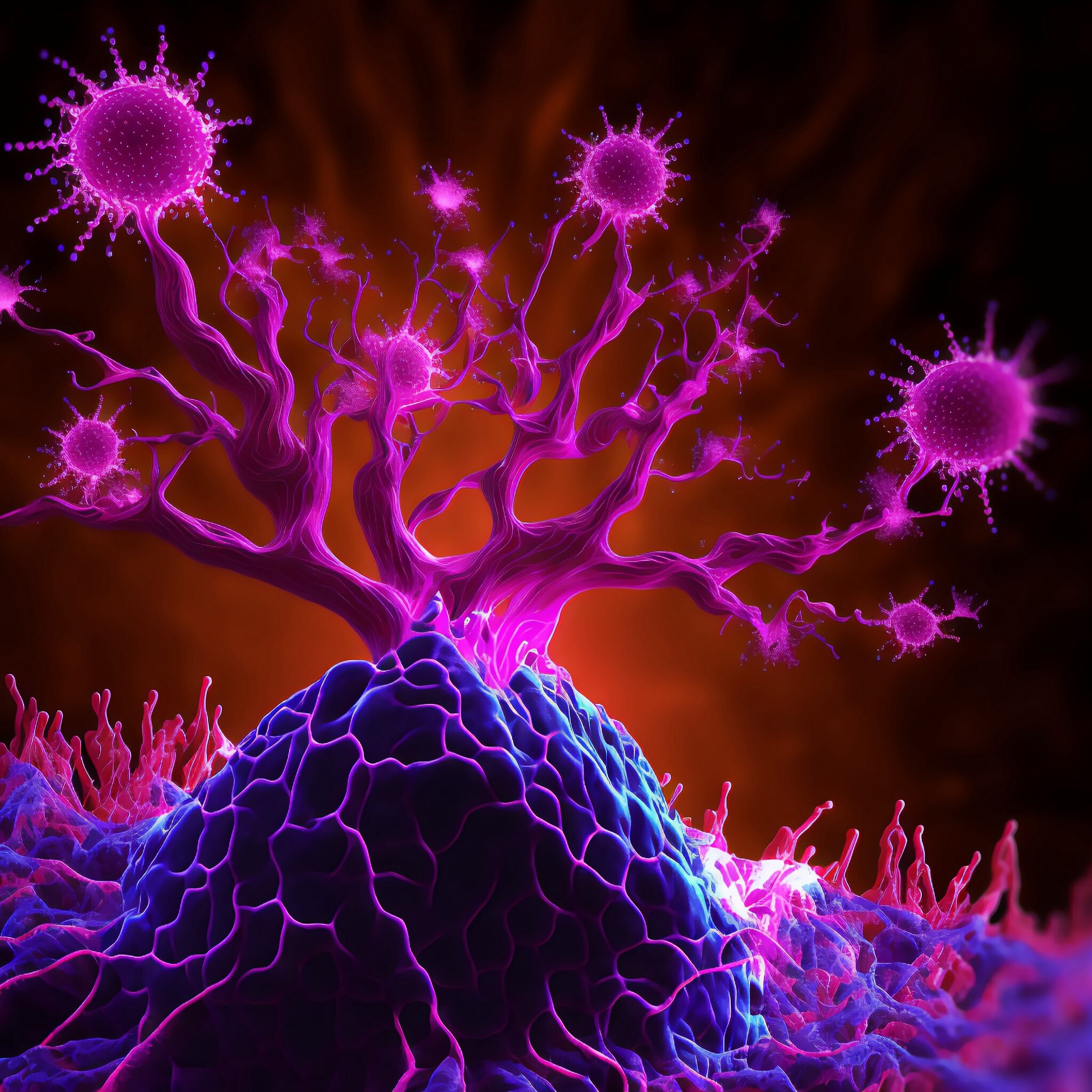
In a collaborative interdisciplinary study, Michael Mitchell of the School of Engineering and Applied Science, Wei Guo of the School of Arts & Sciences, and Drew Weissman of the Perelman School of Medicine show that solid tumors can block drug-delivery mechanisms with a “forcefield-like” effect but certain genetic elements that can effectively “shut down” the forcefield. Their findings hint at new targets for delivering cancer treatments that use the body’s immune system to fight tumors. (Image: iStock / CIPhotos)
The tumor microenvironment—an ad hoc, messy amalgamation of signaling molecules, immune cells, fibroblasts, blood vessels, and the extracellular matrix—acts like a “powerful security system that protects solid tumors from invaders seeking to destroy them,” says Michael Mitchell, a bioengineer at the University of Pennsylvania working on nanoscale therapeutics aimed at targeting cancers.
“A lot like the Death Star with its surrounding fleet of fighter ships and protective shields, solid tumors can use features like immune cells and vasculature to exert force, acting as a physical barrier to rebel forces (nanoparticles) coming in to deliver the payload that destroys it,” Mitchell says.
Now, researchers in the Mitchell lab have teamed up with Wei Guo’s group in the School of Arts & Sciences at Penn and Drew Weissman of the Perelman School of Medicine to figure out the molecular mechanisms that make tumor microenvironments seemingly impenetrable and found that small extracellular vesicles (sEVs) are secreted by tumor cells and act as a “forcefield,” blocking therapeutics. Their findings are published in Nature Materials.
“This discovery reveals how tumors create a robust defense system, making it challenging for nanoparticle-based therapies to reach and effectively target cancer cells,” Guo says. “By understanding the cellular mechanisms driving these responses, we can potentially develop strategies to disable this defense, allowing therapeutics to penetrate and attack the tumor more efficiently.”
The research builds on a prior collaboration between Guo and Mitchell’s labs, wherein the teams focused on how tumor-associated immune cells, known as macrophages, contribute to the suppression of anti-tumor immunity by secreting extracellular vesicles.
Wei Guo is the Hirsch Family President’s Distinguished Professor in the Department of Biology in Penn’s School of Arts & Sciences.
Ningqiang Gong, a former postdoctoral researcher in the Mitchell lab at Penn Engineering, is an assistant professor at the University of Science and Technology of China.
Wenqun Zhong is a reseearch associate in the Guo Laboratory in Penn Arts & Sciences.
Other authors include: Alex G Hamilton, Dongyoon Kim, Junchao Xu, and Lulu Xue of Penn Engineering; Junhyong Kim, Zhiyuan Qin, and Fengyuan Xu of Penn Arts & Sciences; Mohamad-Gabriel Alameh and Drew Weissman of the Perelman School of Medicine; Andrew E. Vaughn and Gan Zhao of the Penn School of Veterinary Medicine; Jinghong Li and Xucong Teng of the University of Beijing; and Xing-Jie Liang of the Chinese Academy of Sciences.
This research received support from the U.S. National Institutes of Health (DP2 TR002776, R35 GM141832, and NCI P50 CA261608), Burroughs Wellcome Fund, U.S. National Science Foundation CAREER Award (CBET-2145491), and an American Cancer Society Research Scholar Grant (RGS-22-1122-01-ET.)
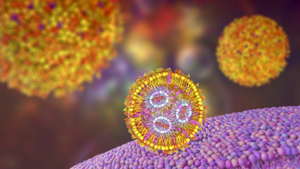
Lipid nanoparticles present one of the most advanced drug delivery platforms to shuttle promising therapeutics such as mRNA but are limited by the time it takes to synthesize cationic lipids, a key component. Now, Michael Mitchell and his team at the School of Engineering and Applied Science have developed a faster way to make cationic lipids that are also more versatile, able to carry different kinds of treatments to target specific organs. (Image: iStock / Dr_Microbe)
Imagine a scenario where a skilled hacker must upload critical software to update a central server and thwart a potentially lethal virus from wreaking havoc across a vast computer network. The programmer, armed with the lifesaving code, must navigate through treacherous territory teeming with adversaries, and success hinges on promptly getting a safe, stealthy delivery vehicle that can place the hacker exactly where they need to be.
In the context of modern medicine, messenger RNA (mRNA) serves as the hacker, carrying genetic instructions to produce specific proteins within cells that can induce desired immune responses or sequester maladaptive cellular elements. Lipid nanoparticles (LNPs) are the stealthy delivery vehicles that transport these fragile mRNA molecules through the bloodstream to their target cells, overcoming the body’s defenses to deliver their payload safely and efficiently.
However, much like building an advanced stealth vehicle, the synthesis of cationic lipids—a type of lipid molecule that’s positively charged and a key component of LNPs—is often a time-consuming process, involving multiple steps of chemical synthesis and purification.
Now, Michael Mitchell and a team at the University of Pennsylvania have addressed this challenge with a novel approach that leverages a compound library fabrication technique known as “click-like chemistry” to create LNPs in a single, simple step. Their findings, published in the journal Nature Chemistry, show that this method not only speeds up the synthesis process but also presents a way to equip these delivery vehicles with a “GPS” to better target specific organs such as the liver, lungs, and spleen, potentially opening new avenues for treating a range of diseases that arise in these organs.
“We’ve developed what we call an amidine-incorporated degradable (AID) lipid, a uniquely structured biodegradable molecule,” Mitchell says. “Think of it as an easy-to-build custom mRNA vehicle with a body kit that informs its navigation system. By adjusting its shape and degradability, we can enhance mRNA delivery into cells in a safe manner. By adjusting the amount of the AID lipid that we incorporate into the LNP, we can also guide it to different organs in the body, much like programming different destinations into a GPS.”
First author Xuexiang Han, a former postdoctoral researcher in the Mitchell Lab, explains that their new approach allows the rapid creation of diverse lipid structures in just an hour, compared to the weekslong process traditionally required.
Congratulations to Alison Pouch, Assistant Professor in Bioengineering in the School of Engineering and Applied Science, and in Radiology in the Perelman School of Medicine, on winning a 2024 Cardiac Center Innovation Award for scientific research from the Children’s Hospital of Philadelphia (CHOP)’s Philly Spin-In. Pouch’s study, titled “Systemic Semilunar Valve Mechanics and Simulated Repair in Congenital Heart Disease,” is a collaboration with Matthew Jolley, Assistant Professor of Anesthesiology and Critical Care at CHOP:
“Through biomechanical assessment, Drs. Matthew Jolley and Alison Pouch are leading an interdisciplinary CHOP-Penn team that plans to determine why current approaches to systemic semilunar valve (SSV) repair fail. They will also investigate methods to design improved repairs before going to the operating room by using computational simulation to iteratively optimize repair.
‘We believe that understanding biomechanics of abnormal SSVs and explorations of simulated repair will markedly improve our ability to characterize, risk stratify, and surgically treat SSV dysfunction, thereby improving long-term outcomes and quality of life in patients with SSV dysfunction,’ Dr. Jolley said.”
Pouch’s lab focuses on 3D/4D segmentation and modeling of heart valves in echocardiographic images with applications to surgical treatment of valvular regurgitation as part of the Penn Image Computing and Science Laboratory.
Cesar de la Fuente (left), Fangping Wan (center), and Marcelo der Torossian Torres (right). Fangping holds a 3D model of a unique ATP synthase fragment, identified by their lab’s deep learning model, APEX, as having potent antibiotic properties.
“Make sure you finish your antibiotics course, even if you start feeling better’ is a medical mantra many hear but ignore,” says Cesar de la Fuente of the University of Pennsylvania.
He explains that this phrase is, however, crucial as noncompliance could hamper the efficacy of a key 20th century discovery, antibiotics. “And in recent decades, this has led to the rise of drug-resistant bacteria, a growing global health crisis causing approximately 4.95 million deaths per year and threatens to make even common infections deadly,” he says.
De la Fuente, a Presidential Assistant Professor, and a team of interdisciplinary researchers have been working on biomedical innovations tackling this looming threat. In a new study, published in Nature Biomedical Engineering, they developed an artificial intelligence tool to mine the vast and largely unexplored biological data—more than 10 million molecules of both modern and extinct organisms— to discover new candidates for antibiotics.
“With traditional methods, it takes around six years to develop new preclinical drug candidates to treat infections and the process is incredibly painstaking and expensive,” de la Fuente says. “Our deep learning approach can dramatically reduce that time, driving down costs as we identified thousands of candidates in just a few hours, and many of them have preclinical potential, as tested in our animal models, signaling a new era in antibiotic discovery.” César de la Fuente holds a 3D model of a unique ATP synthase fragment, identified by his lab’s deep learning model, APEX, as having potent antibiotic properties. This molecular structure, resurrected from ancient genetic data, represents a promising lead in the fight against antibiotic-resistant bacteria.
These latest findings build on methods de la Fuente has been working on since his arrival at Penn in 2019. The team asked a fundamental question: Can machines be used to accelerate antibiotic discovery by mining the world’s biological information? He explains that this idea is based on the notion that biology, at its most basic level, is an information source, which could theoretically be explored with AI to find new useful molecules.
The first few waves of COVID-19 slowed life across the United States, affecting everything from attending school to eating out for dinner and going on vacation. Segments of health care were also affected: Services that were not considered immediately crucial to fighting the virus were slowed or stopped during the pandemic’s first wave.
But once Penn Medicine invited patients back to resume normal health care—including preventive care, like screenings for disease—there was some lag in numbers.
“As we opened up to routine outpatient care, screening rates for situations when patients didn’t have symptoms were not returning back to normal,” said Mitchell Schnall, MD, PhD, FACR, a professor of Radiology, now the senior vice president for Data and Technology Solutions at Penn Medicine, and then the head of a team focused on the “resurgence” efforts to ease patients back into outpatient care. “Although a short delay in health screening is likely not going to cause long-term health problems, we were concerned whether screening rates would stay lower and lead to a long-term impact.”
Top: Axons in female and male subject brains Bottom: damaged axons in male and female brains after injury (Credit: Penn Medicine)
Important brain structures that are key for signaling in the brain are narrower and less dense in females, and more likely to be damaged by brain injuries, such as concussion. Long-term cognitive deficits occur when the signals between brain structures weaken due to the injury. The structural differences in male and female brains might explain why females are more prone to concussions and experience longer recovery from the injury than their male counterparts, according to a preclinical study led by the Perelman School of Medicine at the University of Pennsylvania, published this week in Acta Neuropathologica.
Each year, approximately 50 million individuals worldwide suffer a concussion, also referred to as mild traumatic brain injury (TBI). However, there is nothing “mild” about this condition for the more than 15 percent of individuals who suffer persisting cognitive dysfunction, which includes difficulty concentrating, learning and remembering new information, and making decisions.
Although males make up the majority of emergency department visits for concussion, this has been primarily attributed to their greater exposure to activities with a risk of head impacts compared to females. In contrast, it has recently been observed that female athletes have a higher rate of concussion and appear to have worse outcomes than their male counterparts participating in the same sport.
“Clinicians have observed for a long time that females suffer from concussion at higher rates than males in the same sports, and that they take longer to recover cognitive function, but couldn’t explain the underlying mechanisms of this phenomenon,” said senior author Douglas Smith, MD, a professor of Neurosurgery and director of Penn’s Center for Brain Injury and Repair. “The variances in brain structures of females and males not only illuminate why this disparity exists, but also exposes biomarkers, such as axon protein fragments, that can be measured in the blood to determine injury severity, monitor recovery, and eventually help identify and develop treatments that help patients repair these damaged structures and restore cognitive function.”
The growing threat of antimicrobial resistance demands innovative solutions in drug discovery. Scientists are turning to artificial intelligence (AI) and machine learning (ML) to accelerate the discovery and development of antimicrobial peptides (AMPs). These short strings of amino acids are promising for combating bacterial infections, yet transitioning them into clinical use has been challenging. Leveraging novel AI-driven models, researchers aim to overcome these obstacles, heralding a new era in antimicrobial therapy.
A new article in Nature Reviews Bioengineering illuminates the promises and challenges of using AI for antibiotic discovery. Cesar de la Fuente, Presidential Assistant Professor in Microbiology and Psychiatry in the Perelman School of Medicine, in Bioengineering and Chemical and Biomolecular Engineering in the School of Engineering and Applied Science, and Adjunct Assistant Professor in Chemistry in the School of Arts and Sciences, collaborated with James J. Collins, Termeer Professor of Medical Engineering and Science at MIT, to provide an introduction to this emerging field, outlining both its current limitations and its massive potential.
In the past five years, groundbreaking work in the de la Fuente Lab has dramatically accelerated the discovery of new antibiotics, reducing the timeline from years to mere hours. AI-driven approaches employed in his laboratory have already yielded numerous preclinical candidates, showcasing the transformative potential of AI in antimicrobial research and offering new potential solutions against currently untreatable infections.
Recent advancements in AI and ML are revolutionizing drug discovery by enabling the precise prediction of biomolecular properties and structures. By training ML models on high-quality datasets, researchers can accurately forecast the efficacy, toxicity and other crucial attributes of novel peptides. This predictive power expedites the screening process, identifying promising candidates for further evaluation in a fraction of the time required by conventional methods.
Traditional approaches to AMP development have encountered hurdles such as toxicity and poor stability. AI models help overcome these challenges by designing peptides with enhanced properties, improving stability, efficacy and safety profiles, and fast-tracking the peptides’ clinical application.
While AI-driven drug discovery has made significant strides, challenges remain. The availability of high-quality data is a critical bottleneck, necessitating collaborative efforts to curate comprehensive datasets to train ML models. Furthermore, ensuring the interpretability and transparency of AI-generated results is essential for fostering trust and wider adoption in clinical settings. However, the future is promising, with AI set to revolutionize antimicrobial therapy development and address drug resistance.
Integrating AI and ML into antimicrobial peptide development marks a paradigm shift in drug discovery. By harnessing these cutting-edge technologies, researchers can address longstanding challenges and accelerate the discovery of novel antimicrobial therapies. Continuous innovation in AI-driven approaches is likely to spearhead a new era of precision medicine, augmenting our arsenal against infectious diseases.
The de la Fuente Lab uses use the power of machines to accelerate discoveries in biology and medicine. The lab’s current projects include using AI for antibiotic discovery, molecular de-extinction, reprogramming venom-derived peptides to discover new antibiotics, and developing low-cost diagnostics for bacterial and viral infections. Read more posts featuring de la Fuente’s work in the BE Blog.
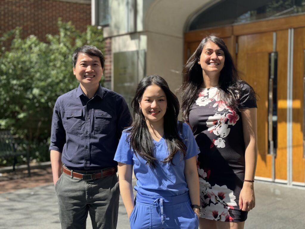
Left to right: Hong-Huy Tran, Chrissie Jaruchotiratanasakul, Manali Mahajan (Photo Courtesy of CiPD)
The Center for Innovation and Precision Dentistry (CiPD), a collaboration between Penn Engineering and Penn Dental Medicine, has partnered with Wharton’s Mack Institute for Innovation Management on a research project which brings robotics to healthcare. More specifically, this project will explore potential uses of nanorobot technology for oral health care. The interdisciplinary partnership brings together three students from different Penn programs to study the commercialization of a new technology that detects and removes harmful dental plaque.
“Our main goal is to bring together dental medicine and engineering for out-of-the-box solutions to address unresolved problems we face in oral health care,” says Hyun (Michel) Koo, Co-Founding Director of CiPD and Professor of Orthodontics. “We are focused on affordable solutions and truly disruptive technologies, which at the same time are feasible and translatable.”