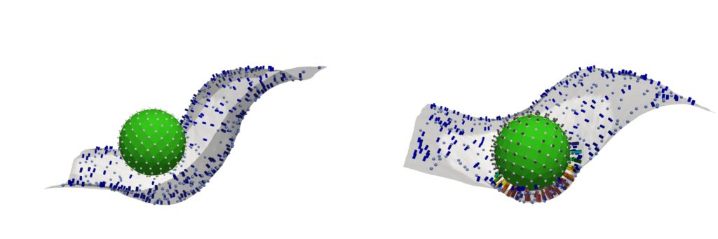
(Left) Pre-ignition (below the activation threshold) Only a handful of immune “tags” (C3b proteins) cover the nanoparticle, so it barely sticks to the white membrane—too few contact points means the immune cell simply can’t grab on. (Right) Post-ignition (above the activation threshold). The nanoparticle is now densely coated with C3b tags, and the immune-cell membrane reaches out with many matching receptors. Dozens of little “hooks” latch on at once, creating a strong, multivalent grip that pulls the particle in for engulfment.(Image: Ravi Radhakrishnan)
How does your body distinguish friendly visitors, like medications and medical devices, from dangerous invaders such as viruses and other infectious agents? The answer lies in a protein network dating back half a billion years—before humans diverged from sea urchins, notes Jake Brenner, a physician-scientist at the University of Pennsylvania.
“The complement system is perhaps the oldest-known part of our extracellular immune system,” says Brenner. “It plays a crucial role in identifying foreign materials like microbes, medical devices, or new drugs—particularly the larger ones like in the COVID vaccine.”
The complement system can, however, simultaneously play friend and foe, offering protection with one hand while backhanding the body with the other. In some cases, this ancient network can significantly exacerbate conditions like stroke by targeting the body’s own tissues. As Brenner explains, leaking blood vessels allow complement proteins to target brain tissue, causing the immune system to mistakenly launch an attack on the body’s own cells and worsen patient outcomes.
Now, using a combination of wet-lab experimentation, coupled differential equations, and computational-based modeling and simulations, an interdisciplinary team from the School of Engineering and Applied Science and the Perelman School of Medicine has decrypted the mathematical language behind the complement network’s “decision” to attack.
Reporting their findings in Cell, the team identifies a molecular tipping point known as the critical percolation threshold, which is based on how densely complement-binding sites are spaced on the surfaces of the model invader they engineered. If spacing between binding sites is too wide—landing above a threshold—complement activation fizzles out; below it, complement network ignites, a chain reaction of immune agent recruitment which spreads like wildfire.
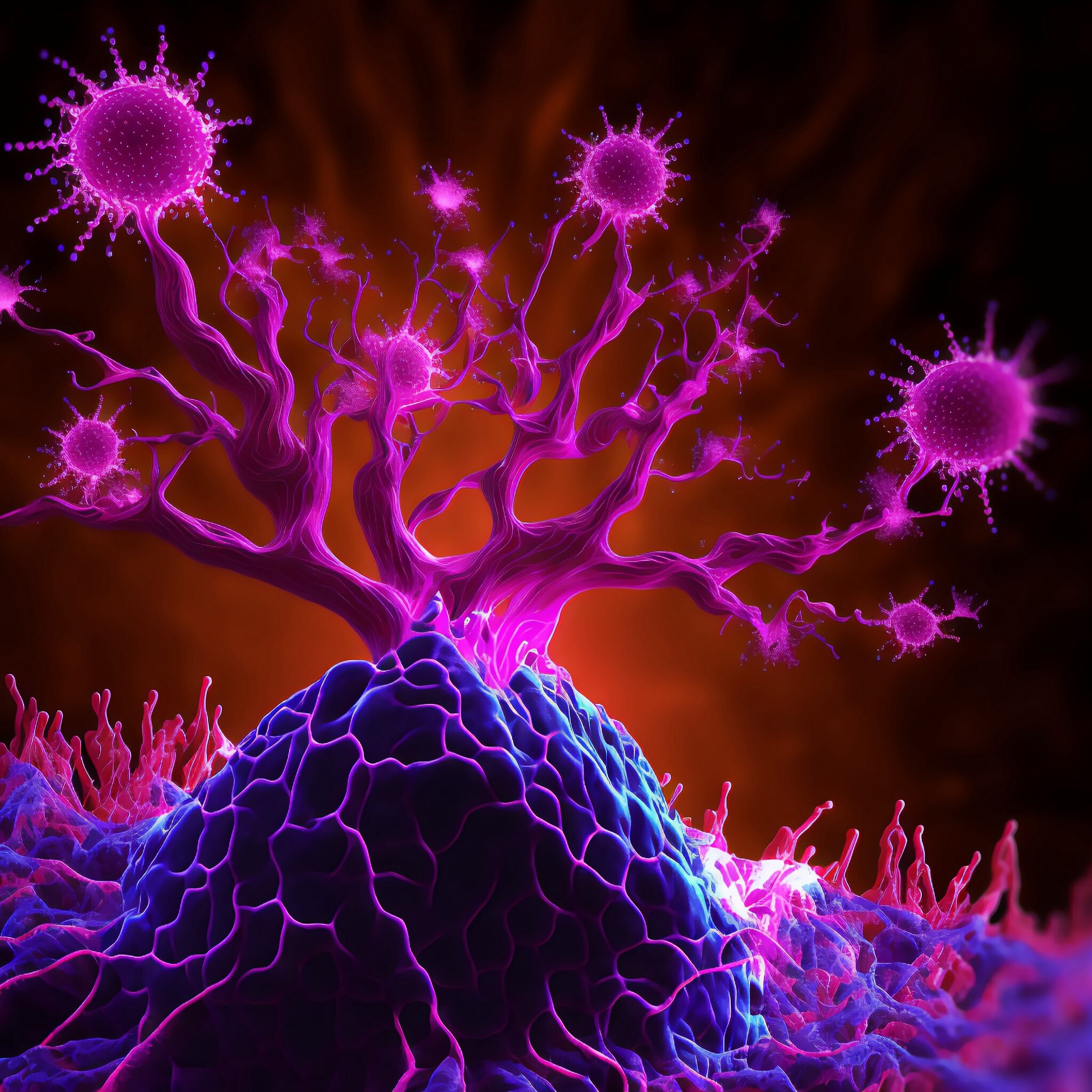
While modern cancer treatments can have tremendous therapeutic impact, formidable obstacles remain. Foremost among these is drug resistance, the ability of cancers to withstand and ultimately progress despite the presence of an anti-cancer drug. However, ongoing research provides hope that these challenges can be overcome, including recent work performed by Penn Engineers.
The lab of Lukasz J. Bugaj, Assistant Professor in the Department of Bioengineering, recently published an article that uncovers new mechanisms of how oncogenes interact with important pathways of cellular signaling that are associated with resistance. This work, titled “Oncogenic EML4-ALK Assemblies Suppress Growth Factor Perception and Modulate Drug Tolerance,” applied a new technique called ‘optogenetic functional profiling’ that allowed measurement of how important molecular signaling pathways respond to precise perturbations applied by the researchers. By applying this technique to many different cell types, the group found important differences in resistance-associated signaling between cancer cells and healthy cells
Specifically, the research showed that an oncogene called EML4-ALK, which activates oncogenic signaling, simultaneously inactivates adjacent pathways that can cause resistance. As a consequence, once an oncogene-blocking drug is applied, the inactivation is relieved, thus boosting activity through these adjacent, resistance-associated pathways. The study also showed that these pathways were not only de-repressed, but were actively stimulated by neighboring cancer cells, further enhancing cell survival in the presence of the drug.
“Our work shows that oncogenes, while driving cell division in cancer cells, simultaneously suppress the cells’ regulation by their environment,” said Dr. Bugaj. “While the work reveals mechanisms of paradoxical responses to drug treatment related to resistance, they may also inspire new ideas for therapies that can more efficiently kill cancer cells while maintaining suppression of resistance signaling. This work was co-led by PhD student David Gonzalez-Martinez and by Lee Roth, PhD, a postdoctoral fellow, and was supported by a grant from the American Cancer Society.
Reliance Industries Term Assistant Professor Claudia Loebel will establish her lab at The University of Pennsylvania’s Department of Bioengineering and the Center for Precision Engineering for Health in January 2025.
Dr. Loebel received her MD from Martin Luther University Halle Wittenberg, Germany and her Ph.D from ETH Zurich, Switzerland.
“My laboratory is developing testable models to investigate how extracellular signals regulate cellular function to direct the development and regeneration of organs, ultimately leading to more effective therapeutic treatments,” said Dr. Loebel in her research statement. “Building upon my K99/R00 and American Lung Association Innovation Awards, a major focus of my group has been on understanding the role of mechanical forces across various states of pulmonary development and regeneration.”
Dr. Loebel’s team is formed with an exciting combination of interdisciplinary scholars including postdoctoral associates, graduate and undergraduate students whose philosophy encourages respect for people’s differences, acknowledging and honoring religious and cultural practices, and foster diverse thinking. Dr. Loebel is also a recent recipient of the 2025 Rising Star Award from BMES CMBE, and also won the CMBE Young Innovators award for her published article, “Magnetoactive, Kirigami- Inspired Hammoks to Probe Lung Epithelial Cell Function.”
The Loebel Lab is funded by the David and Lucile Packard Foundation Fellowship, whose mission is dedicated to further the advancement of people and communities with their three overreaching and interdependent goals: building societies, protecting and restoring the natural world, and investing in families.
Cesar de la Fuente, Presidential Assistant Professor with appointments in the Perelman School of Medicine, School of Engineering and School of Arts & Sciences (Image: Eric Sucar)
In a significant advance against the growing threat of antibiotic-resistant bacteria, researchers have identified a novel class of antimicrobial agents known as encrypted peptides, which may expand the immune system’s arsenal of tools to fight infection. The findings, published in Trends in Biotechnology by Cell Press, reveal that many antimicrobial molecules originate from proteins not traditionally associated with immune responses.
Unlike conventional antibiotics that target specific bacterial processes, these newly discovered peptides disrupt the protective membranes surrounding bacterial cells. By inserting themselves into these membranes—much like breaching a fortress wall—the peptides destabilize and ultimately destroy the bacteria.
“Our findings suggest that these previously overlooked molecules could be key players in the immune system’s response to infection,” says César de la Fuente, presidential assistant professor in bioengineering and in chemical and biomolecular engineering in the School of Engineering and Applied Science, in psychiatry and microbiology in the Perelman School of Medicine, and in chemistry in the School of Arts & Sciences, who led the research team. “This may not only redefine how we understand immunity but also opens up new possibilities for treating drug-resistant infections.”
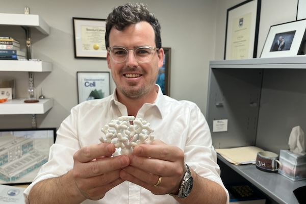
Alex Hughes, Assistant Professor in Bioengineering, holds a model of a developing kidney. (Credit: Bella Ciervo)
To Alex Hughes, Assistant Professor in Bioengineering within Penn Engineering and in Cell and Developmental Biology within Penn Medicine, the kidney is a work of art. “I find the development of the kidney to be a really beautiful process,” says Hughes.
Most people only ever see the organ in cross-section, through textbooks or by dissecting animal kidneys in high school biology class: a bean-shaped slice with lots of tiny tubes. “I think that really undersells how amazing the structure is,” says Hughes, who points out that kidneys grow in utero like forests of pipes, branching exponentially.
Densely packed with tubules clustered in units known as nephrons, kidneys cleanse the blood, maintaining the body’s fluid and electrolyte balance, while also regulating blood pressure. The organ played a crucial role in vertebrates emerging from the ocean: as one paper puts it, kidneys preserve the primordial ocean in all of us.
Unfortunately, kidneys struggle in the modern world. Excessively salty food, being overweight, not exercising enough, drinking too much and smoking can all raise blood pressure, which damages the kidney’s tiny blood vessels, as does diabetes.
In some cases, damage to the kidney’s nephrons can be slowed with lifestyle changes, but, unlike the liver, bones and skin, which can regrow damaged tissue, kidneys have a limited capacity to regenerate. At present, without a transplant, the nephrons we have at birth must last a lifetime.
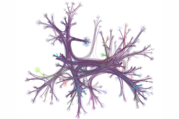
A hyperlink network from English Wikipedia, with only 0.1% of articles (nodes) and their connections (edges) visualized. Seven different reader journeys through this network are highlighted in various colors. The network is organized by topic and displayed using a layout that groups related articles together. (Image: Dale Zhou)
At one point or another, you may have gone online looking for a specific bit of information and found yourself “going down the Wiki rabbit hole” as you discover wholly new, ever-more fascinating related topics — some trivial, some relevant — and you may have gone so far down the hole it’s difficult to piece together what brought you there to begin with.
According to the University of Pennsylvania’s Dani Bassett, who recently worked with a collaborative team of researcher to examine the browsing habits of 482,760 Wikipedia readers from 50 different countries, this style of information acquisition is called the “busybody.” This is someone who goes from one idea or piece of information to another, and the two pieces may not relate to each other much.
“The busybody loves any and all kinds of newness, they’re happy to jump from here to there, with seemingly no rhyme or reason, and this is contrasted by the ‘hunter,’ which is a more goal-oriented, focused person who seeks to solve a problem, find a missing factor, or fill out a model of the world,” says Bassett.
In the research, published in the journal Science Advances, Bassett and colleagues discovered stark differences in browsing habits between countries with more education and gender equality versus less equality, raising key questions about the impact of culture on curiosity and learning.
Dani S. Bassett is the J. Peter Skirkanich Professor at the University of Pennsylvania with a primary appointment in the School of Engineering and Applied Science’s Department of Bioengineering and secondary appointments in the School of Arts & Sciences’ Department of Physics & Astronomy, Penn Engineering’s Department of Electrical and Systems Engineering, and the Perelman School of Medicine’s Departments of Neurology and Psychiatry.
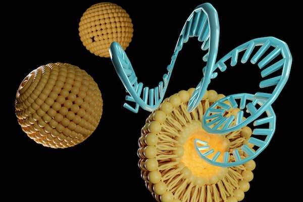
By adjusting the chemical structure of lipid nanoparticles (LNPs), Penn Engineers have discovered how to target specific organs, a major breakthrough in precision medicine. (Love Employee via Getty Images)
Penn Engineers have discovered a novel means of directing lipid nanoparticles (LNPs), the revolutionary molecules that delivered the COVID-19 vaccines, to target specific tissues, presaging a new era in personalized medicine and gene therapy.
While past research — including at Penn Engineering — has screened “libraries” of LNPs to find specific variants that target organs like the lungs, this approach is akin to trial and error. “We’ve never understood how the structure of one key component of the LNP, the ionizable lipid, determines the ultimate destination of LNPs to organs beyond the liver,” says Michael J. Mitchell, Associate Professor in Bioengineering.
In a new paper published in Nature Nanotechnology, Mitchell’s group describes how subtle adjustments to the chemical structure of the ionizable lipid, a key component of the LNP, allows for tissue-specific delivery, in particular to the liver, lungs and spleen.
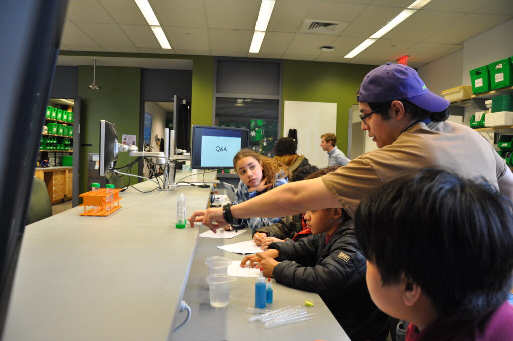
Students learn about bioengineering in the BE Labs at the inaugural BETA Day (credit: Felice Macera)
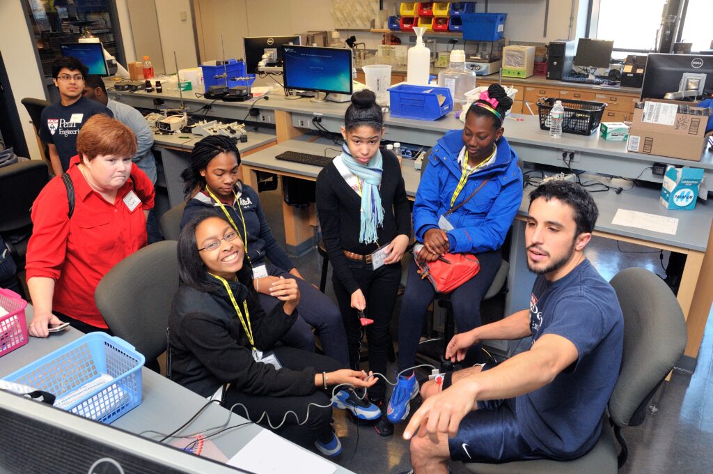
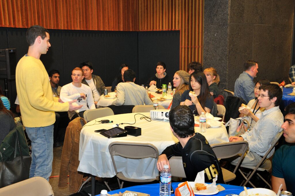
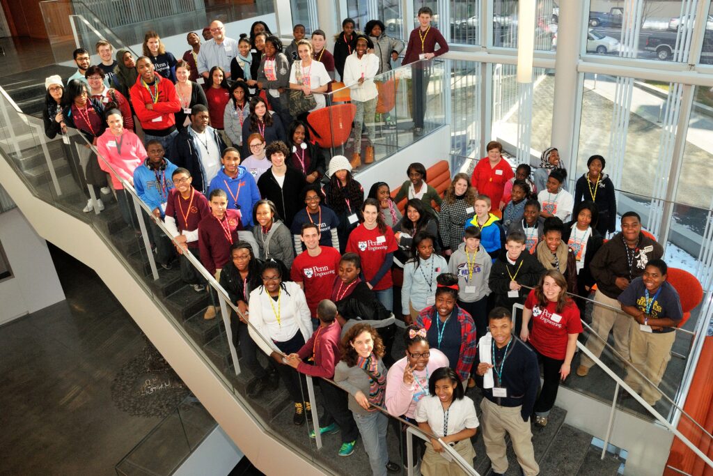
Last year marked not just the 50th anniversary of the Department of Bioengineering (BE) but the 10th anniversary of Bioengineer-Teach-Aspire (BETA) Day, one of the most beloved and impactful programs run by the Graduate Association of Bioengineers (GABE).
BETA Day, an annual event in which a diverse group of Philadelphia middle school students learns about bioengineering and a variety of science, technology, engineering and math (STEM) fields from BE graduate students, has grown into an institution, one whose impact no one could have foreseen.
GABE’s original goal was to provide social opportunities for BE graduate students. While this is still an important function of the group, in the mid-2010s, students and board members found themselves looking for opportunities to provide more formalized outreach and mentorship. They wanted to have an impact on Philadelphia and cultivate the next generation of bioengineers.
The Seeds of BETA Day
Benjamin Freedman, a principal investigator at Beth Israel Deaconess Medical Center, Assistant Professor of Orthopedic Surgery at Harvard Medical School, and founder of biotech startup Limax Biosciences, earned his doctorate in Bioengineering in the lab of Louis Soslowsky, Fairhill Professor in the Department of Orthopaedic Surgery within the Perelman School of Medicine (PSOM) and in Bioengineering within the School of Engineering and Applied Science (Penn Engineering). Freedman played a key role in BETA Day’s founding.
In 2009, Freedman, then an undergraduate at the University of Rochester, attended a talk at the City College of New York (CCNY), which sparked his interest in mentorship. Sheldon Weinbaum, a Distinguished Professor in Biomedical and Mechanical Engineering at CCNY and the Biomedical Engineering Society (BMES) inaugural diversity award winner, spoke about “fulfilling the dream” of mentorship and the struggle for inclusion in STEM fields, echoing the language of Martin Luther King Jr.
Inspired by this encounter, Freedman got involved with a mentorship program during his senior year. He later signed up for a lunch with Weinbaum to talk about mentorship. Freedman recalls that Weinbaum’s face “lit up” when he realized that this student didn’t just want to talk science but was genuinely interested in inclusion, diversity and mentorship.
Arriving at Penn Engineering and PSOM for graduate school in 2011, Freedman joined GABE, bringing this passion and experience with him and helping GABE to shape and clarify their outreach and mentorship programs.
From Campus to Community
Along with other GABE board members, such as Cori Riggin and Shauna Dorsey, Freedman worked over the course of a year and a half to identify the mentorship needs within BE and gauge student interest. David Meaney, Solomon R. Pollack Professor and then Chair of BE, and former BE faculty Susan Margulies, now Professor in the Wallace H. Coulter Department of Biomedical Engineering at Georgia Tech and Emory University, were particularly involved in these discussions.
Benjamin Freedman (left) addresses the first BE mentoring cohort (credit: Felice Macera)
The GABE board reorganized to include mentorship and outreach chairs, and eventually started a formal mentorship program in partnership with the Penn undergraduate Biomedical Engineering Society (BMES). The mentorship program continues to this day, creating opportunities for BE graduate students to engage with undergraduate concerns through one-on-one meetings to discuss career or graduate school advice, summer BBQ’s, roundtable discussions and monthly meetups.
With an internal mentorship program established, the team turned their focus to Philadelphia. Initially, GABE established a partnership with iPraxis, a local STEM education non-profit, to do some outreach activities in middle schools. This partnership resulted in an Outstanding Outreach Award from the national Biomedical Engineering Society in 2014. But with the department’s 40th anniversary approaching, GABE’s members wanted to do something spectacular to celebrate and give back to the community.
Service Learning in Action
By then, Ocek Eke, Director of Graduate Students Programming at Penn Engineering, had been recently appointed Director of Global and Local Service Learning Programs. Eke provided Freedman and GABE advice on setting up effective outreach programs and to determine what resources the School could contribute. “We have a role to play to fulfill our mission,” Eke says, citing Penn’s motto, “Leges Sine Moribus Vanae,” which translates to “Laws without morals are useless.”
GABE’s efforts were part of a “wave” of interest in outreach and community service in both the department and the School, Eke remembers, including the undergraduate group Access Engineering and several service learning courses which took students to Asia, Africa and Central America. He was impressed by the lack of cynicism in the BE student body. “These are students who saw a need, who are passionate about what they want to achieve. They could have just been comfortable but were willing to go and stick their necks out. They used the resources we have here in Penn Engineering to address these needs.”
A (BETA) Day to Remember
The first BETA Day took place at the Singh Center for Nanotechnology, which had only just opened. Held with the enthusiastic participation of around 70 middle schoolers, and almost as many volunteers, the event included a full day of programming, with representation from every Penn Engineering department. There were science talks, workshops, and even a drone demo with Vijay Kumar, Nemirovsky Family Dean of Penn Engineering. The entire day was student-driven and staffed by volunteers, demonstrating the students’ commitment to making a difference.
The first annual BETA Day was held in the Singh Center for Nanotechnology (credit: Felice Macera)
GABE never imagined BETA Day as an annual event, but the first instance was so successful, it became hard to imagine not repeating it. Ten years later, the GABE board continues to introduce bioengineering to a diverse and ambitious group of middle schoolers every spring.
In 2021, during the COVID-19 lockdown, the industrious and creative GABE board even tailored BETA Day activities to be held in an entirely virtual environment. “These types of events are not as successful when they’re only initiated by faculty,” says Freedman. Generating and sustaining student involvement has been a cornerstone of BETA Day’s continued success.
The Legacy of BETA Day
GABE’s mentorship efforts have grown as well, changing to meet evolving student needs. The mentorship program now involves students being placed in “families” of around four undergraduates and two graduate students, spanning a range of class years and experience levels. A third student association, the Master’s Association in Bioengineers (MAB), was established to better foster community and facilitate opportunities for master’s students.
The department also launched an applicant support program in 2020, enhancing BE’s mission of increasing diversity, equity and inclusion by pairing Ph.D. applicants to current doctoral students, who serve as mentors to help navigate the admissions process, giving feedback on application materials and providing other support to prospective students.
Structures of support and outreach activities like BETA Day have become a key emphasis of the department’s graduate student recruitment, helping to attract students who value the department’s core mission and increasing opportunities for underserved or underrepresented communities.
The legacy of that original BETA Day also continues in Freedman’s Lab. After graduating in 2017, having served on the GABE board and as President from 2015-2016, Freedman continued to mentor over 20 students during his postdoctoral research at Harvard. He is now building his own independent lab where diversity, mentorship and outreach are foundational pillars.
A Nebula of Inspiration
Perhaps the most consequential impact of BETA Day is the impression it makes on the middle schoolers who participate each year. “To really get to know what happens on BETA Day and what it’s true impact is, you need to experience it,” says Ravi Radhakrishnan, Herman P. Schwan Chair of the Department of Bioengineering and Professor in Bioengineering and in Chemical and Biomolecular Engineering.
The legacy of BETA Day continues into its second decade. (credit: Afraah Shamim, BE Labs)
“I walked into the Stephenson Foundation Education Lab during BETA Day 2024,” recalls Radhakrishnan, “and what I saw was teams of teenagers tinkering with pipes that were clogged, strategizing on unclogging them without damaging them: an assignment that got them thinking in teams about how to prevent heart attacks.
“Expose these young minds to design thinking, versatile tools, and critical problems in biomedical engineering, and the elegant solutions they brainstorm are truly mind blowing. BETA Day is like the nebula where future biomedical stars are born.”
A multiracial Black and Asian self-described secular humanist, who was raised as one of Jehovah’s Witnesses and is now in an interracial, interfaith marriage, walked into a Passover seder.
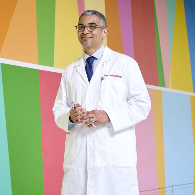
It’s not the setup for a groaner of a joke, or an epic fail of an evening. Rather, as Roy H. Hamilton, MD, tells it, this was his experience this spring as his Jewish in-laws—the family he has loved as his own for over two decades—came together to commemorate the universal human themes of freedom and deliverance from oppression reflected in the Passover narrative. Though he does this every year, this year he had some trepidation. In a time marked by tragic conflict and with tensions both abroad and at home, it seemed like having a frank discussion of these themes might invite acrimony. But what emerged instead was a profound opportunity to listen, to appreciate each other’s perspective, and to “exercise empathy for trauma that’s happening to everyone.”
It was a bit of a revelation for Hamilton, Penn Medicine’s new vice dean for Inclusion, Diversity, and Equity. “In the moment that you would have thought would be the worst to open up certain topics, we all ended up having a great dialogue across differences,” he said. Why? “Because we all felt connected enough to give each other respect, compassion, and grace, even when our thoughts and opinions differed. It made me think about how we can further cultivate a culture of empathy at Penn too.”
Today, as Hamilton begins his third decade on the faculty at Penn’s Perelman School of Medicine, he is devoted to making academia a safe, supportive space for students and colleagues alike. He serves as a professor of Neurology, with secondary appointments in Psychiatry and Physical Medicine and Rehabilitation. Hamilton is also director of both the Laboratory for Cognition and Neural Stimulation; and the Penn Brain Science, Translation and Modulation (BrainSTIM) Center. Previously, he was the Perelman School of Medicine’s assistant dean for Cultural Affairs and Diversity for almost a decade, and launched similar efforts in his field, serving as Penn Neurology’s vice chair for Diversity and Inclusion from 2017 until his recent elevation to the role for Penn Medicine as a whole.
Given his own diverse background and personal life, Hamilton wants everyone—trainees, faculty, patients—to feel valued and included. “I touch enough spaces in my personal life that when groups are being clearly systematically disadvantaged, it often feels like it’s touching on some piece of my own identity,” he said, in discussing his background and hope for his new leadership position. “I bring a lot of myself to this role.”
In a recent discussion, Hamilton shared perspectives on why supporting inclusion, diversity, and equity matters—particularly for an institution training future doctors—and what Penn Medicine is doing in this sphere.
In a collaborative interdisciplinary study, Michael Mitchell of the School of Engineering and Applied Science, Wei Guo of the School of Arts & Sciences, and Drew Weissman of the Perelman School of Medicine show that solid tumors can block drug-delivery mechanisms with a “forcefield-like” effect but certain genetic elements that can effectively “shut down” the forcefield. Their findings hint at new targets for delivering cancer treatments that use the body’s immune system to fight tumors. (Image: iStock / CIPhotos)
The tumor microenvironment—an ad hoc, messy amalgamation of signaling molecules, immune cells, fibroblasts, blood vessels, and the extracellular matrix—acts like a “powerful security system that protects solid tumors from invaders seeking to destroy them,” says Michael Mitchell, a bioengineer at the University of Pennsylvania working on nanoscale therapeutics aimed at targeting cancers.
“A lot like the Death Star with its surrounding fleet of fighter ships and protective shields, solid tumors can use features like immune cells and vasculature to exert force, acting as a physical barrier to rebel forces (nanoparticles) coming in to deliver the payload that destroys it,” Mitchell says.
Now, researchers in the Mitchell lab have teamed up with Wei Guo’s group in the School of Arts & Sciences at Penn and Drew Weissman of the Perelman School of Medicine to figure out the molecular mechanisms that make tumor microenvironments seemingly impenetrable and found that small extracellular vesicles (sEVs) are secreted by tumor cells and act as a “forcefield,” blocking therapeutics. Their findings are published in Nature Materials.
“This discovery reveals how tumors create a robust defense system, making it challenging for nanoparticle-based therapies to reach and effectively target cancer cells,” Guo says. “By understanding the cellular mechanisms driving these responses, we can potentially develop strategies to disable this defense, allowing therapeutics to penetrate and attack the tumor more efficiently.”
The research builds on a prior collaboration between Guo and Mitchell’s labs, wherein the teams focused on how tumor-associated immune cells, known as macrophages, contribute to the suppression of anti-tumor immunity by secreting extracellular vesicles.
Wei Guo is the Hirsch Family President’s Distinguished Professor in the Department of Biology in Penn’s School of Arts & Sciences.
Ningqiang Gong, a former postdoctoral researcher in the Mitchell lab at Penn Engineering, is an assistant professor at the University of Science and Technology of China.
Wenqun Zhong is a reseearch associate in the Guo Laboratory in Penn Arts & Sciences.
Other authors include: Alex G Hamilton, Dongyoon Kim, Junchao Xu, and Lulu Xue of Penn Engineering; Junhyong Kim, Zhiyuan Qin, and Fengyuan Xu of Penn Arts & Sciences; Mohamad-Gabriel Alameh and Drew Weissman of the Perelman School of Medicine; Andrew E. Vaughn and Gan Zhao of the Penn School of Veterinary Medicine; Jinghong Li and Xucong Teng of the University of Beijing; and Xing-Jie Liang of the Chinese Academy of Sciences.
This research received support from the U.S. National Institutes of Health (DP2 TR002776, R35 GM141832, and NCI P50 CA261608), Burroughs Wellcome Fund, U.S. National Science Foundation CAREER Award (CBET-2145491), and an American Cancer Society Research Scholar Grant (RGS-22-1122-01-ET.)