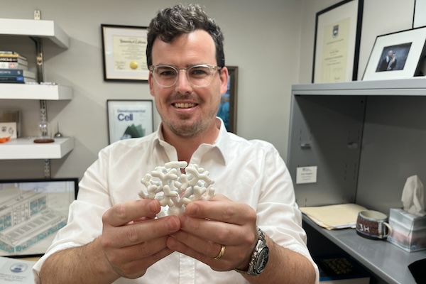
Alex Hughes, Assistant Professor in Bioengineering, holds a model of a developing kidney. (Credit: Bella Ciervo)
To Alex Hughes, Assistant Professor in Bioengineering within Penn Engineering and in Cell and Developmental Biology within Penn Medicine, the kidney is a work of art. “I find the development of the kidney to be a really beautiful process,” says Hughes.
Most people only ever see the organ in cross-section, through textbooks or by dissecting animal kidneys in high school biology class: a bean-shaped slice with lots of tiny tubes. “I think that really undersells how amazing the structure is,” says Hughes, who points out that kidneys grow in utero like forests of pipes, branching exponentially.
Densely packed with tubules clustered in units known as nephrons, kidneys cleanse the blood, maintaining the body’s fluid and electrolyte balance, while also regulating blood pressure. The organ played a crucial role in vertebrates emerging from the ocean: as one paper puts it, kidneys preserve the primordial ocean in all of us.
Unfortunately, kidneys struggle in the modern world. Excessively salty food, being overweight, not exercising enough, drinking too much and smoking can all raise blood pressure, which damages the kidney’s tiny blood vessels, as does diabetes.
In some cases, damage to the kidney’s nephrons can be slowed with lifestyle changes, but, unlike the liver, bones and skin, which can regrow damaged tissue, kidneys have a limited capacity to regenerate. At present, without a transplant, the nephrons we have at birth must last a lifetime.
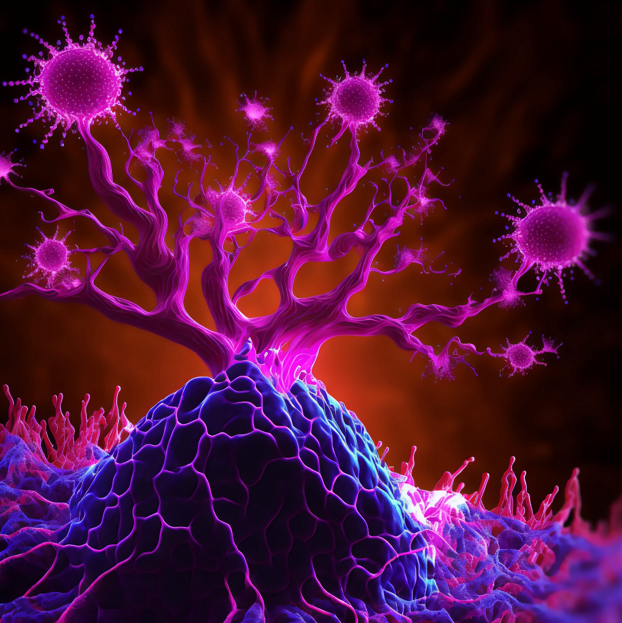
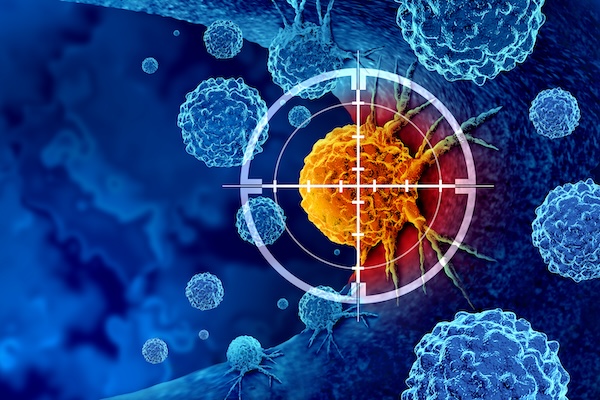
In a collaborative interdisciplinary study, Michael Mitchell of the School of Engineering and Applied Science, Wei Guo of the School of Arts & Sciences, and Drew Weissman of the Perelman School of Medicine show that solid tumors can block drug-delivery mechanisms with a “forcefield-like” effect but certain genetic elements that can effectively “shut down” the forcefield. Their findings hint at new targets for delivering cancer treatments that use the body’s immune system to fight tumors. (Image: iStock / CIPhotos)
The tumor microenvironment—an ad hoc, messy amalgamation of signaling molecules, immune cells, fibroblasts, blood vessels, and the extracellular matrix—acts like a “powerful security system that protects solid tumors from invaders seeking to destroy them,” says Michael Mitchell, a bioengineer at the University of Pennsylvania working on nanoscale therapeutics aimed at targeting cancers.
“A lot like the Death Star with its surrounding fleet of fighter ships and protective shields, solid tumors can use features like immune cells and vasculature to exert force, acting as a physical barrier to rebel forces (nanoparticles) coming in to deliver the payload that destroys it,” Mitchell says.
Now, researchers in the Mitchell lab have teamed up with Wei Guo’s group in the School of Arts & Sciences at Penn and Drew Weissman of the Perelman School of Medicine to figure out the molecular mechanisms that make tumor microenvironments seemingly impenetrable and found that small extracellular vesicles (sEVs) are secreted by tumor cells and act as a “forcefield,” blocking therapeutics. Their findings are published in Nature Materials.
“This discovery reveals how tumors create a robust defense system, making it challenging for nanoparticle-based therapies to reach and effectively target cancer cells,” Guo says. “By understanding the cellular mechanisms driving these responses, we can potentially develop strategies to disable this defense, allowing therapeutics to penetrate and attack the tumor more efficiently.”
The research builds on a prior collaboration between Guo and Mitchell’s labs, wherein the teams focused on how tumor-associated immune cells, known as macrophages, contribute to the suppression of anti-tumor immunity by secreting extracellular vesicles.
Wei Guo is the Hirsch Family President’s Distinguished Professor in the Department of Biology in Penn’s School of Arts & Sciences.
Ningqiang Gong, a former postdoctoral researcher in the Mitchell lab at Penn Engineering, is an assistant professor at the University of Science and Technology of China.
Wenqun Zhong is a reseearch associate in the Guo Laboratory in Penn Arts & Sciences.
Other authors include: Alex G Hamilton, Dongyoon Kim, Junchao Xu, and Lulu Xue of Penn Engineering; Junhyong Kim, Zhiyuan Qin, and Fengyuan Xu of Penn Arts & Sciences; Mohamad-Gabriel Alameh and Drew Weissman of the Perelman School of Medicine; Andrew E. Vaughn and Gan Zhao of the Penn School of Veterinary Medicine; Jinghong Li and Xucong Teng of the University of Beijing; and Xing-Jie Liang of the Chinese Academy of Sciences.
This research received support from the U.S. National Institutes of Health (DP2 TR002776, R35 GM141832, and NCI P50 CA261608), Burroughs Wellcome Fund, U.S. National Science Foundation CAREER Award (CBET-2145491), and an American Cancer Society Research Scholar Grant (RGS-22-1122-01-ET.)
How do we measure chaos and why would we want to? Together, Penn engineers Dani S. Bassett, J. Peter Skirkanich Professor in Bioengineering and in Electrical and Systems Engineering, and postdoctoral researcher Kieran Murphy leverage the power of machine learning to better understand chaotic systems, opening doors for new information analyses in both theoretical modeling and real-world scenarios.
Humans have been trying to understand and predict chaotic systems such as weather patterns, the movement of planets and population ecology for thousands of years. While our models have continued to improve over time, there will always remain a barrier to perfect prediction. That’s because these systems are inherently chaotic. Not in the sense that blue skies and sunshine can turn into thunderstorms and torrential downpours in a second, although that does happen, but in the sense that mathematically, weather patterns and other chaotic systems are governed by physics with nonlinear characteristics.
“This nonlinearity is fundamental to chaotic systems,” says Murphy. “Unlike linear systems, where the information you start with to predict what will happen at timepoints in the future stays consistent over time, information in nonlinear systems can be both lost and generated through time.”
Like a game of telephone where information from the original source gets lost as it travels from person to person while new words and phrases are added to fill in the blanks, outcomes in chaotic systems become harder to predict as time passes. This information decay thwarts our best efforts to accurately forecast the weather more than a few days out.
“You could put millions of probes in the atmosphere to measure wind speed, temperature and precipitation, but you cannot measure every single atom in the system,” says Murphy. “You must have some amount of uncertainty, which will then grow, and grow quickly. So while a prediction for the weather in a few hours might be fairly accurate, that growth in uncertainty over time makes it impossible to predict the weather a month from now.”
In their recent paper published in Physical Review Letters, Murphy and Bassett applied machine learning to classic models of chaos, physicists’ reproductions of chaotic systems that do not contain any external noise or modeling imperfections, to design a near-perfect measurement of chaotic systems to one day improve our understanding of systems including weather patterns.
“These controlled systems are testbeds for our experiments,” says Murphy. “They allow us to compare with theoretical predictions and carefully evaluate our method before moving to real-world systems where things are messy and much less is known. Eventually, our goal is to make ‘information maps’ of real-world systems, indicating where information is created and identifying what pieces of information in a sea of seemingly random data are important.”
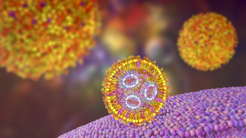
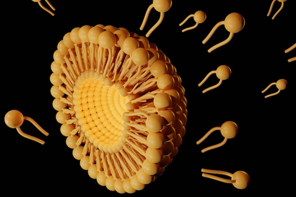
Lipid nanoparticles present one of the most advanced drug delivery platforms to shuttle promising therapeutics such as mRNA but are limited by the time it takes to synthesize cationic lipids, a key component. Now, Michael Mitchell and his team at the School of Engineering and Applied Science have developed a faster way to make cationic lipids that are also more versatile, able to carry different kinds of treatments to target specific organs. (Image: iStock / Dr_Microbe)
Imagine a scenario where a skilled hacker must upload critical software to update a central server and thwart a potentially lethal virus from wreaking havoc across a vast computer network. The programmer, armed with the lifesaving code, must navigate through treacherous territory teeming with adversaries, and success hinges on promptly getting a safe, stealthy delivery vehicle that can place the hacker exactly where they need to be.
In the context of modern medicine, messenger RNA (mRNA) serves as the hacker, carrying genetic instructions to produce specific proteins within cells that can induce desired immune responses or sequester maladaptive cellular elements. Lipid nanoparticles (LNPs) are the stealthy delivery vehicles that transport these fragile mRNA molecules through the bloodstream to their target cells, overcoming the body’s defenses to deliver their payload safely and efficiently.
However, much like building an advanced stealth vehicle, the synthesis of cationic lipids—a type of lipid molecule that’s positively charged and a key component of LNPs—is often a time-consuming process, involving multiple steps of chemical synthesis and purification.
Now, Michael Mitchell and a team at the University of Pennsylvania have addressed this challenge with a novel approach that leverages a compound library fabrication technique known as “click-like chemistry” to create LNPs in a single, simple step. Their findings, published in the journal Nature Chemistry, show that this method not only speeds up the synthesis process but also presents a way to equip these delivery vehicles with a “GPS” to better target specific organs such as the liver, lungs, and spleen, potentially opening new avenues for treating a range of diseases that arise in these organs.
“We’ve developed what we call an amidine-incorporated degradable (AID) lipid, a uniquely structured biodegradable molecule,” Mitchell says. “Think of it as an easy-to-build custom mRNA vehicle with a body kit that informs its navigation system. By adjusting its shape and degradability, we can enhance mRNA delivery into cells in a safe manner. By adjusting the amount of the AID lipid that we incorporate into the LNP, we can also guide it to different organs in the body, much like programming different destinations into a GPS.”
First author Xuexiang Han, a former postdoctoral researcher in the Mitchell Lab, explains that their new approach allows the rapid creation of diverse lipid structures in just an hour, compared to the weekslong process traditionally required.
Cesar de la Fuente (left), Fangping Wan (center), and Marcelo der Torossian Torres (right). Fangping holds a 3D model of a unique ATP synthase fragment, identified by their lab’s deep learning model, APEX, as having potent antibiotic properties.
“Make sure you finish your antibiotics course, even if you start feeling better’ is a medical mantra many hear but ignore,” says Cesar de la Fuente of the University of Pennsylvania.
He explains that this phrase is, however, crucial as noncompliance could hamper the efficacy of a key 20th century discovery, antibiotics. “And in recent decades, this has led to the rise of drug-resistant bacteria, a growing global health crisis causing approximately 4.95 million deaths per year and threatens to make even common infections deadly,” he says.
De la Fuente, a Presidential Assistant Professor, and a team of interdisciplinary researchers have been working on biomedical innovations tackling this looming threat. In a new study, published in Nature Biomedical Engineering, they developed an artificial intelligence tool to mine the vast and largely unexplored biological data—more than 10 million molecules of both modern and extinct organisms— to discover new candidates for antibiotics.
“With traditional methods, it takes around six years to develop new preclinical drug candidates to treat infections and the process is incredibly painstaking and expensive,” de la Fuente says. “Our deep learning approach can dramatically reduce that time, driving down costs as we identified thousands of candidates in just a few hours, and many of them have preclinical potential, as tested in our animal models, signaling a new era in antibiotic discovery.” César de la Fuente holds a 3D model of a unique ATP synthase fragment, identified by his lab’s deep learning model, APEX, as having potent antibiotic properties. This molecular structure, resurrected from ancient genetic data, represents a promising lead in the fight against antibiotic-resistant bacteria.
These latest findings build on methods de la Fuente has been working on since his arrival at Penn in 2019. The team asked a fundamental question: Can machines be used to accelerate antibiotic discovery by mining the world’s biological information? He explains that this idea is based on the notion that biology, at its most basic level, is an information source, which could theoretically be explored with AI to find new useful molecules.
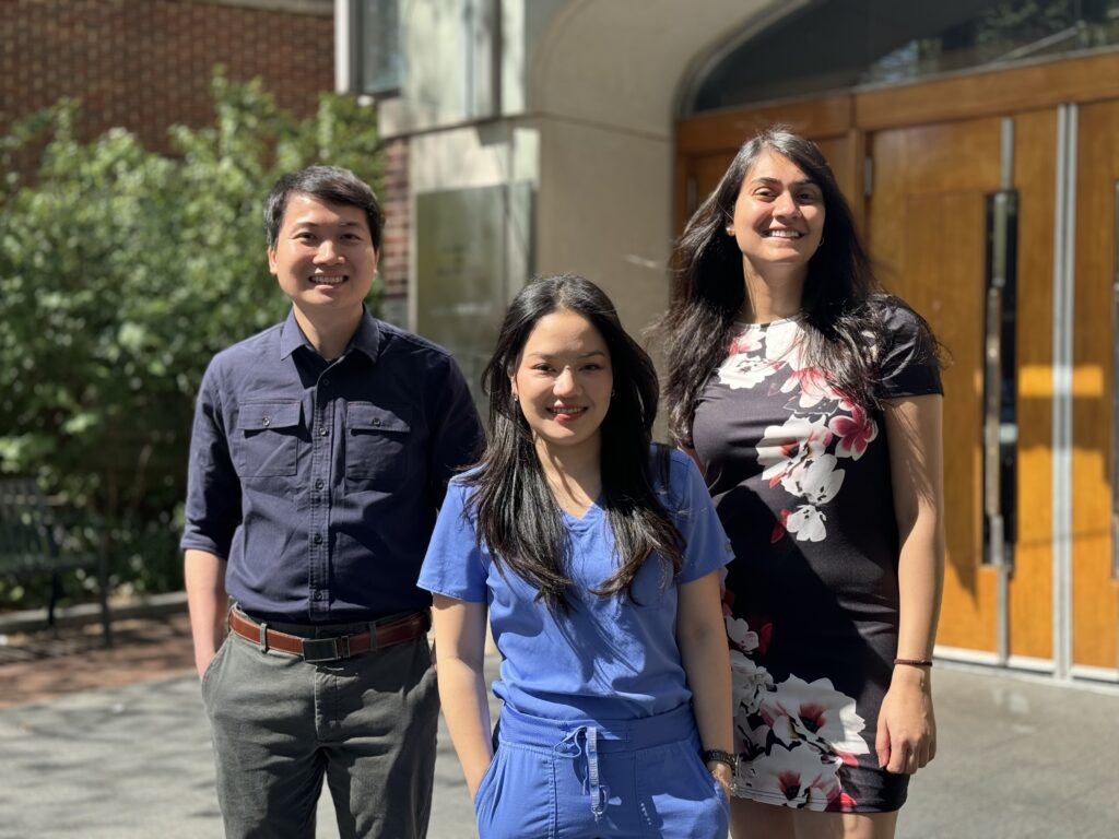
Left to right: Hong-Huy Tran, Chrissie Jaruchotiratanasakul, Manali Mahajan (Photo Courtesy of CiPD)
The Center for Innovation and Precision Dentistry (CiPD), a collaboration between Penn Engineering and Penn Dental Medicine, has partnered with Wharton’s Mack Institute for Innovation Management on a research project which brings robotics to healthcare. More specifically, this project will explore potential uses of nanorobot technology for oral health care. The interdisciplinary partnership brings together three students from different Penn programs to study the commercialization of a new technology that detects and removes harmful dental plaque.
“Our main goal is to bring together dental medicine and engineering for out-of-the-box solutions to address unresolved problems we face in oral health care,” says Hyun (Michel) Koo, Co-Founding Director of CiPD and Professor of Orthodontics. “We are focused on affordable solutions and truly disruptive technologies, which at the same time are feasible and translatable.”
Patients being treated for B-cell non-Hodgkin’s Lymphoma (NHL) who are part of minority populations may not have equal access to cutting-edge CAR T cell therapies, according to a new analysis led by researchers from the Perelman School of Medicine and published in NEJM Evidence.
CAR T cell therapy is a personalized form of cancer therapy that was pioneered at Penn Medicine and has brought hope to thousands of patients who had otherwise run out of treatment options. Six different CAR T cell therapies have been approved since 2017 for a variety of blood cancers, including B-cell NHL that has relapsed or stopped responding to treatment. Image: iStock/PeopleImages
“CAR T cell therapy represents a major leap forward for blood cancer treatment, with many patients living longer than ever before, but its true promise can only be realized if every patient in need has access to these therapies,” says lead author Guido Ghilardi, a postdoctoral fellow in the laboratory of senior author Marco Ruella, an assistant professor of hematology-oncology and scientific director of the Lymphoma Program. “From the scientific perspective, we’re constantly working in the laboratory to make CAR T cell therapy work better, but we also want to make sure that when a groundbreaking treatment like this becomes available, it reaches all patients who might be able to benefit.”
Penn Engineers have developed a way to target lung diseases, including lung cancer, with lipid nanoparticles (LNPs). (wildpixel via Getty Images)
Penn Engineers have developed a new means of targeting the lungs with lipid nanoparticles (LNPs), the miniscule capsules used by the Moderna and Pfizer-BioNTech COVID-19 vaccines to deliver mRNA, opening the door to novel treatments for pulmonary diseases like cystic fibrosis.
In a paper in Nature Communications, Michael J. Mitchell, Associate Professor in the Department of Bioengineering, demonstrates a new method for efficiently determining which LNPs are likely to bind to the lungs, rather than the liver. “The way the liver is designed,” says Mitchell, “LNPs tend to filter into hepatic cells, and struggle to arrive anywhere else. Being able to target the lungs is potentially life-changing for someone with lung cancer or cystic fibrosis.”
Previous studies have shown that cationic lipids — lipids that are positively charged — are more likely to successfully deliver their contents to lung tissue. “However, the commercial cationic lipids are usually highly positively charged and toxic,” says Lulu Xue, a postdoctoral fellow in the Mitchell Lab and the paper’s first author. Since cell membranes are negatively charged, lipids with too strong a positive charge can literally rip apart target cells.
Typically, it would require hundreds of mice to individually test the members of a “library” of LNPs — chemical variants with different structures and properties — to find one with a low charge that has a higher likelihood of delivering a medicinal payload to the lungs.
Instead, Xue, Mitchell and their collaborators used what is known as “barcoded DNA” (b-DNA) to tag each LNP with a unique strand of genetic material, so that they could inject a pool of LNPs into just a handful of animal models. Then, once the LNPs had propagated to different organs, the b-DNA could be scanned, like an item at the supermarket, to determine which LNPs wound up in the lungs.
Like space shuttles using booster rockets to breach the atmosphere, lipid nanoparticles (LNPs) equipped with the new molecule more successfully deliver medicinal payloads. (Love Employee via Getty Images)
Inspired by the design of space shuttles, Penn Engineering researchers have invented a new way to synthesize a key component of lipid nanoparticles (LNPs), the revolutionary delivery vehicle for mRNA treatments including the Pfizer-BioNTech and Moderna COVID-19 vaccines, simplifying the manufacture of LNPs while boosting their efficacy at delivering mRNA to cells for medicinal purposes.
In a paper in Nature Communications, Michael J. Mitchell, Associate Professor in the Department of Bioengineering, describes a new way to synthesize ionizable lipidoids, key chemical components of LNPs that help protect and deliver medicinal payloads. For this paper, Mitchell and his co-authors tested delivery of an mRNA drug for treating obesity and gene-editing tools for treating genetic disease.
Previous experiments have shown that lipidoids with branched tails perform better at delivering mRNA to cells, but the methods for creating these molecules are time- and cost-intensive. “We offer a novel construction strategy for rapid and cost-efficient synthesis of these lipidoids,” says Xuexiang Han, a postdoctoral student in the Mitchell Lab and the paper’s co-first author.
In the human body, the lungs and their vasculature can be likened to a building with an intricate plumbing system. The lungs’ blood vessels are the pipes essential for transporting blood and nutrients for oxygen delivery and carbon dioxide removal. Much like how pipes can get rusty or clogged, disrupting normal water flow, damage from respiratory viruses, like SARS-CoV-2 or influenza, can interfere with this “plumbing system.”
In a recent study, researchers looked at the critical role of vascular endothelial cells in lung repair. Their work, published in Science Translational Medicine, was led by Andrew Vaughan of the University of Pennsylvania’s School of Veterinary Medicine and shows that, by using techniques that deliver vascular endothelial growth factor alpha (VEGFA) via lipid nanoparticles (LNPs), that they were able to greatly enhance modes of repair for these damaged blood vessels, much like how plumbers patch sections of broken pipes and add new ones.
“While our lab and others have previously shown that endothelial cells are among the unsung heroes in repairing the lungs after viral infections like the flu, this tells us more about the story and sheds light on the molecular mechanisms at play,” says Vaughan, assistant professor of biomedical sciences at Penn Vet. “Here we’ve identified and isolated pathways involved in repairing this tissue, delivered mRNA to endothelial cells, and consequently observed enhanced recovery of the damaged tissue. These findings hint at a more efficient way to promote lung recovery after diseases like COVID-19.”
They found VEGFA’s involvement in this recovery, while building on work in which they used single cell RNA sequencing to identify transforming growth factor beta receptor 2 (TGFBR2) as a major signaling pathway. The researchers saw that when TGFBR2 was missing it stopped the activation of VEGFA. This lack of signal made the blood vessel cells less able to multiply and renew themselves, which is vital for the exchange of oxygen and carbon dioxide in the tiny air sacs of the lungs.
“We’d known there was a link between these two pathways, but this motivated us to see if delivering VEGFA mRNA into endothelial cells could improve lung recovery after disease-related injury,” says first author Gan Zhao, a postdoctoral researcher in the Vaughan Lab.
“LNPs have been great for vaccine delivery and have proven incredibly effective delivery vehicles for genetic information. But the challenge here was to get the LNPs into the bloodstream without them heading to the liver, which is where they tend to congregate as its porous structure lends favor to substances passing from the blood into hepatic cells for filtration,” says Mitchell, an associate professor of bioengineering at Penn Engineering and a coauthor of the paper. “So, we had to devise a way to specifically target the endothelial cells in the lungs.”
Lulu Xue, a postdoctoral researcher in the Mitchell Lab and a co-first author of the paper, explains that they engineered the LNP to have an affinity for lung endothelial cells, this is known as extra hepatic delivery, going beyond the liver.