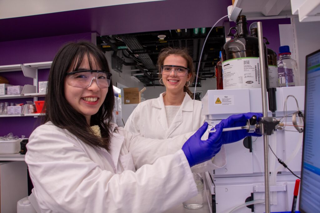
First author Qiuyue Nie and coauthor Maria Zotova, from left, purify samples of the fungus. (Credit: Bella Ciervo)
Penn-led researchers have turned a deadly fungus into a potent cancer-fighting compound. After isolating a new class of molecules from Aspergillus flavus, a toxic crop fungus linked to deaths in the excavations of ancient tombs, the researchers modified the chemicals and tested them against leukemia cells. The result? A promising cancer-killing compound that rivals FDA-approved drugs and opens up new frontiers in the discovery of more fungal medicines.
“Fungi gave us penicillin,” says Sherry Gao, Presidential Penn Compact Associate Professor in Chemical and Biomolecular Engineering (CBE) and in Bioengineering (BE) and senior author of a new paper in Nature Chemical Biology on the findings. “These results show that many more medicines derived from natural products remain to be found.”
From Curse to Cure
A. flavus, named for its yellow spores, has long been a microbial villain. After archaeologists opened King Tutankhamun’s tomb in the 1920s, a series of untimely deaths among the excavation team fueled rumors of a pharaoh’s curse. Decades later, doctors theorized that fungal spores, dormant for millennia, could have played a role.
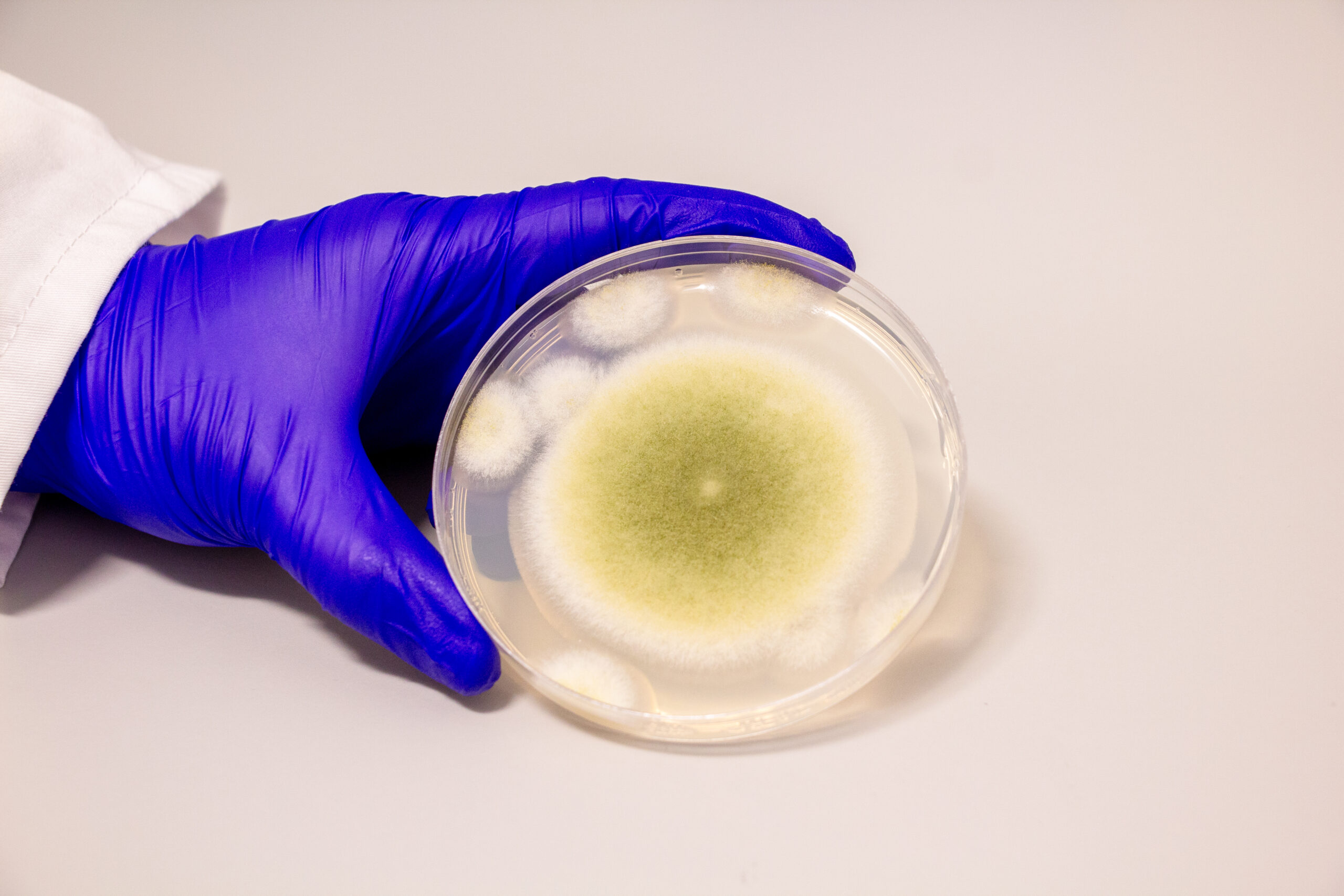
A sample of Aspergillus flavus cultured in the Gao Lab. (Credit: Bella Ciervo)
In the 1970s, a dozen scientists entered the tomb of Casimir IV in Poland. Within weeks, 10 of them died. Later investigations revealed the tomb contained A. flavus, whose toxins can lead to lung infections, especially in people with compromised immune systems.
Now, that same fungus is the unlikely source of a promising new cancer therapy.
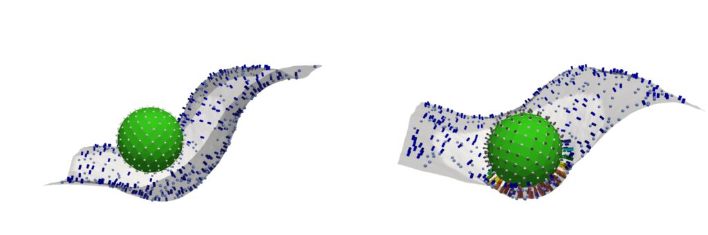
(Left) Pre-ignition (below the activation threshold) Only a handful of immune “tags” (C3b proteins) cover the nanoparticle, so it barely sticks to the white membrane—too few contact points means the immune cell simply can’t grab on. (Right) Post-ignition (above the activation threshold). The nanoparticle is now densely coated with C3b tags, and the immune-cell membrane reaches out with many matching receptors. Dozens of little “hooks” latch on at once, creating a strong, multivalent grip that pulls the particle in for engulfment.(Image: Ravi Radhakrishnan)
How does your body distinguish friendly visitors, like medications and medical devices, from dangerous invaders such as viruses and other infectious agents? The answer lies in a protein network dating back half a billion years—before humans diverged from sea urchins, notes Jake Brenner, a physician-scientist at the University of Pennsylvania.
“The complement system is perhaps the oldest-known part of our extracellular immune system,” says Brenner. “It plays a crucial role in identifying foreign materials like microbes, medical devices, or new drugs—particularly the larger ones like in the COVID vaccine.”
The complement system can, however, simultaneously play friend and foe, offering protection with one hand while backhanding the body with the other. In some cases, this ancient network can significantly exacerbate conditions like stroke by targeting the body’s own tissues. As Brenner explains, leaking blood vessels allow complement proteins to target brain tissue, causing the immune system to mistakenly launch an attack on the body’s own cells and worsen patient outcomes.
Now, using a combination of wet-lab experimentation, coupled differential equations, and computational-based modeling and simulations, an interdisciplinary team from the School of Engineering and Applied Science and the Perelman School of Medicine has decrypted the mathematical language behind the complement network’s “decision” to attack.
Reporting their findings in Cell, the team identifies a molecular tipping point known as the critical percolation threshold, which is based on how densely complement-binding sites are spaced on the surfaces of the model invader they engineered. If spacing between binding sites is too wide—landing above a threshold—complement activation fizzles out; below it, complement network ignites, a chain reaction of immune agent recruitment which spreads like wildfire.
Each year, the Department of Bioengineering at Penn Engineering proudly recognizes outstanding doctoral students whose research exemplifies innovation, impact, and academic excellence. The Solomon R. Pollack Award for Excellence in Graduate Bioengineering Research celebrates the achievements of students who have advanced our understanding of biological systems through engineering.
In 2025, four exceptional Ph.D. students—Nikolas Di Caprio, Harshini Chandrashekar, David Gonzalez-Martinez, and Kelsey Swingle—have been honored with this prestigious award. Their work spans neuroscience, oncology, maternal health, and tissue engineering, reflecting the breadth and interdisciplinary nature of the field.
Nikolas Di Caprio: Engineering Injectable Platforms for Cartilage Repair
Dissertation Title: “Engineering Dynamic Granular Composites for the Repair of Cartilage Tissue”
Nikolas Di Caprio earned his B.S. in Bioengineering with a minor in Chemistry from Temple University in 2019, where he conducted undergraduate research on 3D in-vitro models of adipose tissue.
Building on this foundation, his doctoral work in Dr. Jason Burdick’s lab focused on developing an injectable system using stem cell aggregates and hydrogel microparticles to repair cartilage. His work addressed both the biological and mechanical aspects of tissue regeneration, incorporating automated testing tools he designed to probe particle mechanics. The research offers new strategies for minimally invasive musculoskeletal treatments.
“I would like to thank Jason Burdick for the nomination, the committee for selecting me for the generous award, and my family, friends, and previous advisors for all the support during my Ph.D.“
Nikolas’s research embodies the intersection of material science and regenerative medicine, pushing the boundaries of bioengineering innovation.
Harshini Chandrashekar: Mapping the Genome’s Architecture in Alzheimer’s
Dissertation Title: “Addressing the Genome’s Structure-Function Relationship in Neural Activation and Neurological Disorders”
Harshini Chandrashekar completed her B.Tech. in Biotechnology from Vellore Institute of Technology, India, before moving to the U.S. in 2017 to pursue a master’s degree in Bioinformatics at the Georgia Institute of Technology. In 2019, she joined Dr. Jennifer Cremins lab as a Computational Scientist and began her Ph.D. in Bioengineering at Penn in 2020. In the Cremins lab, she investigated how the three-dimensional genome architecture—particularly chromatin loops—regulates gene expression in the context of familial Alzheimer’s disease (FAD). Her innovative combination of experimental and computational approaches uncovered early-stage disruptions in genome folding that may precede classical disease hallmarks.
Her findings are available on bioRxiv and have already garnered accolades, including the Young Talented and Developing (TAD) Scientist Award from Genome Organization Australia and the Best Research Presentation Award at the 2024 Penn Bioengineering Graduate Research Symposium.
“I’m grateful to my Ph.D. advisor, Dr. Jennifer Cremins, for nominating me for this award and for her support — especially during the challenging moments that are part and parcel of everyday research.. I would also like to thank Dr. Yale Cohen, my thesis committee chair, for his support and encouragement over the years, and Dr. Erin Berlew, whom I had the pleasure of TAing for, for being a generous mentor and role model in teaching. Their support played a meaningful role in both my academic and professional growth at Penn.”
Harshini now works as a Bioinformatics Scientist at the Benaroya Research Institute in Seattle.
David Gonzalez-Martinez: Revealing Drug Tolerance Pathways in Cancer
Dissertation Title: “Dissecting Mechanisms and Consequences of Oncogenic RTK Fusion Signaling”
David Gonzalez-Martinez began his academic journey at Florida State University, where he studied biochemistry. He went on to earn a master’s degree in Microbiology and Cell Science from the University of Florida before joining the Bioengineering Ph.D. program at Penn
Under the mentorship of Dr. Lukasz Bugaj, he used synthetic biology, optogenetics, and live-cell imaging to explore how oncogenic RTK fusions drive aberrant signaling and evade cancer treatment. His research pinpointed key mechanisms underlying drug resistance and identified drug combinations that improve therapeutic response. His recent publication inNature Communications highlights these findings.
“I would really like to thank the department for the recognition and Dr. Lukasz Bugaj, who has been a fantastic mentor throughout my Ph.D. as well as all of my lab mates who have been great friends and collaborators.”
David’s contributions provide crucial insights into the dynamic nature of cancer signaling and potential avenues for more effective treatments.
Kelsey Swingle: Targeting the Placenta to Treat Pre-eclampsia
Dissertation Title: “Engineering Placenta-Tropic VEGF mRNA Lipid Nanoparticles for the Treatment of Pre-eclampsia”
Kelsey Swingle earned her B.S.E. in Biomedical Engineering from Case Western Reserve University in 2020. As a Ph.D. student in the lab of Dr. Mike Mitchell, her research focused on developing new biomaterial strategies for targeted drug delivery to the placenta. Her dissertation work involved engineering VEGF mRNA lipid nanoparticles designed to home specifically to placental tissue, with the goal of treating pre-eclampsia—a major cause of maternal and fetal morbidity for which no clinical treatments currently exist.
“I’m incredibly grateful to my advisor Dr. Mike Mitchell, everyone in the Mitchell Lab, my mentors, friends, and family for their support throughout my PhD. While my PhD was both a rewarding and challenging personal experience, I leaned on and learned from my support system every step of the way.”
Her studies demonstrated the therapeutic potential of this targeted approach in mouse models, offering promising insight into a long-standing challenge in maternal health. In July 2025, Kelsey will begin a faculty position as Assistant Professor in the Department of Bioengineering at Rice University.
Looking Ahead
The 2025 Pollack Award recipients exemplify the best of Penn Bioengineering: rigor, creativity, and a drive to improve human health. Through their diverse research, each has made significant contributions to science and society. We congratulate Nikolas, Harshini, David, and Kelsey on their achievements and look forward to seeing how they will continue to shape the future of bioengineering.
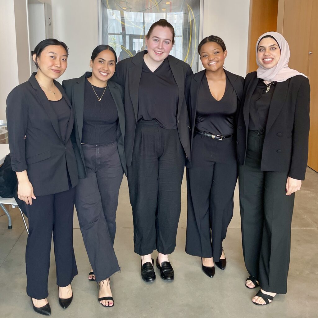
2025Team PRIME – Engineering a Smarter Response to Sepsis
From left to right: Sophie Gu; Shriya Boyapati; Sophie Klessel; McKenzie Davis; Majd Ayyad.
The final feature in the 2025 Senior Design Awards Spotlight highlights Team PRIME, who earned Second Place at the Minnesota Design of Medical Devices Competition.
Team Members: Majd Ayyad, Shriya Boyapati, McKenzie Davis, Sophie Gu, Sophie Klessel
Senior design in Penn Bioengineering is a yearlong capstone experience in which bioengineering seniors identify an unmet bioengineering need, design a solution to address the need, and create a high quality prototype that demonstrates their design. The course consists of BE4950 and BE4960, and was most recently taught by Dr. Erin Berlew, Dr. David Meaney, and Dr. Michael Siedlik.
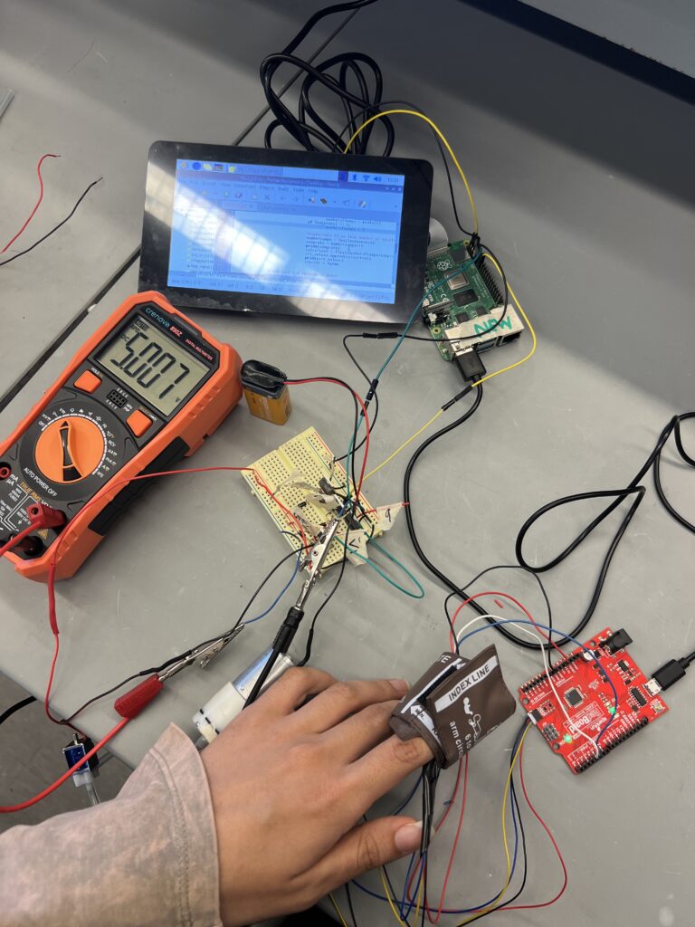
For Team PRIME, the mission was clear: create a tool that could help detect sepsis earlier—when timing can mean the difference between life and death. Their project centers around a device that automatically measures capillary refill time, a simple but powerful indicator of blood perfusion and circulation quality. By providing continuous, automated monitoring, PRIME aims to improve clinical decision-making in intensive care units and emergency settings.
PRIME as an idea; the first prototype of PRIME with all the wires.
The team’s inspiration came from their clinical mentor, Dr. John Greenwood, whose passion for improving sepsis detection was contagious.
Sophie Klessel shared, “We had a great clinical mentor (Dr. John Greenwood) who was really passionate about creating a device for earlier detection of sepsis, and we knew we wanted to work with him. Additionally, sepsis was an issue that resonated with our group and an issue that we were excited about.”
Team PRIME approached the work with a strong sense of collaboration, blending individual strengths across software, hardware, and systems integration. One member led the development of the user interface and application logic, while another focused on designing and assembling the physical and electrical components.
Working on PRIME revealed to the team just how demanding and rewarding bioengineering can be.
“Bioengineers need to understand it all from interviewing clinicians for needs findings, to studying the physiology of the human body, to designing all the technical components including hardware and software and finally towards producing a medical device. It is such a difficult job to be all the engineers at once but the final results are rewarding!” Majd Ayyad explained.
As the project concluded, their work was already gaining traction. Dr. Michael Siedlik, one of the bioengineering senior design instructors, shares, “This technology could greatly surpass the current standard of care, as it provides much needed automation, reproducibility, and clinician-free measurements in hectic medical environments where quick and reliable measurements are critical for preventing the negative outcomes of sepsis.”
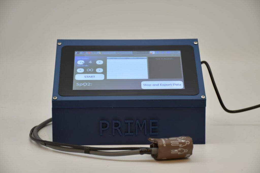
PRIME device.
PRIME earned Second Place at the Minnesota Design of Medical Devices Competition, a national recognition of the team’s thoughtful engineering and strong clinical relevance. Development of the device will continue in partnership with their clinical mentor—bringing them one step closer to impacting real patient care.
2025 Team Prism Optics – Bringing Vision Care Within Reach
Photo Credit: Penn Engineering From left to right: Lyle Brunhofer, Senior Design Project Competition Chairman; Dr. Robert Ghrist, Andrea Mitchell University Professor; Daniel Botros; Fady Fahmy; Daniel Serebrinic Jacobsohn; Danish Mahmood; and Aarush Sahni.
In Part 2 of the 2025 Senior Design Awards Spotlight, we turn to Team Prism Optics, winners of the Leadership Prize at Penn Engineering’s Senior Design Competition.
Team Members: Daniel Botros, Fady Fahmy, Daniel Jacobsohn Serebrinic, Danish Mahmood, Aarush Sahni
Senior design in Penn Bioengineering is a yearlong capstone experience in which bioengineering seniors identify an unmet bioengineering need, design a solution to address the need, and create a high quality prototype that demonstrates their design. The course consists of BE4950 and BE4960, and was most recently taught by Dr. Erin Berlew, Dr. David Meaney, and Dr. Michael Siedlik.
Across the globe, more than a billion people lack access to basic vision care, often simply because there aren’t enough optometrists to perform eye exams (Staff, One billion have preventable vision impairment 2019). Team Prism Optics took on this challenge by building a device that automates the process of determining an eyeglass prescription, offering a low-cost, portable solution that can be used without the need for a trained clinician.
The result is a self-administered vision screening platform that mimics the clinical process of subjective refraction. A user looks through a lens system and responds to a tumbling E eye chart using a joystick, indicating the direction of the letter. This interface, designed to be intuitive for users regardless of literacy, language, or age, was a major innovation in the team’s design. As Danish Mahmood explained, “Realizing the joystick input to indicate the direction of the tumbling E’s is functional for illiterate, non native English speaking, young and old people was our biggest logistical challenge.”
Danish Mahmood is using Prismatic to find his eye prescription by looking through the viewport of the device at a tumbling E’s eye chart located 20 ft away. He uses a joystick to input the direction of the E on the eye chart in response to audio feedback from the device.
The device was designed and built through a deeply collaborative process. Mahmood developed the control software and mechanical precision of the lens adjustment system, while Fady Fahmy handled the acrylic housing and gears. Aarush Sahni envisioned the LCD interface and helped lead algorithm development alongside Daniel Jacobsohn Serebrinic and Daniel Botros, working closely with clinical mentors at Penn Medicine. The system is already being tested with classmates and is set to begin clinical trials with patients this summer.
Throughout the process, the team remained focused on their goal: to make vision care accessible in underserved communities. Their mentor noted that clinical trials beginning just months after graduation are almost unheard of for senior design teams—underscoring just how effectively Prism Optics aligned their design to a global need.
Dr. Michael Siedlik, one of the bioengineering senior design instructors, highlighted just how exceptional this trajectory is for a student team:
“Not many senior design teams are able to start clinical trials a few months after graduation… This is a testament to their ability to identify a bioengineering need that is very well suited to their expertise and to the resources available to them, as well as their ability to execute at a high level as a team.”
“We have prototyped a working self-administered eye exam that mimics the process of subjective refraction with an automated device… Our next step is to ensure many patients can use it successfully and achieve accurate results,” Mahmood shared.
Team Prism Optics earned the Leadership Prize at Penn Engineering’s 2025 Senior Design Competition, a recognition of both their technical achievement and their clear path to real-world deployment.
References: Staff, RO. (2019, October 8). One billion have preventable vision impairment. Review of Optometry. https://www.reviewofoptometry.com/article/one-billion-have-preventable-vision-impairment
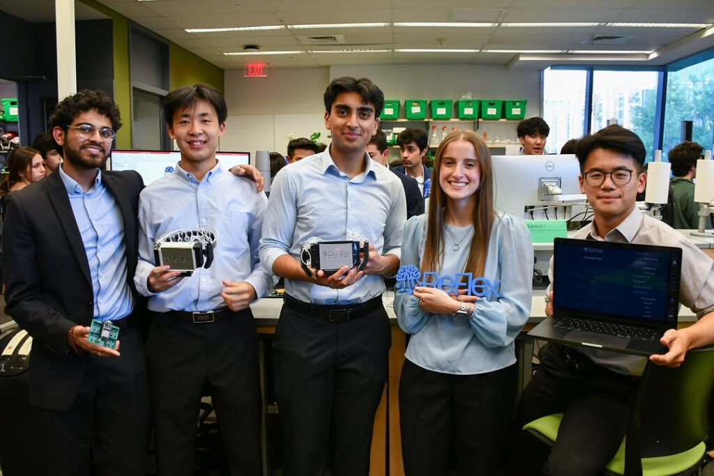
From left to right: Christopher Wun, William Qi, Ryann Joseph, Aditya Gowd, and Udit Garg
This series profiles three Penn Bioengineering senior design teams whose work received recognition at major competitions in 2025. In Part 1, we feature Team ReFlex, recipients of the Judge’s Choice Award at Penn Engineering’s Senior Design Competition.
Team Members: William Qi, Ryann Joseph, Christopher Wun, Udit Garg, Aditya Gowd
Senior design in Penn Bioengineering is a yearlong capstone experience in which bioengineering seniors identify an unmet bioengineering need, design a solution to address the need, and create a high quality prototype that demonstrates their design. The course consists of BE4950 and BE4960, and was most recently taught by Dr. Erin Berlew, Dr. David Meaney, and Dr. Michael Siedlik.
From the beginning, Team ReFlex set out to do something ambitious: create a system that could help stroke patients regain motor function by aligning therapy with the brain’s intent to move. The idea emerged from a shared interest in neurotechnology, combined with a diverse set of technical backgrounds—bioengineering, robotics, computer science, electrical engineering, and data science. After months of conversations with professors, clinicians, and researchers, the concept took shape: an integrated platform that uses EEG signals and artificial intelligence to detect motor intent and trigger functional electrical stimulation (FES).
William Qi, Building and testing the first prototype of ReFlex. Here, the full circuitry of the device is on a breadboard – this image was taken before the team finalized a PCB.
The team’s collaborative spirit was central to their progress. Each member brought complementary skills—some focused on the machine learning algorithms, others on signal processing, printed circuit board (PCB) design, or mechanical fabrication. Together, they built a noninvasive system designed for versatility, comfort, and real-world applicability.
“We knew from the start that we wanted to work on something in neurotechnology, as it was a space where all of our interests came together,” said William Qi. “With teammates in bioengineering, robotics, computer science, electrical engineering, and data science, we felt like we had a unique mix of skills to build something meaningful.”
The path wasn’t without challenges. The interdisciplinary nature of the project meant constantly stepping beyond individual comfort zones. Signal processing became a particular hurdle once the team moved to printed circuit boards—troubleshooting became more complex, but they leaned on strong communication and trust to navigate the setbacks.
As they developed the system, the team connected with a manufacturer of FDA-approved FES devices and successfully integrated one into their prototype—something that Dr. Michael Siedlik, one of the bioengineering senior design instructors, described it as a powerful example of vision meeting execution.
“They are a shining example of how our students can turn a plan that initially seems a little like science fiction into a high-quality biomedical device with the potential to address an important need,” Siedlik noted.
From left to right: Aditya Gowd, William Qi, Udit Garg, Ryann Joseph, and Christopher Wun ReFlex team picture picture after their successful BE demo day.
The result is a modular, user-friendly platform that allows patients to participate in their own rehabilitation more directly and independently. Designed to be compatible with existing clinical tools, ReFlex introduces a new level of personalization and responsiveness to therapy—advancing the potential of brain-computer interfaces in a field that clinicians themselves acknowledge as outdated.
ReFlex received the Judge’s Choice Award at Penn Engineering’s 2025 Senior Design Competition, a recognition not only of their technical achievement, but of their commitment to reshaping what recovery can look like for stroke survivors.
Each year, the American Association of Immunologists hosts its flagship meeting, IMMUNOLOGY, bringing together thousands of scientists from across the globe. Far from being a routine conference, this gathering serves as a critical convergence point for the most current research and thought leadership in the field. IMMUNOLOGY2025, held in Honolulu this year, highlighted the dynamic and rapidly evolving nature of immunology, with topics ranging from tissue-resident memory T cells and systems immunology to the brain-immune interface and cancer immunotherapy.
What sets IMMUNOLOGY2025 apart is its commitment to both scientific excellence and community building. Recognition at this meeting, whether through invited talks, abstract selection, or awards, is not just a personal milestone, but a broader signal of impactful, peer-recognized work. These honors are the result of competitive review and speak to both the scientific rigor and relevance of the selected projects. Oral presentations in block or major symposia place researchers on a global stage, while poster and trainee awards highlight emerging scientists making meaningful contributions to the field.
Reflecting the high regard for his research, Shahab (Shawn) Chizari was selected to present in a Major Symposium, one of the most prestigious forums at the meeting. Xiangcheng (Ison) Chen earned a 2025 AAI Trainee Abstract Award and was chosen for an oral presentation in a Block Symposium. Lingyang (Steven) Kong received a 2025 AAI Trainee Poster Award. Their achievements not only mark personal accomplishments, but also reflect the strength and promise of the next generation of immunologists.
Congratulations to the Penn Bioengineering graduate students who have received awards in the past year.
2025 Schmidt Science Fellow
Serena Omo-Lamai
Serena Omo-Lamai
As a 2025 Schmidt Science Fellow, Serena aims to create gene editing tools that activate only in targeted cells or disease contexts, improving precision and minimizing unintended effects. Her research focuses on safely removing harmful cells in autoimmune conditions like rheumatoid arthritis and type 1 diabetes, with broader potential for diagnostics and treating other diseases.
Solomon R. Pollack Award for Excellence in Graduate Bioengineering Research
Harshini Chandrashekar Niko Di Caprio Kelsey Swingle David Gonzalez-Martinez
The Solomon R. Pollack Award for Excellence in Graduate Bioengineering Research is given annually to the most deserving Bioengineering graduate students who have successfully completed research that is original and recognized as being at the forefront of their field.
NSF Graduate Research Fellowship Winner
Emily Huynh Emily Jacobs Jacqueline Li Emily Lin
Emily Huynh
Emily Jacobs
“I would like to thank Dr Noor Momin and all of my lab mates in the Momin Lab. I would not be able to put in the dedication I do without all of their support, mentorship, and friendship!” -Emily Jacobs
NSF Graduate Research Fellowship Honorable Mention
Awarded a Predoctoral Fellowship for his project, “Neuromodulatory Effects of Social Robot-Assisted Action Observation and Execution Therapy in Stroke Rehabilitation.”
Outstanding Teaching – SEAS Graduate Awards
Alex Hamilton Aoife O’Farrell Nat Thurlow Michael Yao
Alex Hamilton
Nat Thurlow
“I would like to thank Dr. LeAnn Dourte and Dr. Joel Boerckel for their incredible mentorship and support. It has been inspiring to work and teach with mentors who care deeply about students and fostering their growth beyond the curriculum.” -Nat Thurlow
“Many thanks to Dr. Kevin Johnson for being an incredible mentor and teacher! Extremely privileged to have been able to work with him and learn how to be a better educator. A big thank you as well to my co-advisors, Jim Gee and Osbert Bastani, for being fantastic mentors and sources of support!” -Michael Yao
Michael Yao
Outstanding Research – SEAS Graduate Awards
Lysia Cardilla Jiayi Wu
Lysia Cardilla
Outstanding Service – SEAS Graduate Awards
Aoife O’Farrell Ludwig Zhao
Ludwig Zhao
“I would like to thank Drs. Detre, Gottfried, and Tisdall for their nomination and their invaluable support as my mentors. It has been a great privilege to work with them – not only for their academic guidance, but also for their support in enabling me to serve our engineering students.” -Ludwig Zhao
Penn Prize for Excellence in Teaching by Graduate Students
Lasya Sreepada Ajay Thatte
Lasya Sreepada
“Thanks to all my students – high schoolers, undergraduates, and graduates – for engaging so thoughtfully in and out of class. I learned so much from you that has shaped my teaching style and inspired me to strive towards being an even better educator. A special thank you to Professor Paul Yushkevich, who welcomed me on board the teaching team for his Biomedical Image Analysis class and has been an outstanding mentor for me, as an aspiring scientist and professor.” -Lasya Sreepada
“A sincere thank you to Drs. Michael Mitchell, Riccardo Gottardi, Daniel Hammer and Jenny Jiang for giving me the opportunity to TA for them! This award would not have been possible without all of their guidance and support. And a big thank you to all the wonderful students I have had the privilege of teaching for the past 3 years!” -Ajay Thatte
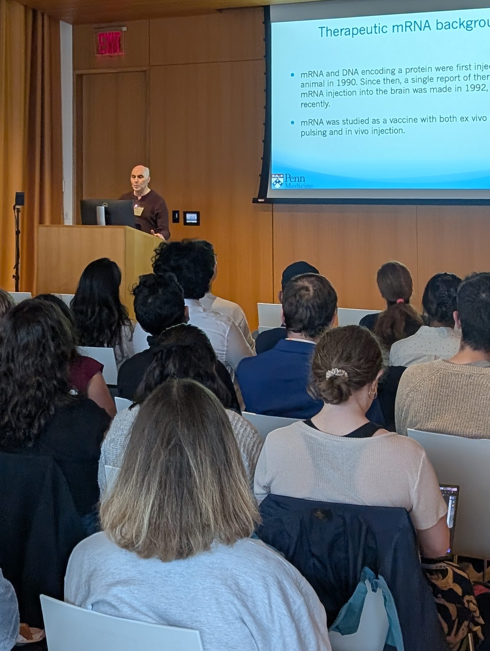
The 2025 Bioengineering Graduate Research Symposium, held on May 9 at the Singh Center for Nanotechnology, was a dynamic showcase of cutting-edge student research and a powerful example of scientific community in action. Organized by the Penn Bioengineering Graduate Group, the event featured a full afternoon of oral and poster presentations highlighting advances in immunotherapy, neuroengineering, regenerative medicine, and more. From engineering CAR-T therapies to decoding brain circuitry in depression and obesity, the symposium demonstrated the ingenuity and collaborative spirit of Penn’s graduate students.
A standout moment of the day was the keynote address by Drew Weissman, MD, PhD, a 2023 Nobel Laureate in Medicine and Director of the Penn Institute for RNA Innovation. Dr. Weissman traced the trajectory of mRNA science, from its early hurdles to its pivotal role in developing life-saving COVID-19 vaccines. He also shared where the field is headed—toward applications in cancer, autoimmune diseases, and beyond. His talk connected fundamental research with global impact, echoing the values at the heart of Penn Bioengineering.
Beyond the research, the symposium fostered a sense of community—welcoming faculty, students, and staff to connect over science and shared curiosity. With two lively poster sessions accompanied by refreshments, attendees had ample opportunity to exchange ideas and forge new collaborations. The event culminated in a reception that reflected the collegial, interdisciplinary ethos that defines Penn Bioengineering.
Whether through high-throughput diagnostic platforms, innovative biomaterials, or neuroimaging technologies, the work presented at the symposium exemplified how research at Penn is not only advancing science but also addressing critical challenges in health and society. The 2025 Symposium was not just a showcase, it was a celebration of a thriving research community, united in its pursuit of innovation and impact.
Each spring, awards are given to undergraduate students in the School of Engineering and Applied Science in recognition of outstanding scholarly achievements and service to the School and University community.
Read the full list of Bioengineering undergraduate award winners below.
The Hugo Otto Wolf Memorial Prize
Joey Wu Christopher Wun
Christopher Wun
This prize is awarded to one or more members of each department’s senior class, distinguishing students who meet with great approval of the professors at large through “thoroughness and originality” in their work.
“Thanks to my BE professors (and lab instructors especially) that made it possible for me to explore just about every facet of engineering!” -Christopher Wun
“I’d like to express my gratitude to the bioengineering professor that supported my work and always believed in me: Dr. LeAnn Dourte. Dr. Dourte has been a personal mentor, academic influence, and professional advisor that has shaped the way I approach the world. She is an incredible teacher, patient mentor, and wonderful friend. Thank you for all that she does!” -Joey Wu
Joey Wu
The Herman P. Schwan Award
Hana Bader
This department award honors a graduating senior who demonstrates the “highest standards of scholarship and academic achievement.”
Hana Bader
The Bioengineering Student Leadership Award
Gregory Datto
This award is given annually to a student in Bioengineering who has demonstrated, through a combination of academic performance, service, leadership, and personal qualities, that they will be a credit to the Department, the School, and the University.
Albert Giandomenico Award
Taken at the Penn Engineering Award Ceremony. From left to right: Gregory Datto (The Bioengineering Student Leadership Award), Jacqueline Li, Hana Bader, Hana Matsuda, and Rudy Whitney—all recipients of the Albert Giandomenico Award.
Hana Bader Jacqueline Li Hana Matsuda Rudy Whitney
The Bioengineering Department also presents a single lab group with the Albert Giandomenico Award which reflects their “teamwork, leadership, creativity, and knowledge applied to discovery-based learning in the laboratory.”
“I would like to extend my heartfelt thanks to my incredible teammates—Hana Bader, Hana Matsuda, and Rudy Whitney—whose collaboration, dedication, and clear communication made this award possible. I am equally grateful to our instructors, Professors David Meaney, David Issadore, and Michael Patterson, for their unwavering support throughout the Bioengineering MADLAB courses. Their technical guidance and encouragement were instrumental to our success and growth as a team.” -Jacqueline Li
Rose Undergraduate Research Award
Ryann Joseph
Awarded by the Center for Undergraduate Research and Fellowships (CURF). Ryann’s project was titled, “Cas9 protein outperforms Cas9 mRNA in CRISPR/Cas9 editing for lipid nanoparticle mediated recovery of CFTR functionality,” and was completed with the assistance of Professor Michael J. Mitchell.
Penn Engineering Exceptional Service Award
Ethan Eisenberg Ryann Joseph Sophie Klessel Brianna Leung
This award recognize students for their outstanding service to the University and their larger communities.
“I have loved working to serve my engineering community as a TA and mentor!” -Ryann Joseph
Ryann Joseph
Ethan Eisenberg
“Thank you very much to my professors and the faculty of the School of Engineering and Applied Science. I am very thankful and appreciative for this recognition.” -Ethan Eisenberg
Ben and Bertha Gomberg Kirsch Award
Victor Maia Portella Dubeux
This competitive award is decided by the SEAS faculty from among the Engineering undergraduate body and distinguishes a member of the B.A.S. senior class who “in applying the flexibility of the program, has created a personal academic experience involving the most creative use of the resources of the University.”
The Wolf-Hallac Award
Kaitlin Mrksich
Kaitlin Mrksich
This award was established in October 2000 to recognize the graduating female senior from across Penn Engineering’s departments who is seen as a role model, has achieved a high GPA (in the top 10% of their class), and who has demonstrated a commitment to school and/or community.