Understanding the Physics of Kidney Development

Abstract image of tubules repelling each other and shifting around.
The model of tubule packing developed by the Hughes Lab shows the tubules repelling each other and shifting around.

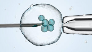
A recent study by Penn Bioengineering researchers sheds new light on the role of physics in kidney development. The kidney uses structures called nephrons and tubules to filter blood and pass urine to the bladder. Nephron number is set at birth and can vary over an order of magnitude (anywhere from 100,000 to over a million nephrons in an individual kidney). While the reasons for this variability remain unclear, low numbers of nephrons predispose patients to hypertension and chronic kidney disease. 

Now, research published in Developmental Cell led by Alex J. Hughes, Assistant Professor in the Department of Bioengineering, demonstrates a new physics-driven approach to better visualize and understand how a healthy kidney develops to avoid organizational defects that would impair its function. While previous efforts have typically approached this problem using molecular genetics and mouse models, the Hughes Lab’s physics-based approach could link particular types of defects to this genetic information and possibly highlight new treatments to prevent or fix congenital defects.

During embryonic development, kidney tubules grow and the tips divide to make a branched tree with clusters of nephron stem cells surrounding each branch tip. In order to build more nephrons, the tree needs to grow more branches. To keep the branches from overlapping, the kidney’s surface grows more crowded as the number of branches increase. “At this point, it’s like adding more people to a crowded elevator,” says Louis Prahl, first author of the paper and Postdoctoral Fellow in the Hughes Lab. “The branches need to keep rearranging to accommodate more until organ growth stops.”

To understand this process, Hughes, Prahl and their team investigated branch organization in mouse kidneys as well as using computer models and a 3D printed model of tubules. Their results show that tubules have to actively restructure – essentially divide at narrower angles – to accommodate more tubules. Computer simulations also identified ‘defective’ packing, in which the simulation parameters caused tubules to either overlap or be forced beneath the kidney surface. The team’s experimentation and analysis of published studies of genetic mouse models of kidney disease confirmed that these defects do occur.

This study represents a unique synthesis of different fields to understand congenital kidney disease. Mathematicians have studied geometric packing problems for decades in other contexts, but the structural features of the kidney present new applications for these models. Previous models of kidney branching have approached these problems from the perspective of individual branches or using purely geometric models that don’t account for tissue mechanics. By contrast, The Hughes Lab’s computer model demonstrates the physics of how tubule families interact with each other, allowing them to identify ‘phases’ of kidney organization that either relate to normal kidney development or organizational defects. Their 3D printed model of tubules shows that these effects can occur even when one sets the biology aside.

Hughes has been widely recognized for his research in the understanding of kidney development. This new publication is the first fruit of his 2021 CAREER Award from the National Science Foundation (NSF) and he was recently named a 2023 Rising Star by the Cellular and Molecular Bioengineering (CMBE) Special Interest Group. In 2020 he became the first Penn Engineering faculty member to receive the Maximizing Investigators’ Research Award (MIRA) from the National Institutes of Health (NIH) for his forward-thinking work in the creation of new tools for tissue engineering.

Pediatric nephrologists have long worked to understand the cause of these childhood kidney defects. These efforts are often confounded by a lack of evidence for a single causative mutation. The Hughes Lab’s approach presents a new and different application of the packing problem and could help answer some of these unsolved questions and open doors to prevention of these diseases. Following this study, Hughes and his lab members will continue to explore the physics of kidney tubule packing, looking for interesting connections between packing organization, mechanical stresses between neighboring tubule tips, and nephron formation while attempting to copy these principles to build stem cell derived tissues to replace damaged or diseased kidney tissue. Mechanical forces play an important role in developmental biology and there is much scope for Hughes, Prahl and their colleagues to learn about these properties in relation to the kidney.

Read The developing murine kidney actively negotiates geometric packing conflicts to avoid defects” in Developmental Cell.

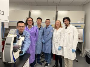
Other authors include Bioengineering Ph.D. students and Hughes Lab members John Viola and Jiageng Liu.

This work was supported by NSF CAREER 2047271, NIH MIRA R35GM133380, Predoctoral Training Program in Developmental Biology T32HD083185, and NIH F32 fellowship DK126385.

Inside the Jiang Lab: An Inventory of Immunity

by

Black and white photo of Jenny Jiang working in her lab on a laptop.
Jenny Jiang, Ph.D.

Engineers in the Center for Precision Engineering for Health (CPE4H) are focusing on innovations in diagnostics and delivery, cellular and tissue engineering, and the development of new devices that integrate novel materials with human tissues. Below is an excerpt from “Going Small to Win Big: Engineering Personalized Medicine,” featuring the research from the laboratory of Jenny Jiang, J. Peter and Geri Skirkanich Associate Professor of Innovation in Bioengineering.

The Challenge

In order to create personalized immune therapies, researchers need to untangle what is happening between an individual patient’s immune cells and the antigens that they interact with on a molecular level. Immune cell-antigen interactions need to be understood in four different areas in order to create a full picture: the unique genetic sequence of the T cell’s antigen receptors, the antigen specificity of that cell, and both the gene and protein expression of the same cell.

The Status Quo

Prior methods of understanding interactions between T cells and antigens could only get a picture of one or two of these four elements because of technology constraints. Other roadblocks included that cells cultured or engineered in a laboratory setting are not in a natural environment so they won’t express genes or proteins in the way T cells would in the body, and technologies that assess the antigen specificity of T cells were not cost-effective for looking at large numbers of antigens.

The Jiang Lab’s Fix

The lab of Jenny Jiang, J. Peter and Geri Skirkanich Associate Professor of Innovation in Bioengineering, developed a technology called TetTCR-SeqHD, which solves these problems. Using this technology, scientists can now simultaneously profile samples of large numbers of single T cells in the four dimensions using high- throughput screening.

The Jiang Lab’s technology is essentially a method for getting a “full-body scan” of an individual’s T cells and creates a catalog of the different types of T cells and the antigens they respond (or don’t respond) to, paving the way for the ability to better target immune therapies to an individual patient.

“Individual T cells are unique, and that’s the challenge of using one treatment to fit all,” says Jiang. “Identifying antigen specificity and creating therapies that target that specificity in an individual’s T cells will be key to truly personalizing immune therapies in the future.”

Read the full story in Penn Engineering magazine.

Penn Scientist Nader Engheta Wins the Benjamin Franklin Medal

Nader Engheta
Nader Engheta (Image: Felice Macera)

by Amanda Mott

University of Pennsylvania scientist Nader Engheta has been selected as a 2023 recipient of the Benjamin Franklin Medal, one of the world’s oldest science and technology awards. The laureates will be honored on April 27 at a ceremony at the Franklin Institute in Philadelphia.

Engheta, H. Nedwill Ramsey Professor in Electrical and Systems Engineering, is among nine outstanding individuals recognized with Benjamin Franklin Medals this year for their achievements in extraordinary scientific, engineering and business leadership.

“As a scientist and a Philadelphian, I am deeply honored and humbled to receive the Franklin Medal. It is the highest compliment to receive an award whose past recipients include some of my scientific heroes such as Albert Einstein, Nikola Tesla, Alexander Graham Bell, and Max Planck. I am very thankful to the Franklin Institute for bestowing this honor upon me.”

Larry Dubinski, President and CEO of The Franklin Institute, says, “We are proud to continue The Franklin Institute’s longtime legacy of recognizing individuals for their contributions to humanity. These extraordinary advancements in areas of such importance as social equity, sustainability, and safety are significantly moving the needle in the direction of positive change and therefore laying the groundwork for a remarkable future.”

The 2023 Benjamin Franklin Medal in Electrical Engineering goes to Engheta for his transformative innovations in engineering novel materials that interact with electromagnetic waves in unprecedented ways, with broad applications in ultrafast computing and communication technologies.

“Professor Engheta’s pioneering work in metamaterials and nano-optics points the way to new and truly revolutionary computing capabilities in the future,” says University of Pennsylvania President Liz Magill. “Penn inaugurated the age of computers by creating the world’s first programmable digital computer in 1945. Professor Engheta’s work continues this tradition of groundbreaking research and discovery that will transform tomorrow. We are thrilled to see him receive the recognition of the Benjamin Franklin Medal.”

Engheta founded the field of optical nanocircuits (“optical metatronics”), which merges nanoelectronics and nanophotonics. He is also known for establishing and& developing the field of near-zero-index optics and epsilon-near-zero (ENZ) materials with near-zero electric permittivity. Through his work he has opened many new frontiers, including optical computation at the nanoscale and scattering control for cloaking and transparency. His work has far-reaching implications in various branches of electrical engineering, materials science, optics, microwaves, and quantum electrodynamics.

“This award recognizes Dr. Engheta’s trailblazing advances in engineering and physics,” says Vijay Kumar, Nemirovsky Family Dean of Penn Engineering.“ The swift and sustainable technologies his research in metamaterials and metatronics offers the world are the result of a lifelong commitment to scientific curiosity. For over 35 years, Nader Engheta has personified Penn Engineering’s mission of inventing the future.”

Nader Engheta is the H. Nedwill Ramsey Professor in the Departments of Electrical and Systems Engineering and Bioengineering in the School of Engineering and Applied Science and professor of physics and astronomy in the School of Arts & Sciences at the University of Pennsylvania.

This story originally appeared in Penn Today.

New Single Cell Analysis Tool

by Nathi Magubane

Researchers at Penn and colleagues have developed a tool to analyze single cells that assesses both the patterns of gene activation within a cell and which sibling cells shared a common progenitor.

3D illustration of a cell held by a pipet and a needle
Arjun Raj of the School of Engineering and Applied Science and the Perelman School of Medicine, former postdoc Lee Richman, now of Brigham and Women’s Hospital, and colleagues have developed a new analysis tool that combines a cell’s unique gene expression data with information about the cell’s origins. The method can be applied to identify new cell subsets throughout development and better understand drug resistance.

Recent advances in analyzing data at the single-cell level have helped biologists make great strides in uncovering new information about cells and their behaviors. One commonly used approach, known as clustering, allows scientists to group cells based on characteristics such as the unique patterns of active or inactive genes or by the progeny of duplicating cells, known as clones, over several generations.

Although single-cell clustering has led to many significant findings, for example, new cancer cell subsets or the way immature stem cells mature into “specialized” cells, researchers to this point had not been able to marry what they knew about gene-activation patterns with what they knew about clone lineages.

Now, research published in Cell Genomics led by University of Pennsylvania professor of bioengineering Arjun Raj has resulted in the development of ClonoCluster, an open-source tool that combines unique patterns of gene activation with clonal information. This produces hybrid cluster data that can quickly identify new cellular traits; that can then be used to better understand resistance to some cancer therapies.

“Before, these were independent modalities, where you would cluster the cells that express the same genes in one lot and cluster the others that share a common ancestor in another,” says Lee Richman, first paper author and a former postdoc in the Raj lab who is now at Brigham and Women’s Hospital in Boston. “What’s exciting is that this tool allows you to draw new lines around your clusters and explore their properties, which could help us identify new cell types, functions, and molecular pathways.”

Researchers in the Raj Lab use a technique known as barcoding to assign labels to cells they are interested in studying, particularly useful for tracking cells, clustering data based on cells’ offspring, and following lineages over time. Believing they could parse more valuable information out of this data by incorporating the cell’s unique patterns of gene activation, the researchers applied ClonoCluster to six experimental datasets that used barcoding to track dividing cells’ offspring. Specifically, they looked at the development of chemotherapy resistance and of stem cells into specialized tissue types.

Read the full story in Penn Today.

Inside the Ko Lab: Tracking Living Tissues

by

Black and white photo of Jina Ko inspecting a clear slide.
Jina Ko, Ph.D.

Engineers in the Center for Precision Engineering for Health (CPE4H) are focusing on innovations in diagnostics and delivery, cellular and tissue engineering, and the development of new devices that integrate novel materials with human tissues. Below is an excerpt from “Going Small to  Win Big: Engineering Personalized Medicine,” featuring the research from the laboratory of Jina Ko, Assistant Professor in Bioengineering and Pathology and Laboratory Medicine.

The Challenge

When scientists create methods to detect disease biomarkers, they give healthcare providers better tools to properly diagnose and treat patients. However, limitations to obtaining this information, especially when using living cells and tissues from patients, prevents a complete picture of what is unique about a case and decreases the chance that the best course of treatment can be identified.

The Status Quo

Several techniques exist for identifying multiple biomarkers in cells, but they are usually not compatible with observing changes over time in living cells or are limited by a set number of biomarkers that can be profiled. The chemicals used to profile multiple (>5) biomarkers are toxic to the cells, preventing live cell monitoring. Due to this limitation, a full understanding of the protein expressions of the living cells could not be obtained and a clear picture of what is actually occurring during the course of cellular changes was out of reach.

The Ko Lab’s Fix

Jina Ko, Assistant Professor in Bioengineering, is working to overcome this limitation with a method known as “scission-accelerated fluorophore exchange” (or SAFE), a new way to detect biomarkers in cells that is highly gentle and allows for high multiplexing via cyclic imaging so that more biomarkers can be identified in a single sample and changes in living cells and tissues can be tracked over time. She first developed this method during her postdoctoral training at Massachusetts General Hospital under the supervision of Jonathan Carlson and Ralph Weissleder.

The method uses “click” chemistry, which is a bioorthogonal, non-toxic and rapid reaction that allows the team to highlight the desired biomarkers in the samples without destroying them each time a microscopy cycle is run.

“You can’t identify a treatment that works for the average person, apply that treatment to everyone and expect the best outcomes,” says Ko. “Using this method, if we want to administer a therapy to a patient, we could remove a sample of their cells and use that sample to try different therapeutic options. After tracking the sample, we could predict if the patient would respond well to therapy A, but not therapy B. Our goal is that this technology will be applied in the clinic to help patients.”

Read the full story in Penn Engineering magazine.

RNA Lipid Nanoparticle Engineering Stops Liver Fibrosis in its Tracks, Reverses Damage

by

Members of the research team include (from left to right) Xuexiang Han, Michael J. Mitchell, Ningqiang Gong, Lulu Xue, Sarah J. Shepherd, and Rakan El-Mayta.

Since the success of the COVID-19 vaccine, RNA therapies have been the object of increasing interest in the biotech world. These therapies work with your body to target the genetic root of diseases and infections, a promising alternative treatment method to that of traditional pharmaceutical drugs.

Lipid nanoparticles (LNPs) have been successfully used in drug delivery for decades. FDA-approved therapies use them as vehicles for delivering messenger RNA (mRNA), which prompts the cell to make new proteins, and small interfering RNA (siRNA), which instruct the cell to silence or inhibit the expression of certain proteins.

The biggest challenge in developing a successful RNA therapy is its targeted delivery. Research is now confronting the current limitations of LNPs, which have left many diseases without an effective RNA therapy.

Liver fibrosis occurs when the liver is repeatedly damaged and the healing process results in the accumulation of scar tissue, impeding healthy liver function. It is a chronic disease characterized by the buildup of excessive collagen-rich extracellular matrix (ECM). Liver fibrosis has remained challenging to treat using RNA therapies due to a lack of delivery systems for targeting activated liver-resident fibroblasts. Both the solid fibroblast structure and the lack of specificity or affinity to target these fibroblasts has impeded current LNPs from entering activated liver-resident fibroblasts, and thus they are unable to deliver RNA therapeutics.

To tackle this issue and help provide a treatment for the millions of people who suffer from this chronic disease, Michael Mitchell, J. Peter and Geri Skirkanich Assistant Professor of Innovation in the Department of Bioengineering, and postdoctoral fellows Xuexiang Han and Ningqiang Gong, found a new way to synthesize ligand-tethered LNPs, increasing their selectivity and allowing them to target liver fibroblasts.

Lulu Xue, Margaret Billingsley, Rakan El-Mayta, Sarah J. Shepherd, Mohamad-Gabriel Alameh and Drew Weissman, Roberts Family Professor in Vaccine Research and Director of the Penn Institute for RNA Innovation at the Perelman School of Medicine, also contributed to this work.

Read the full story in Penn Engineering Today.

ASSET Center Inaugural Seed Grants Will Fund Trustworthy AI Research in Healthcare

by

Illustration credit: Melissa Pappas

Penn Engineering’s newly established ASSET Center aims to make AI-enabled systems more “safe, explainable and trustworthy” by studying the fundamentals of the artificial neural networks that organize and interpret data to solve problems.

ASSET’s first funding collaboration is with Penn’s Perelman School of Medicine (PSOM) and the Penn Institute for Biomedical Informatics (IBI). Together, they have launched a series of seed grants that will fund research at the intersection of AI and healthcare.

Teams featuring faculty members from Penn Engineering, Penn Medicine and the Wharton School applied for these grants, to be funded annually at $100,000. A committee consisting of faculty from both Penn Engineering and PSOM evaluated 18 applications and  judged the proposals based on clinical relevance, AI foundations and potential for impact.

Artificial intelligence and machine learning promise to revolutionize nearly every field, sifting through massive amounts of data to find insights that humans would miss, making faster and more accurate decisions and predictions as a result.

Applying those insights to healthcare could yield life-saving benefits. For example, AI-enabled systems could analyze medical imaging for hard-to-spot tumors, collate multiple streams of disparate patient information for faster diagnoses or more accurately predict the course of disease.

Given the stakes, however, understanding exactly how these technologies arrive at their conclusions is critical. Doctors, nurses and other healthcare providers won’t use such technologies if they don’t trust that their internal logic is sound.

“We are developing techniques that will allow AI-based decision systems to provide both quantifiable guarantees and explanations of their predictions,” says Rajeev Alur, Zisman Family Professor in Computer and Information Science and Director of the ASSET Center. “Transparency and accuracy are key.”

“Development of explainable and trustworthy AI is critical for adoption in the practice of medicine,” adds Marylyn Ritchie, Professor of Genetics and Director of the Penn Institute for Biomedical Informatics. “We are thrilled about this partnership between ASSET and IBI to fund these innovative and exciting projects.”

 Seven projects were selected in the inaugural class, including projects from Dani S. Bassett, J. Peter Skirkanich Professor in the Departments of Bioengineering, Electrical and Systems Engineering, Physics & Astronomy, Neurology, and Psychiatry, and several members of the Penn Bioengineering Graduate Group: Despina Kontos, Matthew J. Wilson Professor of Research Radiology II, Department of Radiology, Penn Medicine and Lyle Ungar, Professor, Department of Computer and Information Science, Penn Engineering; Spyridon Bakas, Assistant Professor, Departments of Pathology and Laboratory Medicine and Radiology, Penn Medicine; and Walter R. Witschey, Associate Professor, Department of Radiology, Penn Medicine.

Optimizing clinical monitoring for delivery room resuscitation using novel interpretable AI

Elizabeth Foglia, Associate Professor, Department of Pediatrics, Penn Medicine and the Children’s Hospital of Philadelphia

Dani S. Bassett, J. Peter Skirkanich Professor, Departments of Bioengineering and Electrical and Systems Engineering, Penn Engineering

 This project will apply a novel interpretable machine learning approach, known as the Distributed Information Bottleneck, to solve pressing problems in identifying and displaying critical information during time-sensitive clinical encounters. This project will develop a framework for the optimal integration of information from multiple physiologic measures that are continuously monitored during delivery room resuscitation. The team’s immediate goal is to detect and display key target respiratory parameters during delivery room resuscitation to prevent acute and chronic lung injury for preterm infants. Because this approach is generalizable to any setting in which complex relations between information-rich variables are predictive of health outcomes, the project will lay the groundwork for future applications to other clinical scenarios.

Read the full list of projects and abstracts in Penn Engineering Today.

Alex Hughes Named CMBE Rising Star

A collage of photos: Alex Hughes presenting, the title slide of his presentation with the title "Interpreting geometric rules of early kidney formation for synthetic morphogenesis," and his acknowledgements slides.
Alex J. Hughes presents at the BMES CMBE conference in January 2023. (Image credit: Riccardo Gottardi, Assistant Professor in Pediatrics and Bioengineering)

Alex J. Hughes, Assistant Professor in the Department of Bioengineering, was one of thirteen recipients of the 2023 Rising Star Award for Junior Faculty by the Cellular and Molecular Bioengineering (CMBE) Special Interest Group. The Rising Star Award recognizes a CMBE member in their early independent career stage that has made an outstanding impact on the field of cellular and molecular bioengineering. CMBE is a special interest group of the Biomedical Engineering Society (BMES), the premier professional organization of bioengineers.

The Hughes Lab in Penn Bioengineering works to “bring developmental processes that operate in vertebrate embryos and regenerating organs under an engineering control framework” in order to “build better tissues.” Hughes’s research interest is in harnessing the developmental principles of organs, allowing him to design medically relevant scaffolds and machines. In 2020 he became the first Penn Engineering faculty member to receive the Maximizing Investigators’ Research Award (MIRA) from the National Institutes of Health (NIH), and he was awarded a prestigious CAREER Award from the National Science Foundation (NSF) in 2021. Most recently, Hughes’s work has focused on understanding the development of cells and tissues in the human kidney via the creation of “organoids”: miniscule organ models that can mimic the biochemical and mechanical properties of the developing kidney. Understanding and engineering how the kidney functions could open doors to more successful regenerative medicine strategies to address highly prevalent congenital and adult diseases.

Hughes and his fellow award recipients were recognized at the annual BMES CBME conference in Indian Wells, CA in January 2023.

Read the full list of 2023 CMBE Award Winners.

OCTOPUS, an Optimized Device for Growing Mini-Organs in a Dish

by Devorah Fischler

With OCTOPUS, Dan Huh’s team has significantly advanced the frontiers of organoid research, providing a platform superior to conventional gel droplets. OCTOPUS splits the soft hydrogel culture material into a tentacled geometry. The thin, radial culture chambers sit on a circular disk the size of a U.S. quarter, allowing organoids to advance to an unprecedented degree of maturity.

When it comes to human bodies, there is no such thing as typical. Variation is the rule. In recent years, the biological sciences have increased their focus on exploring the poignant lack of norms between individuals, and medical and pharmaceutical researchers are asking questions about translating insights concerning biological variation into more precise and compassionate care.

What if therapies could be tailored to each patient? What would happen if we could predict an individual body’s response to a drug before trial-and-error treatment? Is it possible to understand the way a person’s disease begins and develops so we can know exactly how to cure it?

Dan Huh, Associate Professor in the Department of Bioengineering at the University of Pennsylvania’s School of Engineering and Applied Science, seeks answers to these questions by replicating biological systems outside of the body. These external copies of internal systems promise to boost drug efficacy while providing new levels of knowledge about patient health.

An innovator of organ-on-a-chip technology, or miniature copies of bodily systems stored in plastic devices no larger than a thumb drive, Huh has broadened his attention to engineering mini-organs in a dish using a patient’s own cells.

A recent study published in Nature Methods helmed by Huh introduces OCTOPUS, a device that nurtures organs-in-a-dish to unmatched levels of maturity. The study leaders include Estelle Park, doctoral student in Bioengineering, Tatiana Karakasheva, Associate Director of the Gastrointestinal Epithelium Modeling Program at Children’s Hospital of Philadelphia (CHOP), and Kathryn Hamilton, Assistant Professor of Pediatrics in Penn’s Perelman School of Medicine and Co-Director of the Gastrointestinal Epithelial Modeling Program at CHOP.

Read the full story in Penn Engineering Today.

CAR T Cell Therapy Reaches Beyond Cancer

Penn Medicine researchers laud the early results for CAR T therapy in lupus patients, which point to broader horizons for the use of personalized cellular therapies.

Penn Medicine’s Carl June and Daniel Baker.

Engineered immune cells, known as CAR T cells, have shown the world what personalized immunotherapies can do to fight blood cancers. Now, investigators have reported highly promising early results for CAR T therapy in a small set of patients with the autoimmune disease lupus. Penn Medicine CAR T pioneer Carl June and Daniel Baker, a doctoral student in cell and molecular biology in the Perelman School of Medicine, discuss this development in a commentary published in Cell.

“We’ve always known that in principle, CAR T therapies could have broad applications, and it’s very encouraging to see early evidence that this promise is now being realized,” says June, who is the Richard W. Vague Professor in Immunotherapy in the department of Pathology and Laboratory Medicine at Penn Medicine and director of the Center for Cellular Immunotherapies at the Abramson Cancer Center.

T cells are among the immune system’s most powerful weapons. They can bind to, and kill, other cells they recognize as valid targets, including virus-infected cells. CAR T cells are T cells that have been redirected, through genetic engineering, to efficiently kill specifically defined cell types.

CAR T therapies are created out of each patient’s own cells—collected from the patient’s blood, and then engineered and multiplied in the lab before being reinfused into the patient as a “living drug.” The first CAR T therapy, Kymriah, was developed by June and his team at Penn Medicine, and received Food & Drug Administration approval in 2017. There are now six FDA-approved CAR T cell therapies in the United States, for six different cancers.

From the start of CAR T research, experts believed that T cells could be engineered to fight many conditions other than B cell cancers. Dozens of research teams around the world, including teams at Penn Medicine and biotech spinoffs who are working to develop effective treatments from Penn-developed personalized cellular therapy constructs, are examining these potential new applications. Researchers say lupus is an obvious choice for CAR T therapy because it too is driven by B cells, and thus experimental CAR T therapies against it can employ existing anti-B-cell designs. B cells are the immune system’s antibody-producing cells, and, in lupus, B cells arise that attack the patient’s own organs and tissues.

This story is by Meagan Raeke. Read more at Penn Medicine News.

Carl June is a member of the Penn Bioengineering Graduate Group. Read more stories featuring June’s research here.