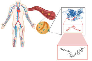
The antimicrobial peptides the researchers studied are “encrypted” in that they are contained within Apolipoprotein B, a blood plasma protein that is not directly involved in the immune response, but are not normally expressed on their own.
The rise of drug-resistant bacteria infections is one of the world’s most severe global health issues, estimated to cause 10 million deaths annually by the year 2050. Some of the most virulent and antibiotic-resistant bacterial pathogens are the leading cause of life-threatening, hospital-acquired infections, particularly dangerous for immunocompromised and critically ill patients. Traditional and continual synthesis of antibiotics will simply not be able to keep up with bacteria evolution.
To avoid the continual process of synthesizing new antibiotics to target bacteria as they evolve, Penn Engineers have looked at a new, natural resource for antibiotic molecules.
César de la Fuente, Ph.D.
A recent study on the search for encrypted peptides with antimicrobial properties in the human proteome has located naturally occurring antibiotics within our own bodies. By using an algorithm to pinpoint specific sequences in our protein code, a team of Penn researchers along with collaborators, led by César de la Fuente, Presidential Assistant Professor in Psychiatry, Bioengineering, Microbiology, and Chemical and Biomolecular Engineering, and Marcelo Torres, a post doc in de la Fuente’s lab, were able to locate novel peptides, or amino acid chains, that when cleaved, indicated their potential to fend off harmful bacteria.
Now, in a new study published in ACS Nano, the team along with Angela Cesaro, the lead author and post doc in de la Fuente’s lab, have identified three distinct antimicrobial peptides derived from a protein in human plasma and demonstrate their abilities in mouse models. Angela Cesaro performed a great part of the activities during her PhD under the supervision of corresponding author, Professor Angela Arciello, from the University of Naples Federico II. The collaborative study also includes Utrecht University in the Netherlands.
“We identified the cardiovascular system as a hot spot for potential antimicrobials using an algorithmic approach,” says de la Fuente. “Then we looked closer at a specific protein in the plasma.”
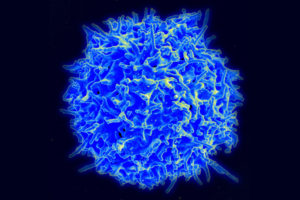
A scanning electron micrograph of a healthy human T cell. A better understanding the wide variety of antigen receptors that appear on the surfaces of these critical components of the immune system is necessary for improving a new class of therapies. (Credit: NIAID)
Our bodies are equipped with specialized white blood cells that protect us from foreign invaders, such as viruses and bacteria. These T cells identify threats using antigen receptors, proteins expressed on the surface of individual T cells that recognize specific amino acid sequences found in or on those invaders. Once a T cell’s antigen receptors bind to the corresponding antigen, it can directly kill infected cells or call for backup from the rest of the immune system.
We have hundreds of billions of T cells, each with unique receptors that recognize unique antigens, so profiling this T cell antigen specificity is essential in our understanding of the immune response. It is especially critical in developing targeted immunotherapies, which equip T cells with custom antigen receptors that recognize threats they would otherwise miss, such as the body’s own mutated cancer cells.
Jenny Jiang, Ph.D.
Jenny Jiang, Peter and Geri Skirkanich Associate Professor of Innovation in Bioengineering, along with lab members and colleagues at the University of Texas, Austin, recently published a study in Nature Immunology that describes their technology, which simultaneously provides information in four dimensions of T cell profiling. Ke-Yue Ma and Yu-Wan Guo, a former post doc and current graduate student in Jiang’s Penn Engineering lab, respectively, also contributed to this study.
This technology, called TetTCR-SeqHD, is the first to provide such detailed information about single T cells in a high-throughput manner, opening doors for personalized immune diagnostics and immunotherapy development.
There are many pieces of information needed to comprehensively understand the immune response of T cells, and gathering all of these measurements simultaneously has been a challenge in the field. Comprehensive profiling of T cells includes sequencing the antigen receptors, understanding how specific those receptors are in their recognition of invading antigens, and understanding T cell gene and protein expression. Current technologies only screen for one or two of these dimensions due to various constraints.
“Current technologies that measure T cell immune response all have limitations,” says Jiang. “Those that use cultured or engineered T cells cannot tell us about their original phenotype, because once you take a cell out of the body to culture, its gene and protein expression will change. The technologies that address T cell and antigen sequencing with mass spectrometry damage genetic information of the sample. And current technologies that do provide information on antigen specificity use a very expensive binding ligand that can cost more than a thousand dollars per antigen, so it is not feasible if we want to look at hundreds of antigens. There is clearly room for advancement here.”
The TetTCR-SeqHD technology combines Jiang’s previously developed T cell receptor sequencing tool, TetTCR-Seq, described in a Nature Biotechnology paper published in 2018, with the new ability of characterizing both gene and protein expression.
Cathy and Marc Lasry Professor Vijay Balasubramanian at Penn’s BioPond.
A study published in PLOS Computational Biology describes a new model for how the olfactory system discerns unique odors. Researchers from the University of Pennsylvania found that a simplified, statistics-based model can explain how individual odors can be perceived as more or less similar from others depending on the context. This model provides a starting point for generating new hypotheses and conducting experiments that can help researchers better understand the olfactory system, a complex, crucial part of the brain.
The sense of smell, while crucial for things like taste and hazard avoidance, is not as well studied as other senses. Study co-author Vijay Balasubramanian, a theoretical physicist with an interest in how living systems process information, says that olfaction is a prime example of a complex information-processing system found in nature, as there are far more types of volatile molecules—on the scale of tens or hundreds of thousands—than there are receptor types in the nose to detect them, on the scale of tens to hundreds depending on the species.
“Every molecule can bind to many receptors, and every receptor can bind to many molecules, so you get this combinatorial mishmash, with the nose encoding smells in a way that involves many receptor types to collectively tell you what a smell is,” says Balasubramanian. “And because there are many fewer receptor types than molecular species, you basically have to compress a very high dimensional olfactory space into a much lower dimensional space of neural responses.”
A cross-disciplinary Penn team is pioneering a new approach to peripheral nerve repair.
In a new publication in the journal npjRegenerative Medicine, a team of Penn researchers from the School of Dental Medicine and the Perelman School of Medicine “coaxed human gingiva-derived mesenchymal stem cells (GMSCs) to grow Schwann-like cells, the pro-regenerative cells of the peripheral nervous system that make myelin and neural growth factors,” addressing the need for regrowing functional nerves involving commercially-available scaffolds to guide nerve growth. The study was led by Anh Le, Chair and Norman Vine Endowed Professor of Oral Rehabilitation in the Department of Oral and Maxillofacial Surgery/Pharmacology at the University of Pennsylvania School of Dental Medicine, and was co-authored by D. Kacy Cullen, Associate Professor in Neurosurgery at the Perelman School of Medicine at Penn and the Philadelphia Veterans Affairs Medical Center and member of the Bioengineering Graduate Group:
D. Kacy Cullen (Image: Eric Sucar)
“To get host Schwann cells all throughout a bioscaffold, you’re basically approximating natural nerve repair,” Cullen says. Indeed, when Le and Cullen’s groups collaborated to implant these grafts into rodents with a facial nerve injury and then tested the results, they saw evidence of a functional repair. The animals had less facial droop than those that received an “empty” graft and nerve conduction was restored. The implanted stem cells also survived in the animals for months following the transplant.
“The animals that received nerve conduits laden with the infused cells had a performance that matched the group that received an autograft for their repair,” he says. “When you’re able to match the performance of the gold-standard procedure without a second surgery to acquire the autograft, that is definitely a technology to pursue further.”
Read the full story and view the full list of collaborators in Penn Today.
George Floyd’s murder had an undeniable emotional impact on people around the world, as evidenced by this memorial mural in Berlin, but quantifying that impact is challenging. Researchers from Penn Engineering and Stanford have used a computational approach on U.S. survey data to break down this emotional toll along racial and geographic lines. Their results show a significantly larger amount of self-reported anger and sadness among Black Americans than their White counterparts. (Photo: Leonhard Lenz)
The murder of George Floyd, an unarmed Black man who was killed by a White police officer, affected the mental well-being of many Americans. The effects were multifaceted as it was an act of police brutality and example of systemic racism that occurred during the uncertainty of a global pandemic, creating an even more complex dynamic and emotional response.
Because poor mental health can lead to a myriad of additional ailments, including poor physical health, inability to hold a job and an overall decrease in quality of life, it is important to understand how certain events affect it. This is especially critical when the emotional burden of these events falls most on demographics affected by systemic racism. However, unlike physical health, mental health is challenging to characterize and measure, and thus, population-level data on mental health has been limited.
To better understand patterns of mental health on a population scale, Penn Engineers Lyle H. Ungar, Professor of Computer and Information Science (CIS), and Sharath Chandra Guntuku, Research Assistant Professor in CIS, take a computational approach to this challenge. Drawing on large-scale surveys as well as language analysis in social media through their work with the World Well-Being Project, they have developed visualizations of these patterns across the U.S.
Their latest study involves tracking changes in emotional and mental health following George Floyd’s murder. Combining polling data from the U.S. Census and Gallup, Guntuku, Ungar and colleagues have shown that Floyd’s murder spiked a wave of unprecedented sadness and anger across the U.S. population, the largest since relevant data began being recorded in 2009.
New collaborative research describes how electrons move through two different configurations of bilayer graphene, the atomically-thin form of carbon. These results provide insights that researchers could use to design more powerful and secure quantum computing platforms in the future.
“Today’s computer chips are based on our knowledge of how electrons move in semiconductors, specifically silicon,” says first and co-corresponding author Zhongwei Dai, a postdoc at Brookhaven. “But the physical properties of silicon are reaching a physical limit in terms of how small transistors can be made and how many can fit on a chip. If we can understand how electrons move at the small scale of a few nanometers in the reduced dimensions of 2-D materials, we may be able to unlock another way to utilize electrons for quantum information science.”
When a material is designed at these small scales, to the size of a few nanometers, it confines the electrons to a space with dimensions that are the same as its own wavelength, causing the material’s overall electronic and optical properties to change in a process called quantum confinement. In this study, the researchers used graphene to study these confinement effects in both electrons and photons, or particles of light.
The work relied upon two advances developed independently at Penn and Brookhaven. Researchers at Penn, including Zhaoli Gao, a former postdoc in the lab of Charlie Johnson who is now at The Chinese University of Hong Kong, used a unique gradient-alloy growth substrate to grow graphene with three different domain structures: single layer, Bernal stacked bilayer, and twisted bilayer. The graphene material was then transferred onto a special substrate developed at Brookhaven that allowed the researchers to probe both electronic and optical resonances of the system.
“This is a very nice piece of collaborative work,” says Johnson. “It brings together exceptional capabilities from Brookhaven and Penn that allow us to make important measurements and discoveries that none of us could do on our own.”
A new study from the Addiction, Health, & Adolescence (AHA!) Lab at the Annenberg School for Communication at the University of Pennsylvania found that men are over-cited and women are under-cited in the field of Communication. The researchers’ findings indicate that this problem is most persistent in papers authored by men.
“Despite known limitations in their use as proxies for research quality, we often turn to citations as a way to measure the impact of someone’s research,” says Professor David Lydon-Staley, “so it matters for individual researchers if one group is being consistently under-cited relative to another group. But it also matters for the field in the sense that if people are not citing women as much as men, then we’re building the field on the work of men and not the work of women. Our field should be representative of all of the excellent research that is being undertaken, and not just that of one group.”
The AHA! Lab is led by David Lydon-Staley, Assistant Professor of Communication and former postdoc in the Complex Systems lab of Danielle Bassett, J. Peter Skirkanich Professor in Bioengineering and in Electrical and Systems Engineering in the School of Engineering and Applied Science. Dr. Bassett and Bassett Lab members Dale Zhou and Jennifer Stiso, graduate students in the Perelman School of Medicine, also contributed to the study.
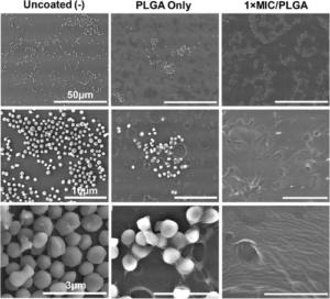
Scanning electron microscope images of endotracheal tubes at three levels of magnification. After 24 hours of Staphylococcus epidermidis exposure, tubes coated with the researchers’ AMPs (right) showed decreased biofilm production, as compared with tubes coated with just polymer (center) and uncoated tubes (left).
Endotracheal tubes are a mainstay of hospital care, as they ensure a patient’s airway is clear when they can’t breathe on their own. However, keeping a foreign object inserted in this highly sensitive part of the anatomy comes is not without risk, such as the possibility of infection, inflammation and a condition known as subglottic stenosis, in which scar tissue narrows the airway.
Broad-spectrum antibiotics are one way to mitigate these risks, but come with risks of their own, including harming beneficial bacteria and contributing to antibiotic resistance.
With this conundrum in mind, Riccardo Gottardi, Assistant Professor of Pediatrics at the Children’s Hospital of Philadelphia (CHOP) and of Bioengineering at Penn Engineering, along with Bioengineering graduate students and lab members Matthew Aronson and Paul Gehret, are developing endotracheal tubes that can provide a more targeted antimicrobial defense.
In a proof-of-concept study published in the journal The Laryngoscope, the team showed how a different type of antimicrobial agent could be incorporated into the tubes’ polymer coating, as well as preliminary results suggesting these devices would better preserve a patient’s microbiome.
Instead, the investigators explored the use of antimicrobial peptides (AMPs), which are small proteins that destabilize bacterial membranes, causing bacterial cells to fall apart and die. This mechanism of action allows them to target specific bacteria and makes them unlikely to promote antimicrobial resistance. Prior studies have shown that it is possible to coat endotracheal tubes with conventional antibiotics, so the research team investigated the possibility of incorporating AMPs into polymer-coated tubes to inhibit bacterial growth and modulate the upper-airway microbiome.
The researchers, led by Matthew Aronson, a graduate student in Penn Engineering’s Department of Bioengineering, tested their theory by creating a polymer coating that would release Lasioglossin-III, an AMP with broad-spectrum antibacterial activity. They found that Lasio released from coated endotracheal tubes, reached the expected effective concentration rapidly and continued to release at the same concentration for a week, which is the typical timeframe that an endotracheal is used before being changed. The investigators also tested their drug-eluting tube against airway microbes, including S. epidermidis, S. pneumoniae, and human microbiome samples and observed significant antibacterial activity, as well as prevention of bacterial adherence to the tube.
Read “CHOP Researchers Develop Coating for Endotracheal Tubes that Releases Antimicrobial Peptides” at CHOP News.
A collaborative study finds that deeper regions of the brain encode visual information more slowly, enabling the brain to identify fast-moving objects and images more accurately and persistently.
by Erica K. Brockmeier
Busy pedestrian crossing at Hong Kong
New research from the University of Pennsylvania, the Scuola Internazionale Superiore de Studi Avanzati (SISSA), and KU Leuven details the time scales of visual information processing across different regions of the brain. Using state-of-the-art experimental and analytical techniques, the researchers found that deeper regions of the brain encode visual information slowly and persistently, which provides a mechanism for explaining how the brain accurately identifies fast-moving objects and images. The findings were published in Nature Communications.
Understanding how the brain works is a major research challenge, with many theories and models developed to explain how complex information is processed and represented. One area of particular interest is vision, a major component of neural activity. In humans, for example, there is evidence that around half of the neurons in the cortex are related to vision.
Researchers are eager to understand how the visual cortex can process and retain information about objects in motion in a way that allows people to take in dynamic scenes while still retaining information about and recognizing the objects around them.
“One of the biggest challenges of all the sensory systems is to maintain a consistent representation of our surroundings, despite the constant changes taking place around us. The same holds true for the visual system,” says Davide Zoccolan, director of SISSA’s Visual Neuroscience Laboratory. “Just look around us: objects, animals, people, all on the move. We ourselves are moving. This triggers rapid fluctuations in the signals acquired by the retina, and until now it was unclear whether the same type of variations apply to the deeper layers of the visual cortex, where information is integrated and processed. If this was the case, we would live in tremendous confusion.”
Experiments using static stimuli, such as photographs, have found that information from the sensory periphery are processed in the visual cortex according to a finely tuned hierarchy. Deeper regions of the brain then translate this information about visual scenes into more complex shapes, objects, and concepts. But how this process works in more dynamic, real-world settings is not well understood.
To shed light on this, the researchers analyzed neural activity patterns in multiple visual cortical areas in rodents while they were being shown dynamic visual stimuli. “We used three distinct datasets: one from SISSA, one from a group in KU Leuven led by Hans Op de Beeck and one from the Allen Institute for Brain Science in Seattle,” says Zoccolan. “The visual stimuli used in each were of different types. In SISSA, we created dedicated video clips showing objects moving at different speeds. The other datasets were acquired using various kinds of clips, including from films.”
Next, the researchers analyzed the signals registered in different areas of the visual cortex through a combination of sophisticated algorithms and models developed by Penn’s Eugenio Pasini and Vijay Balasubramanian. To do this, the researchers developed a theoretical framework to help connect the images in the movies to the activity of specific neurons in order to determine how neural signals evolve over different time scales.
“The art in this science was figuring out an analysis method to show that the processing of visual images is getting slower as you go deeper and deeper in the brain,” says Balasubramanian. “Different levels of the brain process information over different time scales; some things could be more stable, some quicker. It’s very hard to tell if the time scales across the brain are changing, so our contribution was to devise a method for doing this.”
Vijay Balasubramanian is the Cathy and Marc Lasry Professor in the Department of Physics and Astronomy in the School of Arts & Sciences and a member of the Penn Bioengineering Graduate Group at the University of Pennsylvania.
Colin Huber, a Ph.D. candidate in Bioengineering studying head impact biomechanics and concussion in sports at the Center for Injury Research and Prevention (CIRP) at the Children’s Hospital of Philadelphia (CHOP), recently published “Variations in Head Impact Rates in Male and Female High School Soccer” in Medicine & Science in Sports & Exercise with colleagues from CHOP’s Minds Matter Concussion Frontier Program and the CIRP.
Colin’s paper, the goal of which was to compare “to compare head impact exposure rates (head impacts/exposure period) in male and female high school soccer by using multiple methodological approaches,” was recently profiled in the Penn Engineering Research & Innovation Newsletter.