Penn Engineering’s Latest ‘Organ-On-a-Chip’ is a New Way to Study Cancer-related Muscle Wasting

by Melissa Pappas

Bioengineering’s Dan Huh and colleagues have developed a number of organ-on-a-chip devices to simulate how human cells grow and perform in their natural environments. Their latest is a muscle-on-a-chip, which carefully captures the directionality of muscle cells as they anchor themselves within the body. See the full infographic at the bottom of this story. (Illustration by Melissa Pappas).

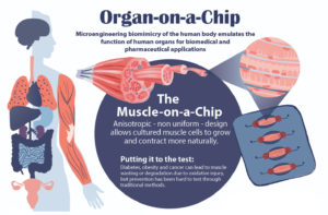
Studying drug effects on human muscles just got easier thanks to a new “muscle-on-a-chip,” developed by a team of researchers from Penn’s School of Engineering and Applied Science and Inha University in Incheon, Korea.

Muscle tissue is essential to almost all of the body’s organs, however, diseases such as cancer and diabetes can cause muscle tissue degradation or “wasting,” severely decreasing organ function and quality of life. Traditional drug testing for treatment and prevention of muscle wasting is limited through animal studies, which do not capture the complexity of the human physiology, and human clinical trials, which are too time consuming to help current patients.

An “organ-on-a-chip” approach can solve these problems. By growing real human cells within microfabricated devices, an organ-on-a-chip provides a way for scientists to study replicas of human organs outside of the body.

Using their new muscle-on-a-chip, the researchers can safely run muscle injury experiments on human tissue, test targeted cancer drugs and supplements, and determine the best preventative treatment for muscle wasting.

organ-on-a-chip
Dan Huh, Ph.D.

This research was published in Science Advances and was led by Dan Huh, Associate Professor in the Department of Bioengineering, and Mark Mondrinos, then a postdoctoral researcher in Huh’s lab and currently an Assistant Professor of Biomedical Engineering at Tulane University. Their co-authors included Cassidy Blundell and Jeongyun Seo, former Ph.D. students in the Huh lab, Alex Yi and Matthew Osborn, then research technicians in the Huh lab, and Vivek Shenoy, Eduardo D. Glandt President’s Distinguished Professor in the Department of Materials Science and Engineering. Lab members Farid Alisafaei and Hossein Ahmadzadeh also contributed to the research. The team collaborated with Insu Lee and professors Sun Min Kim and Tae-Joon Jeon of Inha University.

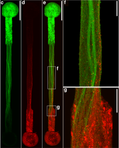
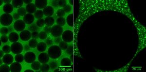
In order to conduct meaningful drug testing with their devices, the research team needed to ensure that cultured structures within the muscle-on-a-chip were as close to the real human tissue as possible. Critically, they needed to capture muscle’s “anisotropic,” or directionally aligned, shape.

“In the human body, muscle cells adhere to specific anchor points due to their location next to ligament tissue, bones or other muscle tissue,” Huh says. “What’s interesting is that this physical constraint at the boundary of the tissue is what sculpts the shape of muscle. During embryonic development, muscle cells pull at these anchors and stretch in the spaces in between, similar to a tent being held up by its poles and anchored down by the stakes. As a result, the muscle tissue extends linearly and aligns between the anchoring points, acquiring its characteristic shape.”

The team mimicked this design using a microfabricated chip that enabled similar anchoring of human muscle cells, sculpting three-dimensional tissue constructs that resembled real human skeletal muscle.

The the full story in Penn Engineering Today.

How HIV Infection Shrinks the Brain’s White Matter

by Katherine Unger Baillie

Researchers from Penn and CHOP detail the mechanism by which HIV infection blocks the maturation process of brain cells that produce myelin, a fatty substance that insulates neurons.

A confocal microscope image shows an oligodendrocyte in cell culture, labeled to show the cell nucleus in blue and myelin proteins in red, green, and yellow. Researchers from Penn and CHOP have shown that HIV infection prevents oligodendrocytes from maturing, leading to a reduction in white matter in the brain. (Image: Raj Putatunda)

It’s long been known that people living with HIV experience a loss of white matter in their brains. As opposed to gray matter, which is composed of the cell bodies of neurons, white matter is made up of a fatty substance called myelin that coats neurons, offering protection and helping them transmit signals quickly and efficiently. A reduction in white matter is associated with motor and cognitive impairment.

Earlier work by a team from the University of Pennsylvania and Children’s Hospital of Philadelphia (CHOP) found that antiretroviral therapy (ART)—the lifesaving suite of drugs that many people with HIV use daily—can reduce white matter, but it wasn’t clear how the virus itself contributed to this loss.

In a new study using both human and rodent cells, the team has hammered out a detailed mechanism, revealing how HIV prevents the myelin-making brain cells called oligodendrocytes from maturing, thus putting a wrench in white matter production. When the researchers applied a compound blocking this process, the cells were once again able to mature.

The work is published in the journal Glia.

“Even when people with HIV have their disease well-controlled by antiretrovirals, they still have the virus present in their bodies, so this study came out of our interest in understanding how HIV infection itself affects white matter,” says Kelly Jordan-Sciutto, a professor in Penn’s School of Dental Medicine and co-senior author on the study. “By understanding those mechanisms, we can take the next step to protect people with HIV infection from these impacts.”

“When people think about the brain, they think of neurons, but they often don’t think about white matter, as important as it is,” says Judith Grinspan, a research scientist at CHOP and the study’s other co-senior author. “But it’s clear that myelination is playing key roles in various stages of life: in infancy, in adolescence, and likely during learning in adulthood too. The more we find out about this biology, the more we can do to prevent white matter loss and the harms that can cause.”

Jordan-Sciutto and Grinspan have been collaborating for several years to elucidate how ART and HIV affect the brain, and specifically oligodendrocytes, a focus of Grinspan’s research. Their previous work on antiretrovirals had shown that commonly used drugs disrupted the function of oligodendrocytes, reducing myelin formation.

In the current study, they aimed to isolate the effect of HIV on this process. Led by Lindsay Roth, who recently earned her doctoral degree within the Biomedical Graduate Studies group at Penn and completed a postdoctoral fellowship working with Jordan-Sciutto and Grinspan, the investigation began by looking at human macrophages, one of the major cell types that HIV infects.

Read the full story in Penn Today.

Kelly Jordan-Sciutto is vice chair and professor in the University of Pennsylvania School of Dental Medicine’s Department of Basic & Translational Sciences and is director of Biomedical Graduate Studies. She is a member of the Penn Bioengineering Graduate Group.

Modified Nanoparticles Can Stop Osteoarthritis Development

Zhiliang Cheng

As we age, the cushioning cartilage between our joints begins to wear down, making it harder and more painful to move. Known as osteoathritis, this extremely common condition has no known cure; if the symptoms can’t be managed, the affected joints must be surgically replaced.

Now, researchers are exploring whether their specially designed nanoparticles can deliver a new inflammation inhibitor to joints, targeting a previously overlooked enzyme called sPLA2.

Zhiliang Cheng, a research associate professor in the Department of Bioengineering, recently collaborated with members of Penn Medicine’s McKay Orthopaedic Research Laboratory, on a study of this approach, published in the journal Science Advances.

The normal function of sPLA2 is to provide lipids (fats) that promote a variety of inflammation processes. The enzyme is always present in cartilage tissue, but typically in low levels. However, when the researchers examined mouse and human cartilage taken from those with osteoarthritis, disproportionately high levels of the enzyme were discovered within the tissue’s structure and cells.

“This marked increase strongly suggests that sPLA2 plays a role in the development of osteoarthritis,” said the study’s corresponding author, Zhiliang Cheng, PhD, a research associate professor of Bioengineering. “Being able to demonstrate this showed that we were on the right track for what could be a potent target for the disease.”

The next step was for the study team – which included lead author Yulong Wei, MD, a researcher in Penn Medicine’s McKay Orthopaedic Research Laboratory – to put together a nanoparticle loaded with an sPLA2 inhibitor. This would block the activity of sPLA2 enzyme and, they believed, inflammation. These nanoparticles were mixed with animal knee cartilage in a lab, then observed as they diffused deeply into the dense cartilage tissue. As time progressed, the team saw that the nanoparticles stayed there and did not degrade significantly or disappear. This was important for the type of treatment the team envisioned.

Continue reading at Penn Medicine News.

Originally posted in Penn Engineering Today.

“New Biosealant Can Stabilize Cartilage, Promote Healing After Injury”

New research from Robert Mauck, Mary Black Ralston Professor in Orthopaedic Surgery and Bioengineering and Director of Penn Medicine’s McKay Orthopaedic Research Laboratory, announces a “new biosealant therapy may help to stabilize injuries that cause cartilage to break down, paving the way for a future fix or – even better – begin working right away with new cells to enhance healing.” Their research was published in Advanced Healthcare Materials. The study’s lead author was Jay Patel, a former postdoctoral fellow in the McKay Lab and now Assistant Professor at Emory University and was contributed to by Claudia Loebel, a postdoctoral research in the Burdick lab and who will begin an appointment as Assistant Professor at the University of Michigan in Fall 2021. In addition, the technology detailed in this publication is at the heart of a new company (Forsagen LLC) spun out of Penn with support from the Penn Center for Innovation (PCI) Ventures Program, which will attempt to spearhead the system’s entry into the clinic. It is co-founded by both Mauck and Patel, along with study co-author Jason Burdick, Professor in Bioengineering, and Ana Peredo, a PhD student in Bioengineering.

Read the story in Penn Medicine News.

Penn, CHOP and Yale Researchers’ Molecular Simulations Uncover How Kinase Mutations Lead to Cancer Progression

by Evan Lerner

A computer model of a mutated anaplastic lymphoma kinase (ALK), a known oncogenic driver in pediatric neuroblastoma.

Kinases are a class of enzymes that are responsible for transferring the main chemical energy source used by the body’s cells. As such, they play important roles in diverse cellular processes, including signaling, differentiation, proliferation and metabolism. But since they are so ubiquitous, mutated versions of kinases are frequently found in cancers. Many cancer treatments involve targeting these mutant kinases with specific inhibitors.

Understanding the exact genetic mutations that lead to these aberrant kinases can therefore be critical in predicting the progression of a given patient’s cancer and tailoring the appropriate response.

To achieve this understanding on a more fundamental level, a team of researchers from the University of Pennsylvania’s School of Engineering and Applied Science and Perelman School of Medicine, the Children’s Hospital of Philadelphia (CHOP) and researchers at the Yale School of Medicine’s Cancer Biology Institute, have constructed molecular simulations of a mutant kinase implicated in pediatric neuroblastoma, a childhood cancer impacting the central nervous system.

Using their computational model to study the relationship between single-point changes in the kinase’s underlying gene and the altered structure of the protein it ultimately produces, the researchers revealed useful commonalities in the mutations that result in tumor formation and growth. Their findings suggest that such computational approaches could outperform existing profiling methods for other cancers and lead to more personalized treatments.

The study, published in the Proceedings of the National Academy of Sciences, was led by Ravi Radhakrishnan, Professor and chair of Penn Engineering’s Department of Bioengineering and professor in its Department of Chemical and Biomolecular Engineering, and Mark A. Lemmon, Professor of Pharmacology at Yale and co-director of Yale’s Cancer Biology Institute. The study’s first authors were Keshav Patil, a graduate student in Penn Engineering’s Department of Chemical and Biomolecular Engineering, along with Earl Joseph Jordan and Jin H. Park, then members of the Graduate Group in Biochemistry and Molecular Biology in Penn’s Perelman School of Medicine. Krishna Suresh, an undergraduate student in Radhakrishnan’s lab, Courtney M. Smith, a graduate student in Lemmon’s lab, and Abigail A. Lemmon, an undergraduate in Lemmon’s lab, contributed to the study. They collaborated with Yaël P. Mossé, Associate Professor of Pediatrics at Penn Medicine and in the division of oncology at CHOP.

“Some cancers rely on the aberrant activation of a single gene product for tumor initiation and progression,” says Radhakrishnan. “This unique mutational signature may hold the key to understanding which patients suffer from aggressive forms of the disease or for whom a given therapeutic drug may yield short- or long-term benefits. Yet, outside of a few commonly occurring ‘hotspot’ mutations, experimental studies of clinically observed mutations are not commonly pursued.”

Read the full post in Penn Engineering Today.

Penn Bioengineers Develop Implantable Living Electrodes

Living Electrode Panels (image courtesy of the Cullen Lab)

Connecting the human brain to electrical devices is a long-standing goal of neuroscientists, bioengineers, and clinicians, with applications ranging from deep brain stimulation (DBS) to treat Parkinson’s disease to more futuristic endeavors such as Elon Musk’s NeuraLink initiative to record and translate brain activity. However, these approaches currently rely on using implantable metallic electrodes that inherently provoke a lasting immune response due to their non-biological nature, generally complicating the reliability and stability of these interfaces over time. To address these challenges, D. Kacy Cullen, Associate Professor in Neurosurgery and Bioengineering, and Dayo Adewole, a doctoral candidate in Bioengineering, worked with a multi-disciplinary team of collaborators to develop the first “living electrodes” as an implantable, biological bridge between the brain and external devices. In a recent article published in Science Advances, the team demonstrated the fabrication of hair-like microtissue comprised of living neuronal networks and bundled tracts of axons the signal sending fibers of the nervous system protected within soft hydrogel cylinders. They showed that these axon-based living electrodes could be fully controlled and monitored with light thus eliminating the need for electrical contact and are capable of surviving and forming synapses with the brain as demonstrated in an adult rat model. While further advancements are necessary prior to clinical use, the current findings provide the foundation for a new class of “living electrodes” as a biological intermediary between humans and devices capable of leveraging natural mechanisms to potentially provide a stable interface for clinical applications.

Cullen has a primary appointment in the Department of Neurosurgery in the Perelman School of Medicine, with a secondary appointment in the Department of Bioengineering in the School of Engineering and Applied Science, and an appointment in the Corporal Michael J. Crescenz VA Medical Center in Philadelphia.

Ravi Radhakrishnan Adapts Multiscale Modeling Course


Ravi Radhakrishnan, PhD

Ravi Radhakrishnan, Professor and Chair of the Department of Bioengineering and Professor in Chemical and Biomolecular Engineering, is among the many faculty who quickly adapted their courses to an online format in the wake of the COVID-19 pandemic. Now, a recent publication in the American Institute of Chemical Engineers (AIChE) Journal reflects one of these revamped courses. The course BE 559: “Multiscale Modeling of Chemical and Biological Systems” provides theoretical, conceptual, and hands-on modeling experience on three different length and time scales: (1) electronic structure (A, ps); (2) molecular mechanics (100A, ns); and (3) deterministic and stochastic approaches for microscale systems (um, sec). During the course, students gained hands-on experience in running codes on real applications together with the following theoretical formalisms: molecular dynamics, Monte Carlo, free energy methods, deterministic and stochastic modeling. The transition to the online format was greatly facilitated by a grant from the Extreme Science and Engineering Discovery Environment (XSEDE) which provided cloud and supercomputing resources to the students facilitating the computational laboratory experience. Radhakrishnan’s article, “A survey of multiscale modeling: Foundations, historical milestones, current status, and future prospects,” reviews the foundations, historical developments, and current paradigms in multiscale modeling (MSM).

Radhakrishnan aspires to modernize computational science, integrating Multiscale Modeling and Data Science for Biological and Biomedical Science & Engineering. His team does so by integrating multiphysics modeling, computing, data science to tackle applications. The integrative approach is pictorially depicted here in terms of modeling different length and timescales using techniques such as molecular dynamics of atomistic systems, Brownian dynamics of coarse-grained systems, and field equations governing continuum scales of macroscopic systems.

Read the full article in the AIChE Journal: https://doi.org/10.1002/aic.17026

Funding source: National Institutes of Health, Grant/Award Number: CA227550

Penn Engineers Devise Easier Way of Sneaking Antibodies into Cells

Getting a complex protein like an antibody through the membrane of a cell without damaging either is a long-standing challenge in the life sciences. Penn Engineers have found a plug-and-play solution that makes antibodies compatible with the delivery vehicles commonly used to ferry nucleic acids across that barrier.

For almost any conceivable protein, corresponding antibodies can be developed to block it from binding or changing shape, which ultimately prevents it from carrying out its normal function. As such, scientists have looked to antibodies as a way of shutting down proteins inside cells for decades, but there is still no consistent way to get them past the cell membrane in meaningful numbers.

Now, Penn Engineering researchers have figured out a way for antibodies to hitch a ride with transfection agents, positively charged bubbles of fat that biologists routinely use to transport DNA and RNA into cells. These delivery vehicles only accept cargo with a highly negative charge, a quality that nucleic acids have but antibodies lack. By designing a negatively charged amino acid chain that can be attached to any antibody without disrupting its function, they have made antibodies broadly compatible with common transfection agents.

Beyond the technique’s usefulness towards studying intracellular dynamics, the researchers conducted functional experiments with antibodies that highlight the technique’s potential for therapeutic applications. One antibody blocked a protein that decreases the efficacy of certain drugs by prematurely ejecting them from cells. Another blocked a protein involved in the transcription process, which could be an even more fundamental way of knocking out proteins with unwanted effects.

Andrew Tsourkas and Hejia Henry Wang

The study, published in the Proceedings of the National Academy of Sciences, was conducted by Andrew Tsourkas, professor in the Department of Bioengineering, and Hejia Henry Wang, a graduate student in his lab.

Read the full story at the Penn Engineering Medium Blog. Media contact Evan Lerner.

Penn Engineers Solve the Paradox of Why Tissue Gets Stiffer When Compressed

The researchers’ experiments involved making synthetic tissues with artificial “cells.” The fibrin network that surrounds these beads pull on them when compressed; by changing the number of beads in their experimental tissues, the researchers could suss out how cell-fiber interplay contributes to the tissue’s overall properties.

Tissue gets stiffer when it’s compressed. That property can become even more pronounced with injury or disease, which is why doctors palpate tissue as part of a diagnosis, such as when they check for lumps in a cancer screening. That stiffening response is a long-standing biomedical paradox, however: tissue consists of cells within a complex network of fibers, and common sense dictates that when you push the ends of a string together, it loosens tension, rather than increasing it.

Now, in a study published in Nature, University of Pennsylvania’s School of Engineering and Applied Science researchers have solved this mystery by better understanding the mechanical interplay between that fiber network and the cells it contains.

The researchers found that when tissue is compressed, the cells inside expand laterally, pulling on attached fibers and putting more overall tension on the network. Targeting the proteins that connect cells to the surrounding fiber network might therefore be the optimal way of reducing overall tissue stiffness, a goal in medical treatments for everything from cancer to obesity.

Headshots of Paul Janmey and Vivek Shenoy

Paul Janmey and Vivek Shenoy

The study was led by Paul Janmey, Professor in the Perelman School of Medicine’s Department of Physiology and in Penn Engineering’s Department of Bioengineering, and Vivek Shenoy, Eduardo D. Glandt President’s Distinguished Professor in Penn Engineering’s Department of Materials Science and Engineering, Mechanical Engineering and Applied Mechanics, and Bioengineering, along with Anne van Oosten and Xingyu Chen, graduate students in Janmey’s and Shenoy’s labs. Van Oosten is now a postdoctoral fellow at Leiden University in The Netherlands.

Shenoy is Director of Penn’s Center for Engineering Mechanobiology, which studies how physical forces influence the behavior of biological systems; Janmey is the co-director of one of the Center’s working groups, organized around the question, “How do cells adapt to and change their mechanical environment?”

Together, they have been interested in solving the paradox surrounding tissue stiffness.

Read the full story on the Penn Engineering Medium Blog.

Penn BE Undergraduates’ Plate Reader Design Published

Microplate reader, Wikimedia Commons

In a paper recently published in Biochemistry, a group of University of Pennsylvania Bioengineering students describe the results of their work designing a new, open-source, low-cost microplate reader. Plate readers are instruments designed to measure light absorption and fluorescence emission from molecules useful for clinical biomarker analyses and assays in a diverse array of fields including synthetic biology, optogenetics, and photosensory biology. This new design costs less than $3500, a significantly lower price than other commercially available alternatives. As described in the paper’s abstract, this design is the latest in a growing trend of open-source  hardware to enhance access to equipment for biology labs. The project originated as part of the annual International Genetically Engineering Machine Competition (iGEM), an annual worldwide competition focusing on “push[ing] the boundaries of synthetic biology by tackling everyday issues facing the world” (iGEM website).

The group consists of current junior Andrew Clark (BSE ’20) and recent graduates Karol Szymula (BSE ’18), who works in the lab of Dr. Danielle Bassett, and Michael Patterson (BSE ’18), a Master’s student in Bioengineering and Engineer of Instructional Laboratories. Assistant Professor of Bioengineering Dr. Brian Chow served as their faculty mentor alongside Director of Instructional Labs Sevile Mannickarottu and Michael Magaraci, a Ph.D. candidate in Bioengineering, all of whom serve as co-authors on the published article. The research and design of the project was conducted in the Stephenson Foundation Bioengineering Educational Laboratory here at the University of Pennsylvania’s Department of Bioengineering.