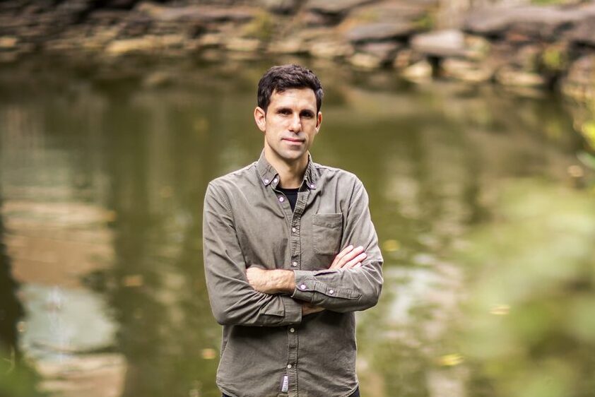
Cesar de la Fuente, Presidential Assistant Professor with appointments in the Perelman School of Medicine, School of Engineering and School of Arts & Sciences (Image: Eric Sucar)
In a significant advance against the growing threat of antibiotic-resistant bacteria, researchers have identified a novel class of antimicrobial agents known as encrypted peptides, which may expand the immune system’s arsenal of tools to fight infection. The findings, published in Trends in Biotechnology by Cell Press, reveal that many antimicrobial molecules originate from proteins not traditionally associated with immune responses.
Unlike conventional antibiotics that target specific bacterial processes, these newly discovered peptides disrupt the protective membranes surrounding bacterial cells. By inserting themselves into these membranes—much like breaching a fortress wall—the peptides destabilize and ultimately destroy the bacteria.
“Our findings suggest that these previously overlooked molecules could be key players in the immune system’s response to infection,” says César de la Fuente, presidential assistant professor in bioengineering and in chemical and biomolecular engineering in the School of Engineering and Applied Science, in psychiatry and microbiology in the Perelman School of Medicine, and in chemistry in the School of Arts & Sciences, who led the research team. “This may not only redefine how we understand immunity but also opens up new possibilities for treating drug-resistant infections.”
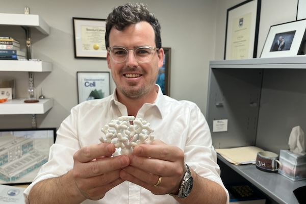
Alex Hughes, Assistant Professor in Bioengineering, holds a model of a developing kidney. (Credit: Bella Ciervo)
To Alex Hughes, Assistant Professor in Bioengineering within Penn Engineering and in Cell and Developmental Biology within Penn Medicine, the kidney is a work of art. “I find the development of the kidney to be a really beautiful process,” says Hughes.
Most people only ever see the organ in cross-section, through textbooks or by dissecting animal kidneys in high school biology class: a bean-shaped slice with lots of tiny tubes. “I think that really undersells how amazing the structure is,” says Hughes, who points out that kidneys grow in utero like forests of pipes, branching exponentially.
Densely packed with tubules clustered in units known as nephrons, kidneys cleanse the blood, maintaining the body’s fluid and electrolyte balance, while also regulating blood pressure. The organ played a crucial role in vertebrates emerging from the ocean: as one paper puts it, kidneys preserve the primordial ocean in all of us.
Unfortunately, kidneys struggle in the modern world. Excessively salty food, being overweight, not exercising enough, drinking too much and smoking can all raise blood pressure, which damages the kidney’s tiny blood vessels, as does diabetes.
In some cases, damage to the kidney’s nephrons can be slowed with lifestyle changes, but, unlike the liver, bones and skin, which can regrow damaged tissue, kidneys have a limited capacity to regenerate. At present, without a transplant, the nephrons we have at birth must last a lifetime.
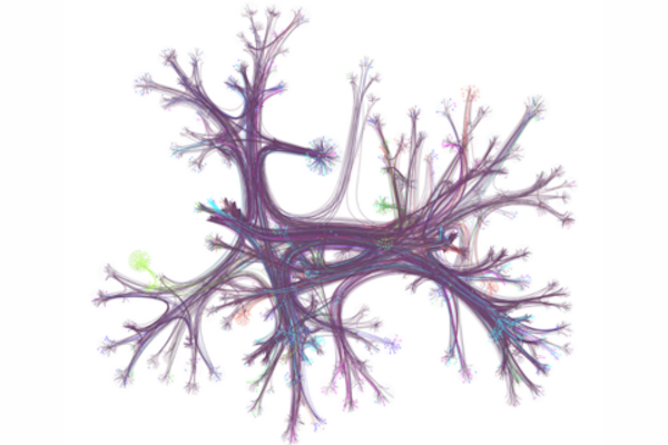
A hyperlink network from English Wikipedia, with only 0.1% of articles (nodes) and their connections (edges) visualized. Seven different reader journeys through this network are highlighted in various colors. The network is organized by topic and displayed using a layout that groups related articles together. (Image: Dale Zhou)
At one point or another, you may have gone online looking for a specific bit of information and found yourself “going down the Wiki rabbit hole” as you discover wholly new, ever-more fascinating related topics — some trivial, some relevant — and you may have gone so far down the hole it’s difficult to piece together what brought you there to begin with.
According to the University of Pennsylvania’s Dani Bassett, who recently worked with a collaborative team of researcher to examine the browsing habits of 482,760 Wikipedia readers from 50 different countries, this style of information acquisition is called the “busybody.” This is someone who goes from one idea or piece of information to another, and the two pieces may not relate to each other much.
“The busybody loves any and all kinds of newness, they’re happy to jump from here to there, with seemingly no rhyme or reason, and this is contrasted by the ‘hunter,’ which is a more goal-oriented, focused person who seeks to solve a problem, find a missing factor, or fill out a model of the world,” says Bassett.
In the research, published in the journal Science Advances, Bassett and colleagues discovered stark differences in browsing habits between countries with more education and gender equality versus less equality, raising key questions about the impact of culture on curiosity and learning.
Dani S. Bassett is the J. Peter Skirkanich Professor at the University of Pennsylvania with a primary appointment in the School of Engineering and Applied Science’s Department of Bioengineering and secondary appointments in the School of Arts & Sciences’ Department of Physics & Astronomy, Penn Engineering’s Department of Electrical and Systems Engineering, and the Perelman School of Medicine’s Departments of Neurology and Psychiatry.
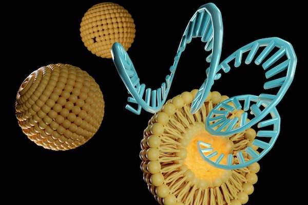
By adjusting the chemical structure of lipid nanoparticles (LNPs), Penn Engineers have discovered how to target specific organs, a major breakthrough in precision medicine. (Love Employee via Getty Images)
Penn Engineers have discovered a novel means of directing lipid nanoparticles (LNPs), the revolutionary molecules that delivered the COVID-19 vaccines, to target specific tissues, presaging a new era in personalized medicine and gene therapy.
While past research — including at Penn Engineering — has screened “libraries” of LNPs to find specific variants that target organs like the lungs, this approach is akin to trial and error. “We’ve never understood how the structure of one key component of the LNP, the ionizable lipid, determines the ultimate destination of LNPs to organs beyond the liver,” says Michael J. Mitchell, Associate Professor in Bioengineering.
In a new paper published in Nature Nanotechnology, Mitchell’s group describes how subtle adjustments to the chemical structure of the ionizable lipid, a key component of the LNP, allows for tissue-specific delivery, in particular to the liver, lungs and spleen.
In a collaborative interdisciplinary study, Michael Mitchell of the School of Engineering and Applied Science, Wei Guo of the School of Arts & Sciences, and Drew Weissman of the Perelman School of Medicine show that solid tumors can block drug-delivery mechanisms with a “forcefield-like” effect but certain genetic elements that can effectively “shut down” the forcefield. Their findings hint at new targets for delivering cancer treatments that use the body’s immune system to fight tumors. (Image: iStock / CIPhotos)
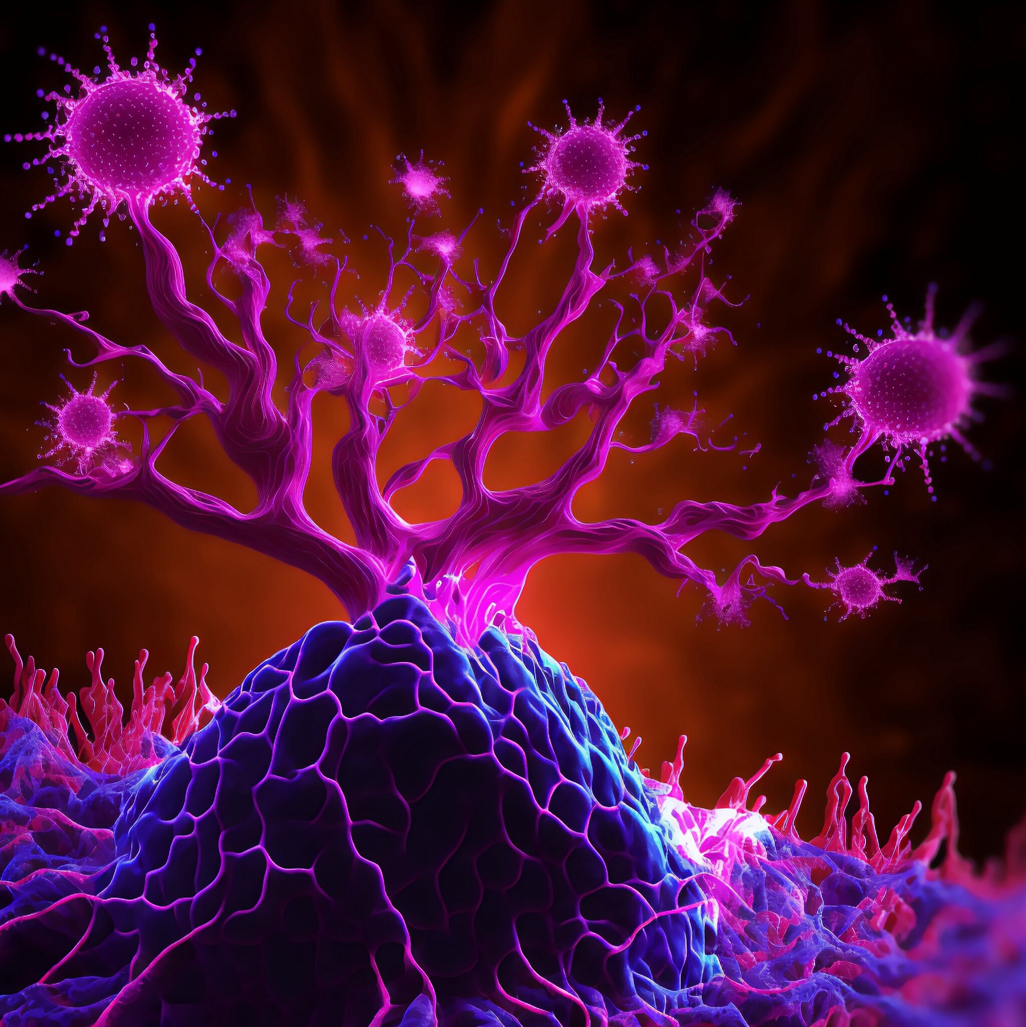
The tumor microenvironment—an ad hoc, messy amalgamation of signaling molecules, immune cells, fibroblasts, blood vessels, and the extracellular matrix—acts like a “powerful security system that protects solid tumors from invaders seeking to destroy them,” says Michael Mitchell, a bioengineer at the University of Pennsylvania working on nanoscale therapeutics aimed at targeting cancers.
“A lot like the Death Star with its surrounding fleet of fighter ships and protective shields, solid tumors can use features like immune cells and vasculature to exert force, acting as a physical barrier to rebel forces (nanoparticles) coming in to deliver the payload that destroys it,” Mitchell says.
Now, researchers in the Mitchell lab have teamed up with Wei Guo’s group in the School of Arts & Sciences at Penn and Drew Weissman of the Perelman School of Medicine to figure out the molecular mechanisms that make tumor microenvironments seemingly impenetrable and found that small extracellular vesicles (sEVs) are secreted by tumor cells and act as a “forcefield,” blocking therapeutics. Their findings are published in Nature Materials.
“This discovery reveals how tumors create a robust defense system, making it challenging for nanoparticle-based therapies to reach and effectively target cancer cells,” Guo says. “By understanding the cellular mechanisms driving these responses, we can potentially develop strategies to disable this defense, allowing therapeutics to penetrate and attack the tumor more efficiently.”
The research builds on a prior collaboration between Guo and Mitchell’s labs, wherein the teams focused on how tumor-associated immune cells, known as macrophages, contribute to the suppression of anti-tumor immunity by secreting extracellular vesicles.
Wei Guo is the Hirsch Family President’s Distinguished Professor in the Department of Biology in Penn’s School of Arts & Sciences.
Ningqiang Gong, a former postdoctoral researcher in the Mitchell lab at Penn Engineering, is an assistant professor at the University of Science and Technology of China.
Wenqun Zhong is a reseearch associate in the Guo Laboratory in Penn Arts & Sciences.
Other authors include: Alex G Hamilton, Dongyoon Kim, Junchao Xu, and Lulu Xue of Penn Engineering; Junhyong Kim, Zhiyuan Qin, and Fengyuan Xu of Penn Arts & Sciences; Mohamad-Gabriel Alameh and Drew Weissman of the Perelman School of Medicine; Andrew E. Vaughn and Gan Zhao of the Penn School of Veterinary Medicine; Jinghong Li and Xucong Teng of the University of Beijing; and Xing-Jie Liang of the Chinese Academy of Sciences.
This research received support from the U.S. National Institutes of Health (DP2 TR002776, R35 GM141832, and NCI P50 CA261608), Burroughs Wellcome Fund, U.S. National Science Foundation CAREER Award (CBET-2145491), and an American Cancer Society Research Scholar Grant (RGS-22-1122-01-ET.)
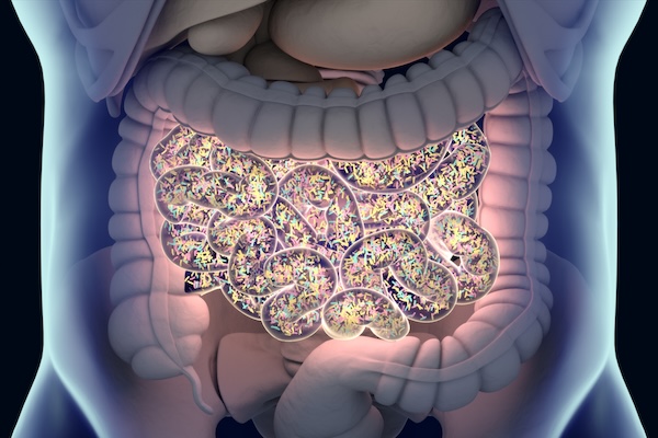
Penn Engineering and Stanford researchers leveraged AI to discover dozens of potential new antibiotics in the human gut microbiome. (ChrisChrisW via Getty Images)
The average human gut contains roughly 100 trillion microbes, many of which are constantly competing for limited resources. “It’s such a harsh environment,” says César de la Fuente, Presidential Assistant Professor in Bioengineering and in Chemical and Biomolecular Engineering within the School of Engineering and Applied Science, in Psychiatry and Microbiology within the Perelman School of Medicine, and in Chemistry within the School of Arts & Sciences. “You have all these bacteria coexisting, but also fighting each other. Such an environment may foster innovation.”
In that conflict, de la Fuente’s lab sees potential for new antibiotics, which may one day contribute to humanity’s own defensive stockpile against drug-resistant bacteria. After all, if the bacteria in the human gut have to develop new tools in the fight against one another to survive, why not use their own weapons against them?
In a new paper in Cell, the labs of de la Fuente and Ami S. Bhatt, Professor in Medicine (Hematology) and Genetics at Stanford, surveyed the gut microbiomes of nearly 2,000 people, discovering dozens of potential new antibiotics. “We think of biology as an information source,” says de la Fuente. “Everything is just code. And if we can come up with algorithms that can sort through that code, we can dramatically accelerate antibiotic discovery.”
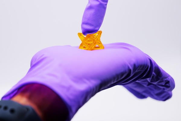
Biomaterials 3D-printed with the new method can be used inside the body and could even serve as bandages on a beating human heart. (Photo by Casey A. Cass/University of Colorado)
In the quest to develop life-like materials to replace and repair human body parts, scientists face a formidable challenge: Real tissues are often both strong and stretchable and vary in shape and size.
A CU Boulder-led team, in collaboration with researchers at the University of Pennsylvania, has taken a critical step toward cracking that code. They’ve developed a new way to 3D print material that is at once elastic enough to withstand a heart’s persistent beating, tough enough to endure the crushing load placed on joints, and easily shapeable to fit a patient’s unique defects.
Their breakthrough, described in the Aug. 2 edition of the journal Science, helps pave the way toward a new generation of biomaterials, from internal bandages that deliver drugs directly to the heart to cartilage patches and needle-free sutures.
“This is a simple 3D processing method that people could ultimately use in their own academic labs as well as in industry to improve the mechanical properties of materials for a wide variety of applications,” says first author Abhishek Dhand, a researcher in the Burdick Lab and doctoral candidate in the Department of Bioengineering at the University of Pennsylvania. “It solves a big problem for 3D printing.”
Jason Burdick is Bowman Endowed Professor in Chemical and Biological Engineering at the University of Colorado Boulder and Adjunct Professor in Bioengineering at Penn Engineering.
The effectiveness of CAR T cell therapy against a variety of cancers, including solid tumors, could be boosted greatly by using CRISPR-Cas9 technology to knock out the gene for CD5, a protein found on the surface of T cells, according to a preclinical study from investigators at the University of Pennsylvania’s Perelman School of Medicine and Abramson Cancer Center.
CAR T cells are T cells that have been engineered to attack specific targets found on cancer cells. They have had remarkable results in some patients with blood cancers. But they have not performed well against other cancers including solid-tumor cancers, such as pancreatic cancer, prostate cancer, and melanoma. Researchers have been searching for techniques to boost the effectiveness of CAR T cell therapy.
The study, published today in Science Immunology, suggests that knocking out CD5 could be a prime technique. Illuminating the protein’s previously murky role, the researchers found that it works as a powerful immune checkpoint, reining in T cell effectiveness. Removing it, they showed, dramatically enhanced CAR T cell anticancer activity in a variety of preclinical cancer models.
“We’ve discovered in preclinical models that CD5 deletion greatly enhances the function of CAR T cells against multiple cancers,” said senior author Marco Ruella, MD, an assistant professor of Hematology-Oncology, researcher with the Center for Cellular Immunotherapies and the scientific director of Penn Medicine’s Lymphoma Program. “The striking effects we observed across preclinical models suggest that CD5 knockout could be a general strategy for enhancing CAR T cell function.”
The study’s first author is Ruchi Patel, PhD, a recent graduate student from the Ruella Laboratory.
How do we measure chaos and why would we want to? Together, Penn engineers Dani S. Bassett, J. Peter Skirkanich Professor in Bioengineering and in Electrical and Systems Engineering, and postdoctoral researcher Kieran Murphy leverage the power of machine learning to better understand chaotic systems, opening doors for new information analyses in both theoretical modeling and real-world scenarios.
Humans have been trying to understand and predict chaotic systems such as weather patterns, the movement of planets and population ecology for thousands of years. While our models have continued to improve over time, there will always remain a barrier to perfect prediction. That’s because these systems are inherently chaotic. Not in the sense that blue skies and sunshine can turn into thunderstorms and torrential downpours in a second, although that does happen, but in the sense that mathematically, weather patterns and other chaotic systems are governed by physics with nonlinear characteristics.
“This nonlinearity is fundamental to chaotic systems,” says Murphy. “Unlike linear systems, where the information you start with to predict what will happen at timepoints in the future stays consistent over time, information in nonlinear systems can be both lost and generated through time.”
Like a game of telephone where information from the original source gets lost as it travels from person to person while new words and phrases are added to fill in the blanks, outcomes in chaotic systems become harder to predict as time passes. This information decay thwarts our best efforts to accurately forecast the weather more than a few days out.
“You could put millions of probes in the atmosphere to measure wind speed, temperature and precipitation, but you cannot measure every single atom in the system,” says Murphy. “You must have some amount of uncertainty, which will then grow, and grow quickly. So while a prediction for the weather in a few hours might be fairly accurate, that growth in uncertainty over time makes it impossible to predict the weather a month from now.”
In their recent paper published in Physical Review Letters, Murphy and Bassett applied machine learning to classic models of chaos, physicists’ reproductions of chaotic systems that do not contain any external noise or modeling imperfections, to design a near-perfect measurement of chaotic systems to one day improve our understanding of systems including weather patterns.
“These controlled systems are testbeds for our experiments,” says Murphy. “They allow us to compare with theoretical predictions and carefully evaluate our method before moving to real-world systems where things are messy and much less is known. Eventually, our goal is to make ‘information maps’ of real-world systems, indicating where information is created and identifying what pieces of information in a sea of seemingly random data are important.”
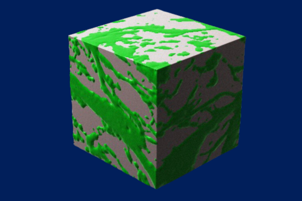
Bicontinuous materials, like this representation of a cube of gelatin and hyaluronic acid, have greater internal surface area, allowing cells to travel faster between two points. (Credit: Karen Xu)
One of the most important but least understood aspects of healing is cell migration, or the process of cells moving from one part of the body to another. “If you are an ambulance out in the woods,” says Karen Xu, an M.D/Ph.D. student in Medicine and Bioengineering, “and there are no paths for you to move forward, it will be a lot harder for you to get to a site that needs you.”
Earlier this year, Xu co-authored a paper in Nature Communications describing a new cue to help cells get to where they need to go: a material made chiefly of hyaluronic acid and gelatin, two gooey substances commonly found outside cells in joints and connective tissue.
“Hundreds of thousands of people tear their meniscus every year,” says Robert Mauck, Mary Black Ralston Professor in Orthopaedic Surgery in Penn Medicine and Professor in Bioengineering at Penn Engineering and one of Xu’s advisors, as well as a senior author on the paper. “This material could potentially speed up their recovery.”
What makes the material — known as a hydrogel due to its blend of gelatinous matter and water — unique is that the combination of hyaluronic acid and gelatin forms a complex network of paths, providing cells many different ways to travel between two points.
This property is known as bicontinuity, and is exemplified by two discrete continuous phases that are each connected throughout the entire volume of the material (for example with a sponge, with phases of cellulose and air; in the hydrogel, this is comprised of gelatin and hyaluronic acid) resulting in a dizzying array of patterns that dramatically increase the surface area inside the material.
To test the hydrogel’s efficacy, Xu and her collaborators — including co-advisor Jason Burdick, formerly the Robert D. Bent Professor in Bioengineering at Penn Engineering and now the Bowman Endowed Professor at the University of Colorado Boulder, and the paper’s other senior author — first created several different versions of the hydrogel to find the sweet spot at which the constituents formed the bicontinuous structure and had the highest internal surface area. “We found that a precise combination of the various hydrogel components and control over their mixing was needed to form the bicontinuous structure,” says Burdick.