A cross-kingdom partnership between bacteria and fungi can result in the two joining to form a “superorganism” with unusual strength and resilience. It may sound like the stuff of science fiction, but these microbial groupings are very much part of the here and now.
Found in the saliva of toddlers with severe childhood tooth decay, these assemblages can effectively colonize teeth. They were stickier, more resistant to antimicrobials, and more difficult to remove from teeth than either the bacteria or the fungi alone, according to the research team, led by University of PennsylvaniaSchool of Dental Medicine scientists.
What’s more, the assemblages unexpectedly sprout “limbs” that propel them to “walk” and “leap” to quickly spread on the tooth surface, despite each microbe on its own being non-motile, the team reported in the journal Proceedings of the National Academy of Sciences.
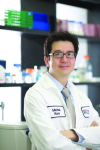
“This started with a very simple, almost accidental discovery, while looking at saliva samples from toddlers who develop aggressive tooth decay,” says Hyun (Michel) Koo, a professor at Penn Dental Medicine and a co-corresponding author on the paper. “Looking under the microscope, we noticed the bacteria and fungi forming these assemblages and developing motions we never thought they would possess: a ‘walking-like’ and ‘leaping-like’ mobility. They have a lot of what we call ‘emergent functions’ that bring new benefits to this assemblage that they could not achieve on their own. It’s almost like a new organism—a superorganism—with new functions.”
Hyun (Michel) Koo is a professor in the Department of Orthodontics and the divisions of Community Oral Health and Pediatric Dentistry in the School of Dental Medicine, co-founder of the Center for Innovation & Precision Dentistry (CiPD) at the University of Pennsylvania, and member of the Penn Bioengineering Graduate Group.
Neuroscientists frequently say that neural activity ‘represents’ certain phenomena, PIK Professor Konrad Kording and postdoc Ben Baker led a study that took a philosophical approach to tease out what the term means.
Neuroscientists use the word “represent” to encompass multifaceted relationships between brain activity, behavior, and the environment.
One of neuroscience’s greatest challenges is to bridge the gaps between the external environment, the brain’s internal electrical activity, and the abstract workings of behavior and cognition. Many neuroscientists rely on the word “representation” to connect these phenomena: A burst of neural activity in the visual cortex may represent the face of a friend or neurons in the brain’s memory centers may represent a childhood memory.
But with the many complex relationships between mind, brain, and environment, it’s not always clear what neuroscientists mean when they say neural activity “represents” something. Lack of clarity around this concept can lead to miscommunication, flawed conclusions, and unnecessary disagreements.
To tackle this issue, an interdisciplinary paper takes a philosophical approach to delineating the many aspects of the word “representation” in neuroscience. The work, published in Trends in Cognitive Sciences, comes from the lab of Konrad Kording, a Penn Integrates Knowledge University Professor and senior author on the study whose research lies at the intersection of neuroscience and machine learning.
“The term ‘representation’ is probably one of the most common words in all of neuroscience,” says Kording, who has appointments in the Perelman School of Medicine and School of Engineering and Applied Science. “But it might mean something very different from one professor to another.”
Also coauthor on the paper is Benjamin Lansdell, a data scientist in the Department of Developmental Neurobiology at St. Jude Children’s Hospital and former postdoctoral researcher in the Kording lab.
Funding for this study came from the National Institutes of Health (awards 1-R01-EB028162-01 and R01EY021579) and the University of Pennsylvania Office of the Vice Provost for Research.
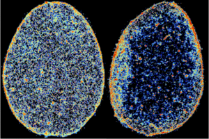
In these super-resolution images of tendon cell nuclei, the color coding represents chromatin density map, from low density in blue to high density in red. Comparing a healthy human tendon cell nucleus (left) to one diagnosed with tendinosis (right) shows that disease alters the spatial localization and compaction of chromatin.
In a recent study published in Nature Biomedical Engineering, the team detailed what they found when they closely observed the nucleus of cells inside connective tissues deteriorating as a result of tendinosis, which is the chronic condition that results from a tendon repeatedly suffering small injuries that don’t heal correctly. Using the latest super-resolution imaging techniques, they found that the tendon cells involved in maintaining the tissue’s structure in a diseased microenvironment improperly reorder their chromatin — the DNA-containing material that chromosomes are composed of — when attempting to repair.
This and other findings highlighted in the report point to the possibility of new treatments, such as small-molecule therapies, that could restore order to the affected cells.
“Interestingly, we were able to explain the role of mechanical forces on the 3-D organization of chromatin by developing a theory that integrates fundamental thermodynamic principles (physics) with the kinetics of epigenetic regulation (biology),” said study co-author and CEMB Director Vivek Shenoy in a news release from Penn Medicine News.
The CEMB, one of 18 active interdisciplinary research centers funded by the National Science Foundation’s Science and Technology Center (STC) program, brings together dozens of researchers from Penn Engineering and the Perelman School of Medicine, as well as others spread across campus and at partner institutions around the world.
With its funding recently renewed for another five years, the CEMB has entered into a new phase of its mission, centered on the nascent concept of “mechanointelligence,” which is exemplified by studies like this one. While mechanobiology is the study of the physical forces that govern the behavior of cells and their communication with their neighbors, mechanointelligence adds another layer of complexity: attempting to understand the forces that allow cells to sense, remember and adapt to their environments.
Ultimately, harnessing these forces would allow researchers to help multicellular organisms — plants, animals and humans — better adapt to their environments as well.
This story originally appeared in Penn Engineering Today.
Vivek Shenoy is Eduardo D. Glandt President’s Distinguished Professor in Materials Science and Engineering, Bioengineering, and in Mechanical Engineering and Applied Mechanics.
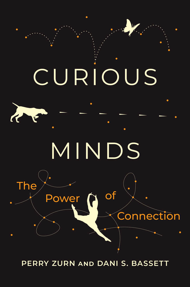
Twin siblings and scholars Dani S. Bassett of Penn and Perry Zurn of American University collaborated over half a dozen years to write “Curious Minds: The Power of Connection.” (Image: Tony and Tracy Wood Photography)
With appointments in the Departments of Bioengineering and Electrical and Systems Engineering, as well as the Department of Physics and Astronomy in Penn Arts & Science, and the Departments of Neuroscience and Psychiatry in Penn Perelman’s School of Medicine, Dani S. Bassett is no stranger to following the thread of an idea, no matter where it might lead.
Those wide-ranging fields and disciplines orbit around an appropriate central question: how does the tangle of neurons in our brains wire itself up to learn new things? Bassett, J. Peter Skirkanich Professor and director of the Complex Systems Lab, studies the relationship between the shape of those networks of neurons and the brain’s abilities, especially the way the shape of the network grows and changes with the addition of new knowledge.
To get at the fundamentals of the question of curiosity, Bassett needed to draw on even more disciplines. Fortunately, they didn’t have to look far; Bassett’s identical twin is Perry Zurn, a professor of philosophy at American University, and the two have investigated the many different ways a person can exhibit curiosity.
Bassett and Zurn have now published a new book on the subject. In Curious Minds: The Power of Connection, the twins draw on their previous research, as well as an expansive network of ideas from philosophy, history, education and art.
“It wasn’t clear at the beginning of our careers that we would even ever have a chance to write a book together because our areas were so wildly different,” Bassett says – but then, as postgraduates, Zurn was studying the philosophy of curiosity while Bassett was working on the neuroscience of learning. “And so that’s when we started talking. That talking led to seven years of doing research together,” Bassett says. “This book is a culmination of that.”
How exactly do philosophy and neuroscience complement each other? It all starts with the book’s first, and most deceptively simple question: what is curiosity? “Several investigators in science have underscored that perhaps the field isn’t even ready to define curiosity and how it’s different from other cognitive processes,” says Bassett. The ambiguity in the neuroscience literature motivated Bassett to turn to philosophy, “where there are really rich historical definitions and styles and subtypes that we can then put back into neuroscience and ask: ‘Can we see these in the brain?’”
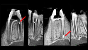
Controlled and actuated by magnetic fields, these mircrorobots are capable of precisely targeting the apical region — the opening where blood vessels and nerve enter the tooth — in a root canal.
With its irregularities and anatomical complexities, the root canal system is one of the most clinically challenging spaces in the oral cavity. As a result, biofilm not fully cleared from the nooks and crannies of the canals remains a leading cause of treatment failure and persistent endodontic infections, and there are limited means to diagnose or assess the efficacy of disinfection. One day, clinicians may have a new tool to overcome these challenges in the form of microrobots.
In a proof-of-concept study, researchers from Penn Dental Medicine and its Center for Innovation & Precision Dentistry (CiPD), have shown that microrobots can access the difficult to reach surfaces of the root canal with controlled precision, treating and disrupting biofilms and even retrieving samples for diagnostics, enabling a more personalized treatment plan. The Penn team shared their findings on the use of two different microrobotic platforms for endodontic therapy in the August issue of the Journal of Dental Research;the work was selected for the issue’s cover.
“The technology could enable multimodal functionalities to achieve controlled, precision targeting of biofilms in hard-to-reach spaces, obtain microbiological samples, and perform targeted drug delivery, ” says Dr. Alaa Babeer, lead author of the study and a Penn Dental Medicine Doctor of Science in Dentistry (DScD) and endodontics graduate, who is now within the lab of Dr. Michel Koo, co-director of the CiPD .
In both platforms, the building blocks for the microrobots are iron oxide nanoparticles (NPs) that have both catalytic and magnetic activity and have been FDA approved for other uses. In the first platform, a magnetic field is used to concentrate the NPs in aggregated microswarms and magnetically control them to the apical area of the tooth to disrupt and retrieve biofilms through a catalytic reaction. The second platform uses 3D printing to create miniaturized helix-shaped robots embedded with iron oxide NPs. These helicoids are guided by magnetic fields to move within the root canal, transporting bioactives or drugs that can be released on site.
“This technology offers the potential to advance clinical care on a variety of levels,” says Dr. Koo, co-corresponding author of the study with Dr. Edward Steager, a senior research investigator in Penn’s School of Engineering and Applied Science. “One important aspect is the ability to have diagnostic as well as therapeutic applications. In the microswarm platform, we can not only remove the biofilm, but also retrieve it, enabling us identify what microorganisms caused the infection. In addition, the ability to conform to the narrow and difficult-to-reach spaces within the root canal allows for a more effective disinfection in comparison to the files and instrumentation techniques presently used.”
Michel Koo is a professor in the Department of Orthodontics and divisions of Community Oral Health and Pediatric Dentistry in Penn Dental Medicine and co-director of the Center for Innovation & Precision Dentistry. He is a member of the Penn Bioengineering Graduate Group.
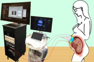
Combining optical measurements with ultrasound, an interdisciplinary team from the School of Arts & Sciences, Perelman School of Medicine, and CHOP developed a device to better measure blood flow and oxygenation in the placenta. (Image: Lin Wang)
A study published in Nature Biomedical Engineering details a novel method for imaging the placenta in pregnant patients as well as the results of a pilot clinical study. By combining optical measurements with ultrasound, the findings show how oxygen levels can be monitored noninvasively and provides a new way to generate a better understanding of this complex, crucial organ. This research was the result of a collaboration of the groups of the University of Pennsylvania’s Arjun Yodh and Nadav Schwartz with colleagues from the Children’s Hospital of Philadelphia (CHOP) and was led by postdoc Lin Wang.
Schwartz describes the placenta as the “engine” of pregnancy, an organ that plays a crucial role in delivering nutrients and oxygen to the fetus. Placental dysfunction can lead to complications such as fetal growth restriction, preeclampsia, and stillbirth. To increase knowledge about this crucial organ, the National Institute of Child Health and Human Development launched the Human Placenta Project in 2014. One focus of the program is to develop tools to assess human placental structure and function in real time, including optical devices.
For three years, the researchers optimized the design of their instrument and tested it in preclinical settings. The process involved integrating optical fibers with ultrasound probes, exploring various ultrasound transducers, and improving the multimodal technology so that measurements were stable, accurate, and reproducible while collecting data at the bedside. The resulting instrumentation now enables researchers to study the anatomy of the placenta while also collecting detailed functional information about placenta blood flow and oxygenation, capabilities that existing commercially devices do not have, the researchers say.
Because the placenta is located far below the body’s surface, one of the key technical challenges addressed by Wang, a postdoc in Yodh’s lab, was reducing background noise in the opto-electronic system. Light is scattered and absorbed when it travels through thick tissues, Yodh says, and the key for success was to reduce background interference so that the small amount of light that penetrates deep into the placenta and then returns is still large enough for a high-quality measurement.
“We’re sending a light signal that goes through the same deep tissues as the ultrasound. The extremely small amount of light that returns to the surface probe is then used to accurately assess tissue properties, which is only possible with very stable lasers, optics, and detectors,” says Yodh. “Lin had to overcome many barriers to improve the signal-to-noise ratio to the point where we trusted our data.”
The authors are Lin Wang, Jeffrey M. Cochran, Kenneth Abramson, Lian He, Venki Kavuri, Samuel Parry, Arjun G. Yodh, and Nadav Schwartz from Penn; Tiffany Ko, Wesley B. Baker, and Rebecca L. Linn from the Children’s Hospital of Philadelphia, and David R. Busch, previously a research associate at Penn and now at the University of Texas Southwestern Medical School.
This research was supported by National Institutes of Health grants F31HD085731, R01NS113945, R01NS060653, P41EB015893, P41EB015893, T32HL007915, and U01HD087180.
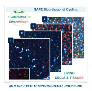
Cells in complex organisms undergo frequent changes, and researchers have struggled to monitor these changes and create a comprehensive profile for living cells and tissues. Historically researchers have been limited to only 3-5 markers due to spectral overlaps in fluorescence microscopy, an essential tool required for imaging cells. With only this small handful of markers, it is difficult to monitor protein expressions of live cells and a comprehensive profile of cellular dynamics cannot be created. However, a new study in Nature Biotechnology addresses these limitations by demonstrating a new method for comprehensive profiling of living cells.
Jina Ko, PhD
Jina Ko, Assistant Professor in Bioengineering in the School of Engineering and Applied Science and in Pathology and Laboratory Medicine in the Perelman School of Medicine, conducted postdoctoral research at Massachusetts General Hospital (MGH) and the Wyss Institute at Harvard University, and the work for this study was done under the supervision of Jonathan Carlson M.D., Ph.D. and Ralph Weissleder M.D., Ph.D. of MGH. Ko’s lab at Penn develops novel technologies using bioengineering, molecular biology, and chemistry to address diagnostic challenges for precision medicine.
To address these limitations in microscopy, the team developed a new chemistry tool which was highly gentle to cells. This “scission-accelerated fluorophore exchange (or SAFE)” method utilizes “click” chemistry, a type of chemistry that follows examples found in nature to create fast and simple reactions. This new SAFE method functions with non-toxic conditions to living cells and tissues, whereas previous methods have used harsh chemicals that would strip off fluorophores and consequently would not work with living cells and tissues.
With the development of SAFE, the authors demonstrated that researchers can now effectively perform multiple cycles of cell profiling and can monitor cellular changes over the course of their observations. Instead of the previous limitation of 3-5 markers total, SAFE allows for many more cycles and can keep track of almost as many markers as the researcher wants. One can now stain cells and quench/release fluorophores and repeat the cycle multiple times for multiplexing on living cells. Each cycle can profile 3 markers, and so someone interested in profiling 15 markers could easily perform 5 cycles to achieve this much more comprehensive cell profile. With this breakthrough in more detailed imaging of cells, SAFE demonstrates broad applicability for allowing researchers to better investigate the physiologic dynamics in living systems.
This study was supported by the Schmidt Science Fellows in Partnership with the Rhodes Trust and National Institutes of Health, National Cancer Institute (K99CA256353).
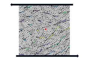
A suspension of particles of different sizes during shearing experiments conducted in the lab of Paulo Arratia, with arrows indicating particle “flow” and trajectories. In a new study published in Nature Physics, researchers detail the relationship between a disordered material’s individual particle arrangement and how it reacts to external stressors. The study also found that these materials have “memory” that can be used to predict how and when they will flow. (Image: Arratia lab)
New research published in Nature Physics details the relationship between a disordered material’s individual particle arrangement and how it reacts to external stressors. The study also found that these materials have “memory” that can be used to predict how and when they will flow. The study was led by Larry Galloway, a Ph.D. student in the lab of Paulo Arratia, and Xiaoguang Ma, a former postdoc in the lab of Arjun Yodh, in collaboration with researchers in the labs of Douglas Jerolmack and Celia Reina.
A disordered material is randomly arranged at the particle-scale, e.g. atoms or grains, instead of being systematically distributed—think of a pile of sand instead of a neatly stacked brick wall. Researchers in the Arratia lab are studying this class of materials as part of Penn’s Materials Research Science & Engineering Center, where one of the program’s focuses is on understanding the organization and proliferation of particle-scale rearrangements in disordered, amorphous materials.
The key question in this study was whether one could observe the structure of a disordered material and have some indication as to how stable it is or when it might begin to break apart. This is known as the yield point, or when the material “flows” and begins to move in response to external forces. “For example, if you look at the grains of a sand castle and how they are arranged, can I tell you whether the wind can blow it over or if it has to be hit hard to fall over?” says Arratia. “We want to know, just by looking at the way the particles are arranged, if we can say anything about the way they’re going to flow or if they are going to flow at all.”
While it has been known that individual particle distribution influences yield point, or flow, in disordered materials, it has been challenging to study this phenomenon since the field lacks ways to “quantify” disorder in such materials. To address this challenge, the researchers collaborated with colleagues from across campus to combine expertise across the fields of experimentation, theory, and simulations.
The authors are Larry Galloway, Erin Teich, Christoph Kammer, Ian Graham, Celia Reina, Douglas Jerolmack, Arjun Yodh, and Paulo Arratia from Penn; Xiaoguang Ma, previously a postdoc at Penn and now at the Southern University of Science and Technology in Shenzhen, China; and Nathan Keim, previously a postdoc at Penn and now at Pennsylvania State University.
A processed image representative of the types of images used in this study. Natural landscapes were transformed into binary images, ones made of black and white pixels, that were decomposed into different textures defined by specific statistics. (Image: Eugenio Piasini)
The human brain uses more energy than any other organ in the body, requiring as much as 20% of the body’s total energy. While this may sound like a lot, the amount of energy would be even higher if the brain were not equipped with an efficient way to represent only the most essential information within the vast, constant stream of stimuli taken in by the five senses. The hypothesis for how this works, known as efficient coding, was first proposed in the 1960s by vision scientist Horace Barlow.
Now, new research from the Scuola Internazionale Superiore di Studi Avanzati (SISSA) and the University of Pennsylvania provides evidence of efficient visual information coding in the rodent brain, adding support to this theory and its role in sensory perception. Published in eLife, these results also pave the way for experiments that can help understand how the brain works and can aid in developing novel artificial intelligence (AI) systems based on similar principles.
According to information theory—the study of how information is quantified, stored, and communicated—an efficient sensory system should only allocate resources to how it represents, or encodes, the features of the environment that are the most informative. For visual information, this means encoding only the most useful features that our eyes detect while surveying the world around us.
Vijay Balasubramanian, a computational neuroscientist at Penn, has been working on this topic for the past decade. “We analyzed thousands of images of natural landscapes by transforming them into binary images, made up of black and white pixels, and decomposing them into different textures defined by specific statistics,” he says. “We noticed that different kinds of textures have different variability in nature, and human subjects are better at recognizing those which vary the most. It is as if our brains assign resources where they are most necessary.”
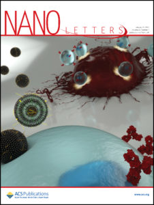
The Penn researchers’ latest paper on the design of lipid nanoparticles was featured on the cover of the most recent edition of the journal Nano Letters.
From COVID vaccines to cancer immunotherapies to the potential for correcting developmental disorders in utero, mRNA-based approaches are a promising tool in the fight against a wide range of diseases. These treatments all depend on providing a patient’s cells with genetic instructions for custom proteins and other small molecules, meaning that getting those instructions inside the target cells is of critical importance.
The current delivery method of choice uses lipid nanoparticles (LNPs). Thanks to surfaces customized with binding and signaling molecules, they encapsulate mRNA sequences and smuggle them through the cell membrane. But with a practically unlimited number of variables in the makeup of those surfaces and molecules, figuring out how to design the most effective LNP is a fundamental challenge.
Now, in a study featured on the cover of the journal Nano Letters, researchers from the University of Pennsylvania’s School of Engineering and Applied Science and Perelman School of Medicine have now shown how to computationally optimize the design of these delivery vehicles.
Using an established methodology for comparing a wide range of variables known as “orthogonal design of experiments,” the researchers simultaneously tested 256 candidate LNPs. They found the frontrunner was three times better at delivering mRNA sequences into T cells than the current standard LNP formulation for mRNA delivery.
The study was led by Michael Mitchell, Skirkanich Assistant Professor of Innovation in the Department of Bioengineering in Penn’s School of Engineering and Applied Science, and Margaret Billingsley, a graduate student in his lab.