Researchers from Penn and CHOP detail the mechanism by which HIV infection blocks the maturation process of brain cells that produce myelin, a fatty substance that insulates neurons.
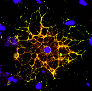
A confocal microscope image shows an oligodendrocyte in cell culture, labeled to show the cell nucleus in blue and myelin proteins in red, green, and yellow. Researchers from Penn and CHOP have shown that HIV infection prevents oligodendrocytes from maturing, leading to a reduction in white matter in the brain. (Image: Raj Putatunda)
It’s long been known that people living with HIV experience a loss of white matter in their brains. As opposed to gray matter, which is composed of the cell bodies of neurons, white matter is made up of a fatty substance called myelin that coats neurons, offering protection and helping them transmit signals quickly and efficiently. A reduction in white matter is associated with motor and cognitive impairment.
Earlier work by a team from the University of Pennsylvania and Children’s Hospital of Philadelphia (CHOP) found that antiretroviral therapy (ART)—the lifesaving suite of drugs that many people with HIV use daily—can reduce white matter, but it wasn’t clear how the virus itself contributed to this loss.
In a new study using both human and rodent cells, the team has hammered out a detailed mechanism, revealing how HIV prevents the myelin-making brain cells called oligodendrocytes from maturing, thus putting a wrench in white matter production. When the researchers applied a compound blocking this process, the cells were once again able to mature.
“Even when people with HIV have their disease well-controlled by antiretrovirals, they still have the virus present in their bodies, so this study came out of our interest in understanding how HIV infection itself affects white matter,” says Kelly Jordan-Sciutto, a professor in Penn’s School of Dental Medicine and co-senior author on the study. “By understanding those mechanisms, we can take the next step to protect people with HIV infection from these impacts.”
“When people think about the brain, they think of neurons, but they often don’t think about white matter, as important as it is,” says Judith Grinspan, a research scientist at CHOP and the study’s other co-senior author. “But it’s clear that myelination is playing key roles in various stages of life: in infancy, in adolescence, and likely during learning in adulthood too. The more we find out about this biology, the more we can do to prevent white matter loss and the harms that can cause.”
Jordan-Sciutto and Grinspan have been collaborating for several years to elucidate how ART and HIV affect the brain, and specifically oligodendrocytes, a focus of Grinspan’s research. Their previous work on antiretrovirals had shown that commonly used drugs disrupted the function of oligodendrocytes, reducing myelin formation.
In the current study, they aimed to isolate the effect of HIV on this process. Led by Lindsay Roth, who recently earned her doctoral degree within the Biomedical Graduate Studies group at Penn and completed a postdoctoral fellowship working with Jordan-Sciutto and Grinspan, the investigation began by looking at human macrophages, one of the major cell types that HIV infects.
Kelly Jordan-Sciutto is vice chair and professor in the University of Pennsylvania School of Dental Medicine’s Department of Basic & Translational Sciences and is director of Biomedical Graduate Studies. She is a member of the Penn Bioengineering Graduate Group.
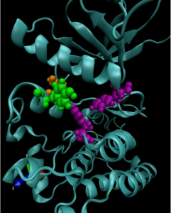
A computer model of a mutated anaplastic lymphoma kinase (ALK), a known oncogenic driver in pediatric neuroblastoma.
Kinases are a class of enzymes that are responsible for transferring the main chemical energy source used by the body’s cells. As such, they play important roles in diverse cellular processes, including signaling, differentiation, proliferation and metabolism. But since they are so ubiquitous, mutated versions of kinases are frequently found in cancers. Many cancer treatments involve targeting these mutant kinases with specific inhibitors.
Understanding the exact genetic mutations that lead to these aberrant kinases can therefore be critical in predicting the progression of a given patient’s cancer and tailoring the appropriate response.
To achieve this understanding on a more fundamental level, a team of researchers from the University of Pennsylvania’s School of Engineering and Applied Science and Perelman School of Medicine, the Children’s Hospital of Philadelphia (CHOP) and researchers at the Yale School of Medicine’s Cancer Biology Institute, have constructed molecular simulations of a mutant kinase implicated in pediatric neuroblastoma, a childhood cancer impacting the central nervous system.
Using their computational model to study the relationship between single-point changes in the kinase’s underlying gene and the altered structure of the protein it ultimately produces, the researchers revealed useful commonalities in the mutations that result in tumor formation and growth. Their findings suggest that such computational approaches could outperform existing profiling methods for other cancers and lead to more personalized treatments.
The study, published in the Proceedings of the National Academy of Sciences, was led by Ravi Radhakrishnan, Professor and chair of Penn Engineering’s Department of Bioengineering and professor in its Department of Chemical and Biomolecular Engineering, and Mark A. Lemmon, Professor of Pharmacology at Yale and co-director of Yale’s Cancer Biology Institute. The study’s first authors were Keshav Patil, a graduate student in Penn Engineering’s Department of Chemical and Biomolecular Engineering, along with Earl Joseph Jordan and Jin H. Park, then members of the Graduate Group in Biochemistry and Molecular Biology in Penn’s Perelman School of Medicine. Krishna Suresh, an undergraduate student in Radhakrishnan’s lab, Courtney M. Smith, a graduate student in Lemmon’s lab, and Abigail A. Lemmon, an undergraduate in Lemmon’s lab, contributed to the study. They collaborated with Yaël P. Mossé, Associate Professor of Pediatrics at Penn Medicine and in the division of oncology at CHOP.
“Some cancers rely on the aberrant activation of a single gene product for tumor initiation and progression,” says Radhakrishnan. “This unique mutational signature may hold the key to understanding which patients suffer from aggressive forms of the disease or for whom a given therapeutic drug may yield short- or long-term benefits. Yet, outside of a few commonly occurring ‘hotspot’ mutations, experimental studies of clinically observed mutations are not commonly pursued.”
“Kevin Johnson is a gifted physician-scientist,” Gutmann said, “who has harnessed and aligned the power of medicine, engineering, and technology to improve the health of individuals and communities. He has championed the development and implementation of clinical information systems and artificial intelligence to drive medical research, encouraged the effective use of technology at the bedside, and empowered patients to use new tools to better understand how medications and supplements may affect their health. He is a board-certified pediatrician, and his commitment to patient health and welfare knows no age limits. In so many different settings, Kevin’s work is driving progress in patient care and improving our health care system. He is a perfect fit for Penn, where our goal is to create a maximally inclusive and integrated academic community to spur unprecedented global impact.”
Johnson is currently the Cornelius Vanderbilt Professor and chair of the Department of Biomedical Informatics at the Vanderbilt University School of Medicine, where he has taught since 2002. He is a world-renowned innovator in developing clinical information systems that improve best practices in patient safety and compliance with medical practice guidelines, especially the use of computer-based documentation systems and other digital technologies. His research bridges biomedical informatics, bioengineering, and computer science. As senior vice president for health information technology at the Vanderbilt University Medical Center from 2014 to 2019, he led the development of clinical systems that enabled doctors to make better treatment and care decisions for individual patients, in part by alerting patients as to how medications or supplements might affect their body chemistry, as well as new systems to integrate artificial intelligence into patient care workflows and to unify and simplify all the Medical Center’s clinical and administrative systems.
The author of more than 150 publications, books, or book chapters, Johnson has held numerous leadership positions in the American Medical Informatics Association and the American Academy of Pediatrics, leads the American Board of Pediatrics Informatics Advisory Committee, directs the Board of Scientific Counselors of the National Library of Medicine, and is a member of the NIH Council of Councils. He has been elected to the National Academy of Medicine (Institute of Medicine), American College of Medical Informatics, and Academic Pediatric Society and has received awards from the Robert Wood Johnson Foundation and American Academy of Pediatrics, among many others.
“Kevin Johnson exemplifies our most profound Penn values,” Pritchett said. “He is a brilliant innovator committed to bringing together disciplines across traditional boundaries. Yet he always does so in the service of helping others, finding technological solutions that can make a tangible impact on improving people’s lives. He will be an extraordinary colleague, teacher and mentor across multiple areas of our campus in the years to come.”
Johnson earned an M.D. from the Johns Hopkins University School of Medicine, an M.S. in medical informatics from Stanford University, and a B.S. with honors in biology from Dickinson College. He became the first Black chief resident in pediatrics at Johns Hopkins in 1992, and was a faculty member in both pediatrics and biomedical information sciences at Johns Hopkins until 2002.
The Penn Integrates Knowledge program was launched by Gutmann in 2005 as a University-wide initiative to recruit exceptional faculty members whose research and teaching exemplify the integration of knowledge across disciplines and who are appointed in at least two Schools at Penn.
William H. Peranteau, Michael J. Mitchell, Margaret Billingsley, Meghana Kashyap, and Rachel Riley (Clockwise from top left)
As COVID-19 vaccines roll out, the concept of using mRNA to fend off viruses has become a part of the public dialogue. However, scientists have been researching how mRNA can be used to in life-saving medical treatments well before the pandemic.
The “m” in “mRNA” is for “messenger.” A single-stranded counterpart to DNA, it translates the genetic code into the production of proteins, the building blocks of life. The Moderna and Pfizer COVID-19 vaccines work by introducing mRNA sequences that act as a set of instructions for the body to produce proteins that mimic parts of the virus itself. This prepares the body’s immune response to recognize the real virus and fight it off.
Because it can spur the production of proteins that the body can’t make on its own, mRNA therapies also have the potential to slow or prevent genetic diseases that develop before birth, such as cystic fibrosis and sickle-cell anemia.
However, because mRNA is a relatively unstable molecule that degrades quickly, it needs to be packaged in a way that maintains its integrity as its delivered to the cells of a developing fetus.
To solve this challenge, Michael J. Mitchell, Skirkanich Assistant Professor of Innovation in the Department of Bioengineering, is researching the use of lipid nanoparticles as packages that transport mRNA into the cell. He and William H. Peranteau, an attending surgeon in the Division of General, Thoracic and Fetal Surgery and the Adzick-McCausland Distinguished Chair in Fetal and Pediatric Surgery at Children’s Hospital of Philadelphia, recently co-authored a “proof-of-concept” paper investigating this technique.
In this study, published in Science Advances, Mitchel examined which nanoparticles were optimal in the transport of mRNA to fetal mice. Although no disease or organ was targeted in this study, the ability to administer mRNA to a mouse while still in the womb was demonstrated, and the results are promising for the next stages of targeted disease prevention in humans.
Mitchel spoke with Tom Avril at The Philadelphia Inquirer about the mouse study and its implications for treatment of rare infant diseases through the use of mRNA, ‘the messenger of life.’
Penn bioengineering professor Michael J. Mitchell, the other senior author of the mouse study, tested various combinations of lipids to see which would work best.
The appeal of the fatty substances is that they are biocompatible. In the vaccines, for example, two of the four lipids used to make the delivery spheres are identical to lipids found in the membranes of human cells — including plain old cholesterol.
When injected, the spheres, called nanoparticles, are engulfed by the person’s cells and then deposit their cargo, the RNA molecules, inside. The cells respond by making the proteins, just as they make proteins by following the instructions in the person’s own RNA. (Important reminder: The RNA in the vaccines cannot become part of your DNA.)
Among the different lipid combinations that Mitchell and his lab members tested, some were better at delivering their cargo to specific organs, such as the liver and lungs, meaning they could be a good vehicle for treating disease in those tissues.
William H. Peranteau, Michael J. Mitchell, Margaret Billingsley, Meghana Kashyap, and Rachel Riley (Clockwise from top left)
Researchers at Children’s Hospital of Philadelphia and the School of Engineering and Applied Science at the University of Pennsylvania have identified ionizable lipid nanoparticles that could be used to deliver mRNA as part of fetal therapy. The proof-of-concept study, published today in Science Advances, engineered and screened a number of lipid nanoparticle formulations for targeting mouse fetal organs and has laid the groundwork for testing potential therapies to treat genetic diseases before birth.
“This is an important first step in identifying nonviral mediated approaches for delivering cutting-edge therapies before birth,” said co-senior author William H. Peranteau, MD, an attending surgeon in the Division of General, Thoracic and Fetal Surgery and the Adzick-McCausland Distinguished Chair in Fetal and Pediatric Surgery at CHOP. “These lipid nanoparticles may provide a platform for in utero mRNA delivery, which would be used in therapies like fetal protein replacement and gene editing.”
Michael J. Mitchell, Skirkanich Assistant Professor of Innovation in Penn Engineering’s Department of Bioengineering, is the other co-senior author of the study. The co-first authors are Mitchell Lab members Rachel Riley, a postdoctoral fellow, and Margaret Billingsley, a graduate student, and Peranteau Lab member Meghana Kashyap, a research fellow.
Recent advances in DNA sequencing technology and prenatal diagnostics have made it possible to diagnose many genetic diseases before birth. Some of these diseases are treated by protein or enzyme replacement therapies after birth, but by then, some of the damaging effects of the disease have taken hold. Thus, applying therapies while the patient is still in the womb has the potential to be more effective for some conditions. The small fetal size allows for maximal therapeutic dosing, and the immature fetal immune system may be more tolerant of replacement therapy.
A collaborative study conducted by researchers at the Children’s Hospital of Philadelphia (CHOP), Penn Engineering and Pennsylvania State University has uncovered new information about how chromosomal material in cell nuclei reorganizes itself after cell division.
While a deep understanding of the cell cycle is a cornerstone of biology and health sciences, research into the complex relationship between three-dimensional chromatin structure and gene transcription is still in its infancy. The results of this study will contribute to a more robust understanding of chromatin rebuilding after mitosis and potentially aid in the treatment of genetic diseases.
Jennifer E. Phillips-Cremins, Ph.D.
Jennifer E. Phillips-Cremins, Assistant Professor in the Department of Bioengineering, contributed to the study alongside Gerd A. Blobel, Frank E. Weise III Endowed Chair in Pediatric Hematology at CHOP and Ross C. Hardison, an expert in gene regulation at Penn State.
Phillips-Cremins’ research uses genetic engineering approaches to discover the mechanisms regulating chromatin organizing principles in cells, as well as computational approaches to investigate cellular function. Her lab’s techniques provide ways of mapping the three-dimensional organization of genes while they are folded together in the genome and how those spatial relationships impact gene expression.
The research team performed their experiments in blood-forming cells from a well-established mouse model. They used sophisticated techniques called high throughput chromosome conformation capture (Hi-C) that detect and map interactions across three-dimensional space between specific sites in chromosomal DNA. These maps also allowed the scientists to measure such interactions at different time points in the cell cycle. In all, the tools detected roughly 2 billion interactions during mitosis and thereafter, when the daughter nuclei are rebuilt.
Members of the Cremins Lab, Daniel J. Emerson, Thomas G. Gilgenast and Katelyn R. Titus, also contributed to the study, which was published in Nature.
New Vascularized Patches Could Help Patient Recovery from Heart Attacks
Heart attacks are the result of a stoppage of blood flow to the heart – an interruption to normal function that can result in severe tissue damage, or even tissue death. This loss of healthy tissue function is one of the biggest challenges in treating patients that undergo heart attacks, as the damaged tissue increases their risk of having future attacks. One of the main solutions to this issue right now is the creation of cardiac tissue scaffolds using stem cells to create a platform for new and healthy tissue to grow in vivo. A group of biomedical engineers at Michigan Technological University hopes to expand on this basis by focusing not just on cellular alignment in the scaffold but on that of microvessels too. Led by Feng Zhao, Ph.D., Associate Professor of Biomedical Engineering, the team hopes that this new attention on microvessel organization will improve the vasculature of the scaffolds, and thus improve the success of the scaffolds in vivo, allowing for a better recovery from heart attacks.
Some Stem Cells May Be More Fit Than Others
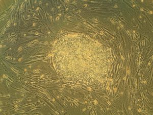
Stem cells are one of the hottest research areas in the field of bioengineering today. Widely known as the cells in the human embryo that have the ability to eventually transform into specific cells for the brain, lung, and every other organ, stem cells are also of recent interest because researchers found ways to reverse this process, transforming organ-specific cells back to the pluripotent stem cell level. This achievement however, is mostly applicable to individual stem cells, and doesn’t fully encapsulate the way this process might work on a larger population level. So Peter Zandstra, Ph. D., a bioengineering faculty member at the University of British Columbia, decided to research just that.
Using mouse embryonic fibroblasts (MEFs), Zandstra and his lab attempted to track the cells throughout their reprogramming, to more clearly trace each back to its respective parent population. Surprisingly, they found that after only one week of reprogramming, nearly 80% of the original cell population had been removed, meaning that most of the parent generation was not “fit” enough to undergo the process of reprogramming, indicating that perhaps some stem cells will have a better chance of survival in this process than others. This research may suggest that not all cells have the capacity to undergo reprogramming, as many researchers originally thought.
A New Microdevice Will Help Model Bronchial Spasms
The difficulty in breathing associated with asthma is the result of bronchial spasms, which are a kind of muscle contraction in the airways. But little was known about just how these spasms occurred in patients, so Andre Levchenko, Ph.D., Professor of Biomedical Engineering at Johns Hopkins, and his lab created a microdevice to model them. Calling the device a “bronchi on a chip,” Levchenko and his team used a microphysiological model to look at some of the biochemical and mechanical signals associated with these kinds of muscle contractions. They found that the contractions operate in a positive feedback system, so that those caused by disturbance from allergens will subsequently cause even more contractions to occur. But surprisingly, they also found that a second contraction, if triggered at the right time during the initial contraction, could actually stop the process and allow the muscles to relax. Because asthma is a notoriously difficult disease to translate from animal to human models, this new device opens the door to understanding different mechanisms of asthma before taking research to clinical trials.
New CHOP Research Center to Focus Research on Pediatric Airway Disorders
A new bioengineering lab at the Children’s Hospital of Philadelphia called the Center for Pediatric Airway Disorders will specialize in a variety of airway procedures for pediatric patients such as tracheal reconstruction and recurrent laryngeal nerve reinnervation. This new lab will be one of the first to give a unique focus to the application of bioengineering to pediatric laryngology. The interdisciplinary center brings together students and researchers from all different fields, including materials science and microbiology, to find new ways of repairing tissue and regenerating organs related to respiratory disorders. Specific areas of research will involve the modeling of children’s vocal cords, understanding the mechanisms of fibrosis, and improving surgical procedures.
Deeper Understanding of Sickle Cell Anemia Could Lead to New Treatments
Though sickle cell anemia is a common and well-known disease, a new study of its causes at the nanoscale level might reveal previously unknown information about the assembly of hemoglobin fibers. Using microscopes with the ability to visualize these molecules at such a small level, researchers at the University of Minnesota found that the beginning organizations that lead to sickle cell anemia are much less ordered than originally thought. Led by Associate Professor of Biomedical Engineering David Wood, Ph.D., the team of researchers used this higher level of microscopy to find that hemoglobin self-assembly process, which was originally thought to be 96% efficient, is actually only 4% efficient. Wood hopes that this new knowledge will help allow for the development of new and better treatments for patients with sickle cell anemia, as there are currently only two FDA-approved ones on the market.
People & Places
Penn Today asked five Penn researchers about the women in STEM who have been a source of inspiration and encouragement throughout their own careers. Their responses include active researchers who have paved the way for better inclusion in STEM and famous female scientists from the past who broke boundaries as they made strides with their research.
Joel Boerckel, Ph.D, Assistant Professor of Orthopaedic Surgery and Bioengineering
This week, we want to congratulate Joel Boerckel, Ph.D., Assistant Professor of Orthopaedic Surgery and Bioengineering, and his lab on receiving a second R01 Grant from the National Institute of Arthritis and and Musculoskeletal Skin Diseases for their work on defining the roles of YAP and TAZ in embryonic bone morphogenesis and mechanoregulation of fracture repair. Dr. Boerckel is a member of the McKay Orthopaedic Research Laboratory.
We would also like to congratulate Christopher Yip, Ph. D., on being appointed as the new dean of the University of Toronto’s Faculty of Applied Science and Engineering. A professor in both the Department of Chemical Engineering and Applied Chemistry the Institute of Biomaterials and Biomedical Engineering, Dr. Yip’s research involves the use of molecular imaging to understand the self-assembly of proteins.


 Heart attacks are the result of a stoppage of blood flow to the heart – an interruption to normal function that can result in severe tissue damage, or even tissue death. This loss of healthy tissue function is one of the biggest challenges in treating patients that undergo heart attacks, as the damaged tissue increases their risk of having future attacks. One of the main solutions to this issue right now is the creation of cardiac tissue scaffolds using stem cells to create a platform for new and healthy tissue to grow in vivo. A group of biomedical engineers at Michigan Technological University hopes to