A Novel Method for Monitoring the ‘Engine’ of Pregnancy

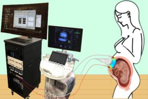
Combining optical measurements with ultrasound, an interdisciplinary team from the School of Arts & Sciences, Perelman School of Medicine, and CHOP developed a device to better measure blood flow and oxygenation in the placenta. (Image: Lin Wang)

A study published in Nature Biomedical Engineering details a novel method for imaging the placenta in pregnant patients as well as the results of a pilot clinical study. By combining optical measurements with ultrasound, the findings show how oxygen levels can be monitored noninvasively and provides a new way to generate a better understanding of this complex, crucial organ. This research was the result of a collaboration of the groups of the University of Pennsylvania’s Arjun Yodh and Nadav Schwartz with colleagues from the Children’s Hospital of Philadelphia (CHOP) and was led by postdoc Lin Wang.

Schwartz describes the placenta as the “engine” of pregnancy, an organ that plays a crucial role in delivering nutrients and oxygen to the fetus. Placental dysfunction can lead to complications such as fetal growth restriction, preeclampsia, and stillbirth. To increase knowledge about this crucial organ, the National Institute of Child Health and Human Development launched the Human Placenta Project in 2014. One focus of the program is to develop tools to assess human placental structure and function in real time, including optical devices.

For three years, the researchers optimized the design of their instrument and tested it in preclinical settings. The process involved integrating optical fibers with ultrasound probes, exploring various ultrasound transducers, and improving the multimodal technology so that measurements were stable, accurate, and reproducible while collecting data at the bedside. The resulting instrumentation now enables researchers to study the anatomy of the placenta while also collecting detailed functional information about placenta blood flow and oxygenation, capabilities that existing commercially devices do not have, the researchers say.

Because the placenta is located far below the body’s surface, one of the key technical challenges addressed by Wang, a postdoc in Yodh’s lab, was reducing background noise in the opto-electronic system. Light is scattered and absorbed when it travels through thick tissues, Yodh says, and the key for success was to reduce background interference so that the small amount of light that penetrates deep into the placenta and then returns is still large enough for a high-quality measurement.

“We’re sending a light signal that goes through the same deep tissues as the ultrasound. The extremely small amount of light that returns to the surface probe is then used to accurately assess tissue properties, which is only possible with very stable lasers, optics, and detectors,” says Yodh. “Lin had to overcome many barriers to improve the signal-to-noise ratio to the point where we trusted our data.”

Read the full story in Penn Today.

The authors are Lin Wang, Jeffrey M. Cochran, Kenneth Abramson, Lian He, Venki Kavuri, Samuel Parry, Arjun G. Yodh, and Nadav Schwartz from Penn; Tiffany Ko, Wesley B. Baker, and Rebecca L. Linn from the Children’s Hospital of Philadelphia, and David R. Busch, previously a research associate at Penn and now at the University of Texas Southwestern Medical School.

Arjun Yodh is the James M. Skinner Professor of Science in the Department of Physics and Astronomy in the School of Arts & Sciences at the University of Pennsylvania. He is a member of the Penn Bioengineering Graduate Group.

Nadav Schwartz is an Associate Professor in the Department of Obstetrics and Gynecology in Penn’s Perelman School of Medicine.

Lin Wang is a postdoc in the Department of Physics and Astronomy in Penn’s School of Arts & Sciences.

This research was supported by National Institutes of Health grants F31HD085731, R01NS113945, R01NS060653, P41EB015893, P41EB015893, T32HL007915, and U01HD087180.

Penn Medicine and Children’s Hospital of Philadelphia Announce Partnership with Costa Rica for CAR T Cell Therapy

Carl June, MD

Carl June, MD, Professor in the Perelman School of Medicine and member of the Penn Bioengineering Graduate Group, was quoted in a recent press release  announcing a new international partnership between Penn Medicine (PSOM), the Children’s Hospital of Pennsylvania (CHOP), and Costa Rica’s CCSS, or the Caja Costarricense de Seguro Social (Social Security Program), to develop CAR T research in Costa Rica. June is a world renowned cancer cell therapy pioneer whose research led to the initial development and FDA approval of CAR T cell therapy:

“‘At least 15,000 patients across the world have received CAR T cells, and dozens more clinical trials using this approach are in progress, for almost every major tumor type, but people in many parts of the globe still do not have access to treatment with these transformative therapies,’ said Carl H. June, MD, the Richard W. Vague Professor in Immunotherapy and director of the Center for Cellular Immunotherapies in Penn’s Perelman School of Medicine. “We are honored to work with our colleagues in Costa Rica in hopes of building a path for patients in underserved areas to have the opportunity to benefit from clinical research programs offering this personalized therapy.’”

Read the the announcement in Penn Medicine News.


Kevin Johnson Discusses the Future of the Electronic Health Record

Kevin B. Johnson, M.D., Ph.D.

Kevin B. Johnson, M.D., M.S., was featured in Cincinnati Children’s Hospital’s “Envisioning Our Future for Children” speaker series, discussing “the evolution of the EHR and its future directions.” An electronic health record, or EHR, is a digital record of a patient’s chart, recording health information and data, coordinating orders, tracking results, and providing patient support. Johnson “predicts a new wave of transformation in digital health technologies that could make rapid progress” in several areas of medicine, including reducing cost and improving patience outcomes. Johnson is Vice President for Applied Informatics at the University of Pennsylvania Health System and the David L. Cohen University Professor with appointments in Biostatistics, Epidemiology and Informatics and Computer and Information Science and secondary appointments in the Annenberg School for Communication, Pediatrics, and Bioengineering.

Read “What Will It Take to Make EHR a Partner Instead of a Burden?” in the Cincinnati Children’s Hospital Research Horizons blog. View Johnson’s seminar talk on the Envisioning Our Future website.

Decade-long Remission After CAR T Cell Therapy

Bill Ludwig, left, was the first patient to receive CAR T cells as part of clinical trials at Abramson Cancer Center. Carl June, right, has played a pioneering roll in the therapeutic use of CAR T cells. (Image: Penn Medicine)

Carl H. June, the Richard W. Vague Professor in Immunotherapy in Pathology and Laboratory Medicine at Penn Medicine, director of the Center for Cellular Immunotherapies and the Parker Institute for Cancer Immunotherapy, and member of the Penn Bioengineering Graduate Group at the University of Pennsylvania, has led a new analytical study published in Nature that explains the longest persistence of CAR T cell therapy recorded to date against chronic lymphocytic leukemia (CLL), and shows that the CAR T cells remained detectable at least a decade after infusion, with sustained remission in both patients. June’s pioneering work in gene therapy led to the FDA approval for the CAR T therapy (sold by Novartis as Kymriah) for treating leukemia and transforming the fight against cancer. His lab develops new forms of T cell based therapies.

Read the story in Penn Today

“You get what you put in”: A First-generation Penn Bioengineering Graduate Student Discusses His Journey

Joseph Lance Casila

Joseph Lance Casila, a doctoral student and Fontaine Fellow in Bioengineering, was profiled by his alma mater, the University of Guam (UOG. Casila was the first person in his family to graduate from a U.S.-accredited university and is now studying tissue engineering and regenerative medicine in the Bioengineering and Biomaterials Laboratory of Riccardo Gottardi, Assistant Professor in Bioengineering in Penn Engineering and Pediatrics in Penn Medicine and the Children’s Hospital of Philadelphia (CHOP). His research in the Gottardi lab employs “tissue engineering and drug delivery for biomedical problems relating to knees, ears, nose, and throat but specifically to pediatric airway disorders.” The article discusses Casila’s journey from valedictorian of his high school, to a first-generation undergraduate interested bioengineering, and now a graduate student studying at Penn on a full scholarship. After completing his degree, Casila hopes to bring what he’s learned back home to advance health care in Guam.

“My mentors, and especially my friends, helped me make the most of what UOG had to offer, and it paid off rewardingly,” he said. “You get what you put in.”

Read “A first-generation student’s path to an Ivy League Ph.D. program” in the University of Guam News & Announcements.

Developing Endotracheal Tubes that Release Antimicrobial Peptides

by Evan Lerner

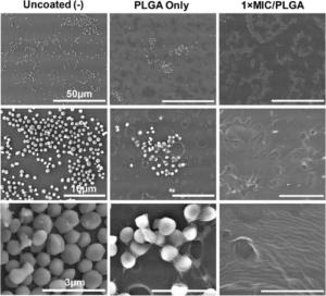
Scanning electron microscope images of endotracheal tubes at three levels of magnification. After 24 hours of Staphylococcus epidermidis exposure, tubes coated with the researchers’ AMPs (right) showed decreased biofilm production, as compared with tubes coated with just polymer (center) and uncoated tubes (left).

Endotracheal tubes are a mainstay of hospital care, as they ensure a patient’s airway is clear when they can’t breathe on their own. However, keeping a foreign object inserted in this highly sensitive part of the anatomy comes is not without risk, such as the possibility of infection, inflammation and a condition known as subglottic stenosis, in which scar tissue narrows the airway.

Broad-spectrum antibiotics are one way to mitigate these risks, but come with risks of their own, including harming beneficial bacteria and contributing to antibiotic resistance.

With this conundrum in mind, Riccardo Gottardi, Assistant Professor of Pediatrics at the Children’s Hospital of Philadelphia (CHOP) and of Bioengineering at Penn Engineering, along with Bioengineering graduate students and lab members Matthew Aronson and Paul Gehret, are developing endotracheal tubes that can provide a more targeted antimicrobial defense.

In a proof-of-concept study published in the journal The Laryngoscope, the team showed how a different type of antimicrobial agent could be incorporated into the tubes’ polymer coating, as well as preliminary results suggesting these devices would better preserve a patient’s microbiome.

Instead, the investigators explored the use of antimicrobial peptides (AMPs), which are small proteins that destabilize bacterial membranes, causing bacterial cells to fall apart and die. This mechanism of action allows them to target specific bacteria and makes them unlikely to promote antimicrobial resistance. Prior studies have shown that it is possible to coat endotracheal tubes with conventional antibiotics, so the research team investigated the possibility of incorporating AMPs into polymer-coated tubes to inhibit bacterial growth and modulate the upper-airway microbiome.

The researchers, led by Matthew Aronson, a graduate student in Penn Engineering’s Department of Bioengineering, tested their theory by creating a polymer coating that would release Lasioglossin-III, an AMP with broad-spectrum antibacterial activity. They found that Lasio released from coated endotracheal tubes, reached the expected effective concentration rapidly and continued to release at the same concentration for a week, which is the typical timeframe that an endotracheal is used before being changed. The investigators also tested their drug-eluting tube against airway microbes, including S. epidermidis, S. pneumoniae, and human microbiome samples and observed significant antibacterial activity, as well as prevention of bacterial adherence to the tube.

Read “CHOP Researchers Develop Coating for Endotracheal Tubes that Releases Antimicrobial Peptides” at CHOP News.

This post originally appeared in Penn Engineering Today.

Student Research Highlight: Colin Huber

Colin Huber, Ph.D. student

Colin Huber, a Ph.D. candidate in Bioengineering studying head impact biomechanics and concussion in sports at the Center for Injury Research and Prevention (CIRP) at the Children’s Hospital of Philadelphia (CHOP), recently published “Variations in Head Impact Rates in Male and Female High School Soccer” in Medicine & Science in Sports & Exercise with colleagues from CHOP’s Minds Matter Concussion Frontier Program and the CIRP.

Colin’s paper, the goal of which was to compare “to compare head impact exposure rates (head impacts/exposure period) in male and female high school soccer by using multiple methodological approaches,” was recently profiled in the Penn Engineering Research & Innovation Newsletter.

Read the full story in the ADRO Newsletter.

How HIV Infection Shrinks the Brain’s White Matter

by Katherine Unger Baillie

Researchers from Penn and CHOP detail the mechanism by which HIV infection blocks the maturation process of brain cells that produce myelin, a fatty substance that insulates neurons.

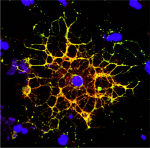
A confocal microscope image shows an oligodendrocyte in cell culture, labeled to show the cell nucleus in blue and myelin proteins in red, green, and yellow. Researchers from Penn and CHOP have shown that HIV infection prevents oligodendrocytes from maturing, leading to a reduction in white matter in the brain. (Image: Raj Putatunda)

It’s long been known that people living with HIV experience a loss of white matter in their brains. As opposed to gray matter, which is composed of the cell bodies of neurons, white matter is made up of a fatty substance called myelin that coats neurons, offering protection and helping them transmit signals quickly and efficiently. A reduction in white matter is associated with motor and cognitive impairment.

Earlier work by a team from the University of Pennsylvania and Children’s Hospital of Philadelphia (CHOP) found that antiretroviral therapy (ART)—the lifesaving suite of drugs that many people with HIV use daily—can reduce white matter, but it wasn’t clear how the virus itself contributed to this loss.

In a new study using both human and rodent cells, the team has hammered out a detailed mechanism, revealing how HIV prevents the myelin-making brain cells called oligodendrocytes from maturing, thus putting a wrench in white matter production. When the researchers applied a compound blocking this process, the cells were once again able to mature.

The work is published in the journal Glia.

“Even when people with HIV have their disease well-controlled by antiretrovirals, they still have the virus present in their bodies, so this study came out of our interest in understanding how HIV infection itself affects white matter,” says Kelly Jordan-Sciutto, a professor in Penn’s School of Dental Medicine and co-senior author on the study. “By understanding those mechanisms, we can take the next step to protect people with HIV infection from these impacts.”

“When people think about the brain, they think of neurons, but they often don’t think about white matter, as important as it is,” says Judith Grinspan, a research scientist at CHOP and the study’s other co-senior author. “But it’s clear that myelination is playing key roles in various stages of life: in infancy, in adolescence, and likely during learning in adulthood too. The more we find out about this biology, the more we can do to prevent white matter loss and the harms that can cause.”

Jordan-Sciutto and Grinspan have been collaborating for several years to elucidate how ART and HIV affect the brain, and specifically oligodendrocytes, a focus of Grinspan’s research. Their previous work on antiretrovirals had shown that commonly used drugs disrupted the function of oligodendrocytes, reducing myelin formation.

In the current study, they aimed to isolate the effect of HIV on this process. Led by Lindsay Roth, who recently earned her doctoral degree within the Biomedical Graduate Studies group at Penn and completed a postdoctoral fellowship working with Jordan-Sciutto and Grinspan, the investigation began by looking at human macrophages, one of the major cell types that HIV infects.

Read the full story in Penn Today.

Kelly Jordan-Sciutto is vice chair and professor in the University of Pennsylvania School of Dental Medicine’s Department of Basic & Translational Sciences and is director of Biomedical Graduate Studies. She is a member of the Penn Bioengineering Graduate Group.

Penn, CHOP and Yale Researchers’ Molecular Simulations Uncover How Kinase Mutations Lead to Cancer Progression

by Evan Lerner

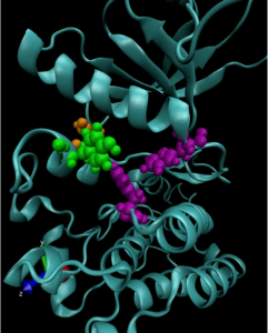
A computer model of a mutated anaplastic lymphoma kinase (ALK), a known oncogenic driver in pediatric neuroblastoma.

Kinases are a class of enzymes that are responsible for transferring the main chemical energy source used by the body’s cells. As such, they play important roles in diverse cellular processes, including signaling, differentiation, proliferation and metabolism. But since they are so ubiquitous, mutated versions of kinases are frequently found in cancers. Many cancer treatments involve targeting these mutant kinases with specific inhibitors.

Understanding the exact genetic mutations that lead to these aberrant kinases can therefore be critical in predicting the progression of a given patient’s cancer and tailoring the appropriate response.

To achieve this understanding on a more fundamental level, a team of researchers from the University of Pennsylvania’s School of Engineering and Applied Science and Perelman School of Medicine, the Children’s Hospital of Philadelphia (CHOP) and researchers at the Yale School of Medicine’s Cancer Biology Institute, have constructed molecular simulations of a mutant kinase implicated in pediatric neuroblastoma, a childhood cancer impacting the central nervous system.

Using their computational model to study the relationship between single-point changes in the kinase’s underlying gene and the altered structure of the protein it ultimately produces, the researchers revealed useful commonalities in the mutations that result in tumor formation and growth. Their findings suggest that such computational approaches could outperform existing profiling methods for other cancers and lead to more personalized treatments.

The study, published in the Proceedings of the National Academy of Sciences, was led by Ravi Radhakrishnan, Professor and chair of Penn Engineering’s Department of Bioengineering and professor in its Department of Chemical and Biomolecular Engineering, and Mark A. Lemmon, Professor of Pharmacology at Yale and co-director of Yale’s Cancer Biology Institute. The study’s first authors were Keshav Patil, a graduate student in Penn Engineering’s Department of Chemical and Biomolecular Engineering, along with Earl Joseph Jordan and Jin H. Park, then members of the Graduate Group in Biochemistry and Molecular Biology in Penn’s Perelman School of Medicine. Krishna Suresh, an undergraduate student in Radhakrishnan’s lab, Courtney M. Smith, a graduate student in Lemmon’s lab, and Abigail A. Lemmon, an undergraduate in Lemmon’s lab, contributed to the study. They collaborated with Yaël P. Mossé, Associate Professor of Pediatrics at Penn Medicine and in the division of oncology at CHOP.

“Some cancers rely on the aberrant activation of a single gene product for tumor initiation and progression,” says Radhakrishnan. “This unique mutational signature may hold the key to understanding which patients suffer from aggressive forms of the disease or for whom a given therapeutic drug may yield short- or long-term benefits. Yet, outside of a few commonly occurring ‘hotspot’ mutations, experimental studies of clinically observed mutations are not commonly pursued.”

Read the full post in Penn Engineering Today.

Kevin Johnson Appointed Penn Integrates Knowledge University Professor

Ron Ozio

Kevin Johnson, Penn’s 27th Penn Integrates Knowledge University Professor.

Kevin Johnson has been named the University of Pennsylvania’s 27th Penn Integrates Knowledge University Professor.

The announcement was made by Penn President Amy Gutmann and Provost Wendell Pritchett.

A pioneer of medical information technologies to improve patient safety, Johnson will hold joint appointments in the Department of Biostatistics, Epidemiology, and Informatics in the Perelman School of Medicine and the Department of Computer and Information Science in the School of Engineering and Applied Science, with secondary appointments in the Annenberg School for Communication and in the Department of Bioengineering. He will also serve as vice president for applied informatics in the University of Pennsylvania Health System and professor of pediatrics at the Children’s Hospital of Philadelphia.

“Kevin Johnson is a gifted physician-scientist,” Gutmann said, “who has harnessed and aligned the power of medicine, engineering, and technology to improve the health of individuals and communities. He has championed the development and implementation of clinical information systems and artificial intelligence to drive medical research, encouraged the effective use of technology at the bedside, and empowered patients to use new tools to better understand how medications and supplements may affect their health. He is a board-certified pediatrician, and his commitment to patient health and welfare knows no age limits. In so many different settings, Kevin’s work is driving progress in patient care and improving our health care system. He is a perfect fit for Penn, where our goal is to create a maximally inclusive and integrated academic community to spur unprecedented global impact.”

Johnson is currently the Cornelius Vanderbilt Professor and chair of the Department of Biomedical Informatics at the Vanderbilt University School of Medicine, where he has taught since 2002. He is a world-renowned innovator in developing clinical information systems that improve best practices in patient safety and compliance with medical practice guidelines, especially the use of computer-based documentation systems and other digital technologies. His research bridges biomedical informatics, bioengineering, and computer science. As senior vice president for health information technology at the Vanderbilt University Medical Center from 2014 to 2019, he led the development of clinical systems that enabled doctors to make better treatment and care decisions for individual patients, in part by alerting patients as to how medications or supplements might affect their body chemistry, as well as new systems to integrate artificial intelligence into patient care workflows and to unify and simplify all the Medical Center’s clinical and administrative systems.

The author of more than 150 publications, books, or book chapters, Johnson has held numerous leadership positions in the American Medical Informatics Association and the American Academy of Pediatrics, leads the American Board of Pediatrics Informatics Advisory Committee, directs the Board of Scientific Counselors of the National Library of Medicine, and is a member of the NIH Council of Councils. He has been elected to the National Academy of Medicine (Institute of Medicine), American College of Medical Informatics, and Academic Pediatric Society and has received awards from the Robert Wood Johnson Foundation and American Academy of Pediatrics, among many others.

“Kevin Johnson exemplifies our most profound Penn values,” Pritchett said. “He is a brilliant innovator committed to bringing together disciplines across traditional boundaries. Yet he always does so in the service of helping others, finding technological solutions that can make a tangible impact on improving people’s lives. He will be an extraordinary colleague, teacher and mentor across multiple areas of our campus in the years to come.”

Johnson earned an M.D. from the Johns Hopkins University School of Medicine, an M.S. in medical informatics from Stanford University, and a B.S. with honors in biology from Dickinson College. He became the first Black chief resident in pediatrics at Johns Hopkins in 1992, and was a faculty member in both pediatrics and biomedical information sciences at Johns Hopkins until 2002.

The Penn Integrates Knowledge program was launched by Gutmann in 2005 as a University-wide initiative to recruit exceptional faculty members whose research and teaching exemplify the integration of knowledge across disciplines and who are appointed in at least two Schools at Penn.

Originally posted in Penn Today.