Save the date for the first Penn Bioengineering seminar of the fall 2021 semester! This year’s seminars will be hybrids, held virtually on zoom and live on campus!
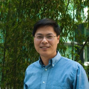
Michael Lin, Ph.D.
Speaker: Michael Lin, Ph.D.
Associate Professor
Neurobiology, Bioengineering, and by courtesy Chemical and Systems Biology
Stanford Medicine, Stanford University
Date: Thursday, September 2, 2021
Time: 3:30-4:30 PM EDT
Zoom – check email for link or contact ksas@seas.upenn.edu
Location: Moore Room 216, 200 S. 33rd Street
Abstract: The most effective medicines are those that target the earliest causes of disease, rather than later manifestations. Engineering of biomolecules is a promising but underexplored approach to precisely detecting or targeting disease causes. I will present our work to develop a novel approach to treating cancer by detecting the signaling abnormalities that give rise to cancer. Interestingly, this effort involves biomolecular engineering at multiple scales: proteins, pathways, and viruses. I will also discuss how our work has translated serenditously to developing treatments for SARSCoV2.
Michael Lin Bio: Michael Z. Lin received an A.B. summa cum laude in Biochemistry from Harvard, an M.D. from UCLA, and a Ph.D. from Harvard Medical School. After training in biochemistry and neurobiology as a PhD student with Michael Greenberg at Harvard Medical School, Dr. Lin performed postdoctoral research in fluorescent protein engineering with Chemistry Nobel Laureate Roger Y. Tsien at UCSD. Dr. Lin is a recipient of a Burroughs Wellcome Career Award for Medical Scientists, a Rita Allen Scholar Award, a Damon Runyon-Rachleff Innovation Award, and a NIH Pioneer Award.
Jenny Jiang, the Peter & Geri Skirkanich Associate Professor of Innovation in the department of Bioengineering, has received a Lloyd J. Old STAR Program grant from the Cancer Research Institute (CRI), which is a major supporter of cancer immunotherapy research and clinical trials with the goal of curing all types of cancer.
The CRI Lloyd J. Old Scientists Taking Risks (STAR) Program “provides long-term funding to mid-career scientists, giving them the freedom and flexibility to pursue high-risk, high-reward research at the forefront of discovery and innovation in cancer immunotherapy.” This prestigious grant was give to six awardees this year, chosen from a pool of hundreds of applicants, and recognizes “future leaders in the field of cancer immunotherapy [who are expected to] carry out transformational research.”
The Old STAR Program Grant comes with $1.25 million in funding over 5 years to support the awardees’ cancer immunology research.
Jiang, who recently joined Penn Bioengineering, is a pioneer in developing tools in genomics, biophysics, immunology, and informatics and applying them to study systems immunology and immune engineering in human diseases. She was also inducted into the American Institute for Medical and Biological Engineering (AIMBE) College of Fellows in March 2021 for her outstanding contributions to the field of systems immunology and immunoengineering and devotion to the success of women in engineering. Jiang’s research focuses on systems immunology by developing technologies that enable high-throughput, high-content, single cell profiling of T cells in health and disease and she is recognized as one of the leading authorities in systems immunology and immunoengineering.
“The STAR Award from CRI allows my lab to answer some of the fundamental questions in T cell biology, such as is the T cell repertoire complete to cover all possible cancer antigens, as well as to improve the efficacy of T cell based cancer immunotherapies,” says Jiang.
A.T. Charlie Johnson, Rebecca W. Bushnell Professor of Physics and Astronomy at the Penn School of Arts & Sciences, and member of the Penn Bioengineering Graduate Group has been working with a team of researchers on a new “electronic nose” that could help track the spread of COVID-19 based on the disease’s unique odor profile. Now, similar research shows that vapors emanating from blood samples can be tested to distinguish between benign and cancerous pancreatic and ovarian cells. Johnson presented the results at the annual American Society of Clinical Oncology meeting on June 4 (Abstract # 5544):
“It’s an early study but the results are very promising,” Johnson said. “The data shows we can identify these tumors at both advanced and the earliest stages, which is exciting. If developed appropriately for the clinical setting, this could potentially be a test that’s done on a standard blood draw that may be part of your annual physical.”
Carl June, MD, the Richard W. Vague Professor in Immunotherapy in the department of Pathology and Laboratory Medicine in the Perelman School of Medicine at the University of Pennsylvania, director of the Center for Cellular Immunotherapies at Penn’s Abramson Cancer Center, and member of the Penn Bioengineering Graduate Group, received the $1 million Sanford Lorraine Cross Award for his groundbreaking work in developing chimeric antigen receptor (CAR) T cell therapy. June is a world renowned cancer cell therapy pioneer.
“Sanford Health, the only health system in the country to award a $1 million prize for achievements in the medical sciences, announced the award on April 13 at a special ceremony in Sioux Falls, South Dakota. The biennial award recognizes life-changing breakthroughs and bringing emerging transformative medical innovations to patients.
‘This is a well-deserved and exciting award for one of Penn’s most distinguished faculty members, whose pioneering research has reshaped the fight against cancer and brought fresh hope for both adults and children with the disease,’ said J. Larry Jameson, MD, PhD, Executive Vice President of the University of Pennsylvania for the Health System and Dean of the Perelman School of Medicine. ‘His contributions truly have been transformative for patients across the globe and taken the field of oncology in new and powerful directions.'”
Speaker: Xiling Shen, Ph.D.
Hawkins Family Associate Professor
Biomedical Engineering
Duke University
Date: Thursday, April 15, 2021
Time: 3:00-4:00 PM EDT
Zoom – check email for link or contact ksas@seas.upenn.edu
Abstract:
Bodily cells undergo transformations in space and time during development, disease progression, and therapeutic treatment. A holistic approach that combines engineering tools, patient-derived models, and analytical methods is needed to map cellular reprogramming and expose new therapeutic opportunities. The talk will cover our effort across the entire spectrum from bench to bedside, including organogenesis during embryonic development, epigenetic and metabolic reprogramming of cancer metastasis and COVID-19 patients, and organoid technology to guide precision- and immune-oncology.
Xiling Shen Bio:
Dr. Shen is the Hawkins Family Associate Professor in the Department of Biomedical Engineering at Duke University. He is also the director of the Woo Center for Big Data and Precision Health. He received his BS, MS, and PhD degrees from Stanford University and the NSF career award at Cornell University. He is the steering committee chair of the NCI Patient-Derived Model of Cancer Consortium. His lab studies precision medicine from a systems biology perspective. Areas of interests include cancer, stem cells, the but-brain axis, and infectious diseases.
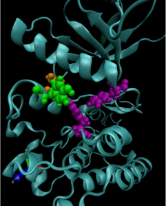
A computer model of a mutated anaplastic lymphoma kinase (ALK), a known oncogenic driver in pediatric neuroblastoma.
Kinases are a class of enzymes that are responsible for transferring the main chemical energy source used by the body’s cells. As such, they play important roles in diverse cellular processes, including signaling, differentiation, proliferation and metabolism. But since they are so ubiquitous, mutated versions of kinases are frequently found in cancers. Many cancer treatments involve targeting these mutant kinases with specific inhibitors.
Understanding the exact genetic mutations that lead to these aberrant kinases can therefore be critical in predicting the progression of a given patient’s cancer and tailoring the appropriate response.
To achieve this understanding on a more fundamental level, a team of researchers from the University of Pennsylvania’s School of Engineering and Applied Science and Perelman School of Medicine, the Children’s Hospital of Philadelphia (CHOP) and researchers at the Yale School of Medicine’s Cancer Biology Institute, have constructed molecular simulations of a mutant kinase implicated in pediatric neuroblastoma, a childhood cancer impacting the central nervous system.
Using their computational model to study the relationship between single-point changes in the kinase’s underlying gene and the altered structure of the protein it ultimately produces, the researchers revealed useful commonalities in the mutations that result in tumor formation and growth. Their findings suggest that such computational approaches could outperform existing profiling methods for other cancers and lead to more personalized treatments.
The study, published in the Proceedings of the National Academy of Sciences, was led by Ravi Radhakrishnan, Professor and chair of Penn Engineering’s Department of Bioengineering and professor in its Department of Chemical and Biomolecular Engineering, and Mark A. Lemmon, Professor of Pharmacology at Yale and co-director of Yale’s Cancer Biology Institute. The study’s first authors were Keshav Patil, a graduate student in Penn Engineering’s Department of Chemical and Biomolecular Engineering, along with Earl Joseph Jordan and Jin H. Park, then members of the Graduate Group in Biochemistry and Molecular Biology in Penn’s Perelman School of Medicine. Krishna Suresh, an undergraduate student in Radhakrishnan’s lab, Courtney M. Smith, a graduate student in Lemmon’s lab, and Abigail A. Lemmon, an undergraduate in Lemmon’s lab, contributed to the study. They collaborated with Yaël P. Mossé, Associate Professor of Pediatrics at Penn Medicine and in the division of oncology at CHOP.
“Some cancers rely on the aberrant activation of a single gene product for tumor initiation and progression,” says Radhakrishnan. “This unique mutational signature may hold the key to understanding which patients suffer from aggressive forms of the disease or for whom a given therapeutic drug may yield short- or long-term benefits. Yet, outside of a few commonly occurring ‘hotspot’ mutations, experimental studies of clinically observed mutations are not commonly pursued.”
William H. Peranteau, Michael J. Mitchell, Margaret Billingsley, Meghana Kashyap, and Rachel Riley (Clockwise from top left)
As COVID-19 vaccines roll out, the concept of using mRNA to fend off viruses has become a part of the public dialogue. However, scientists have been researching how mRNA can be used to in life-saving medical treatments well before the pandemic.
The “m” in “mRNA” is for “messenger.” A single-stranded counterpart to DNA, it translates the genetic code into the production of proteins, the building blocks of life. The Moderna and Pfizer COVID-19 vaccines work by introducing mRNA sequences that act as a set of instructions for the body to produce proteins that mimic parts of the virus itself. This prepares the body’s immune response to recognize the real virus and fight it off.
Because it can spur the production of proteins that the body can’t make on its own, mRNA therapies also have the potential to slow or prevent genetic diseases that develop before birth, such as cystic fibrosis and sickle-cell anemia.
However, because mRNA is a relatively unstable molecule that degrades quickly, it needs to be packaged in a way that maintains its integrity as its delivered to the cells of a developing fetus.
To solve this challenge, Michael J. Mitchell, Skirkanich Assistant Professor of Innovation in the Department of Bioengineering, is researching the use of lipid nanoparticles as packages that transport mRNA into the cell. He and William H. Peranteau, an attending surgeon in the Division of General, Thoracic and Fetal Surgery and the Adzick-McCausland Distinguished Chair in Fetal and Pediatric Surgery at Children’s Hospital of Philadelphia, recently co-authored a “proof-of-concept” paper investigating this technique.
In this study, published in Science Advances, Mitchel examined which nanoparticles were optimal in the transport of mRNA to fetal mice. Although no disease or organ was targeted in this study, the ability to administer mRNA to a mouse while still in the womb was demonstrated, and the results are promising for the next stages of targeted disease prevention in humans.
Mitchel spoke with Tom Avril at The Philadelphia Inquirer about the mouse study and its implications for treatment of rare infant diseases through the use of mRNA, ‘the messenger of life.’
Penn bioengineering professor Michael J. Mitchell, the other senior author of the mouse study, tested various combinations of lipids to see which would work best.
The appeal of the fatty substances is that they are biocompatible. In the vaccines, for example, two of the four lipids used to make the delivery spheres are identical to lipids found in the membranes of human cells — including plain old cholesterol.
When injected, the spheres, called nanoparticles, are engulfed by the person’s cells and then deposit their cargo, the RNA molecules, inside. The cells respond by making the proteins, just as they make proteins by following the instructions in the person’s own RNA. (Important reminder: The RNA in the vaccines cannot become part of your DNA.)
Among the different lipid combinations that Mitchell and his lab members tested, some were better at delivering their cargo to specific organs, such as the liver and lungs, meaning they could be a good vehicle for treating disease in those tissues.
Christian Figueroa-Espada, a Penn Bioengineering Ph.D. student and National Science Foundation (NSF) Fellow, was selected as a Hispanic Scholarship Fund (HSF) Scholar from a highly-competitive pool of 85,000 applicants for their 2020-2021 program. One of only 5,100 awardees, Figueroa-Espada’s scholarship comes from the Toyota Motor North America Program. As an HSF Scholar, he has access to a full range of Scholar Support Services, such as career coaching, internship, and full-time employment opportunities, mentoring, leadership development, and wellness resources, including tools for self-advocacy, well-being, and knowledge building.
Born and raised in the Island of Enchantment, Puerto Rico, Figueroa-Espada received his B.S. in Mechanical Engineering from the University of Puerto Rico at Mayagüez, and is currently a second-year Ph.D. student in the lab of Michael J. Mitchell, Skirkanich Assistant Professor of Innovation in Bioengineering, where he is funded by the National Science Foundation Graduate Research Fellowship Program (NSF GRFP), the Graduate Education for Minorities (GEM) Fellowship Program, and the William Fontaine Fellowship. His research interests lie in the interface of biomaterials, drug delivery, and immunology – designing RNAi therapeutics for the reprogramming of the tumor microenvironment. His current project focuses on polymer-lipid drug delivery systems to study potential strategies to prevent homing and proliferation of multiple myeloma cancer within the bone marrow microenvironment. This project is part of the Mitchell lab’s recent National Institutes of Health (NIH) New Innovator Award.
“Chris has really hit the ground running on his Ph.D. studies at Penn Bioengineering, developing a new bone marrow-targeted nanoparticle platform to disrupt the spread of multiple myeloma throughout the body,” says Mitchell. “I’m very hopeful that this prestigious fellowship from HSF will permit him to make important contributions to nanomedicine and cancer research.”
“This fellowship, along with my NSF Graduate Research Fellowship, GEM Fellowship, and William Fontaine Fellowship through the University of Pennsylvania, make my research on nanoparticle-based RNA therapeutics for the reprogramming of the tumor microenvironment to treat malignancies and overcome drug resistance possible,” says Figueroa-Espada. “While my professional goal is to stay in academia and lead a research lab, my personal goal is to become whom I needed: a role model within the Latino STEM community, hoping to address many of the difficulties that impede Latino students’ success in higher education, and thanks to Toyota Motor/HSF, NSF, and GEM, I am one step closer to meeting these goals.”
Audrey Bowden, PhD, Associate Professor of Biomedical Engineering. (Vanderbilt University / Steve Green)
Speaker: Audrey Bowden, Ph.D.
Dorothy J. Wingfield Phillips Chancellor’s Faculty Fellow and Associate Professor of Biomedical Engineering and Electrical Engineering & Computer Science
Vanderbilt University
Date: Thursday, November 19, 2020
Time: 3:00-4:00 PM EST
Zoom – check email for link or contact ksas@seas.upenn.edu
Title: “Emerging Technologies for Detection of Early Stage Bladder Cancer”
Abstract:
Bladder cancer (BC) — the 4th most common cancer in men and the most expensive cancer to treat over a patient’s lifetime — is a lifelong burden to BC patients and a significant economic burden to the U.S. healthcare system. The high cost of BC stems largely from its high recurrence rate (>50%); hence, BC management involves frequent surveillance. Unfortunately, the current in-office standard-of-care tool for BC surveillance, white light cystoscopy (WLC), is limited by low sensitivity and specificity for carcinoma in situ (CIS), a high-grade carcinoma with high potential to metastasize. Early detection and complete eradication of CIS are critical to improve treatment outcomes and to minimize recurrence. The most promising macroscopic technique to improve sensitivity to CIS detection, blue light cystoscopy (BLC), is costly, time-intensive, has low availability and a high false-positive rate. Given the limitations of WLC, we aim to change the paradigm around how BC surveillance is performed by validating new tools with high sensitivity and specificity for CIS that are appropriate for in-office use. In this seminar, I discuss our innovative solutions to improve mapping the bladder for longitudinal tracking of suspicious lesions and to create miniature tools for optical detection based on optical coherence tomography (OCT). OCT and its functional variant, cross-polarized OCT, can detect early-stage BC with better sensitivity and specificity than WLC. We discuss the critical technical innovations necessary to make OCT and CP-OCT a practical tool for in-office use, and new results from recent explorations of human bladder samples that speak to the promise of this approach to change the management of patient care.
Bio:
Audrey K. Bowden is the Dorothy J. Wingfield Phillips Chancellor Faculty Fellow and Associate Professor of Biomedical Engineering (BME) and of Electrical Engineering and Computer Science (EECS) at Vanderbilt University. Prior to this, she served as Assistant and later Associate Professor of Electrical Engineering and Bioengineering at Stanford University. Dr. Bowden received her BSE in Electrical Engineering from Princeton University, her PhD in BME from Duke University and completed her postdoctoral training in Chemistry and Chemical Biology at Harvard University. During her career, Dr. Bowden served as an International Fellow at Ngee Ann Polytechnic in Singapore. From 2007-2008, she was the Arthur H. Guenther Congressional Fellow sponsored by the OSA and SPIE and served as a Legislative Assistant in the United States Senate through the AAAS Science and Technology Policy Fellows Program. Dr. Bowden is a Fellow of SPIE, a Fellow of AIMBE and is the recipient of numerous awards, including the Air Force Young Investigator Award, the NSF Career Award, the Hellman Faculty Scholars Award, the Phi Beta Kappa Teaching Award, Ford Foundation Postdoctoral Fellowship, and the NSBE Golden Torch Award. She is a former Associate Editor of IEEE Photonics Journal, former Lead Guest Editor of a Biomedical Optics Express Special Issue and is a member of numerous professional committees. Her research interests include biomedical optics – particularly optical coherence tomography and near infrared spectroscopy – microfluidics, and point of care diagnostics.
Title: “Engineering Next-Generation CAR-T Cells for Cancer Immunotherapy”
Abstract:
The adoptive transfer of T cells expressing chimeric antigen receptors (CARs) has demonstrated clinical efficacy in the treatment of advanced cancers, with anti-CD19 CAR-T cells achieving up to 90% complete remission among patients with relapsed B-cell malignancies. However, challenges such as antigen escape and immunosuppression limit the long-term efficacy of adoptive T-cell therapy. Here, I will discuss the development of next-generation T cells that can target multiple cancer antigens and resist immunosuppression, thereby increasing the robustness of therapeutic T cells against tumor defense mechanisms. Specifically, I will discuss the development of multi-input receptors and T cells that can interrogate intracellular antigens. I will also discuss the engineering of T cells that can effectively convert TGF-beta from a potent immunosuppressive cytokine into a T-cell stimulant. This presentation will highlight the potential of synthetic biology in generating novel mammalian cell systems with multifunctional outputs for therapeutic applications.
Bio:
Dr. Yvonne Chen is an Associate Professor of Microbiology, Immunology, and Molecular Genetics at the University of California, Los Angeles. She is also a faculty, by courtesy, in the Department of Chemical and Biomolecular Engineering. The Chen Laboratory focuses on applying synthetic biology and biomolecular engineering techniques to the development of novel mammalian-cell systems. The Chen Lab’s work on engineering next-generation T-cell therapies for cancer has been recognized by the NIH Director’s Early Independence Award, the NSF CAREER Award, the Hellman Fellowship, the ACGT Young Investigator Award in Cell and Gene Therapy for Cancer, the Mark Foundation Emerging Leader Award, and the Cancer Research Institute Lloyd J. Old STAR Award. Prior to joining UCLA in 2013, Yvonne was a Junior Fellow in the Harvard Society of Fellows. She received postdoctoral training at the Center for Childhood Cancer Research within the Seattle Children’s Research Institute, and in the Department of Systems Biology at Harvard Medical School. Yvonne received her B.S. in Chemical Engineering from Stanford University and her Ph.D. in Chemical Engineering from the California Institute of Technology.