The first lecture in the Fall 2020 Penn Bioengineering Seminar Series will be held Thursday, September 10th. All seminars this semester will be held virtually on Zoom.
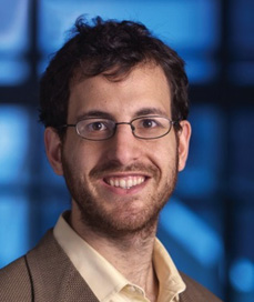
Quinton Smith, PhD
Speaker: Quinton Smith, Ph.D.
Postdoctoral Fellow
Laboratory for Multiscale Regenerative Technologies
Massachusetts Institute of Technology
Date: Thursday, September 10, 2020
Time: 3:00-4:00 pm
Zoom – check email for link or contact ksas@seas.upenn.edu
Title: “Stem Cell Fate is a Touchy Subject”
Abstract:
The success of regenerative cell therapy relies on the integration of a functional vascular system within the redeveloping tissue, to mediate the exchange of oxygen, nutrients and waste. Although the advent of human induced pluripotent stem cells (hiPSCs) has accelerated progress towards this goal, owing to their potential to generate clinically relevant scales of patient-specific cells, techniques to drive their specification mainly rely on chemical cues. In this seminar, I will discuss engineering strategies to control the complex stem cell extracellular milieu, emphasizing the importance of mechanical cues during hiPSC development, specification and downstream functionality as it relates to vascular differentiation.
Bio:
Quinton Smith received his PhD in Chemical and Biomolecular Engineering from Johns Hopkins University in 2017 after completing his bachelor’s degree in Chemical Engineering from the University of New Mexico. As a graduate student under the guidance of Dr. Sharon Gerecht, Quinton implemented various engineering tools to explore the roles of physical and chemical cues on stem cell lineage specification and downstream maturation. Dr. Smith is currently a postdoctoral fellow under the mentorship of Dr. Sangeeta Bhatia at MIT’s Koch Institute for Integrative Cancer Research, where he is investigating the role biliary epithelium in liver regeneration. Dr. Smith’s predoctoral work was supported by an NIH/NHLBI F-31 and NSF Graduate Research Fellowship. He is a recipient of the 2017 Siebel Scholar award, and most recently joined the class of 2018 HHMI Hanna Gray Fellows.
See the full list of upcoming Penn Bioengineering fall seminars here.
Speaker: Paul Macklin, Ph.D.
Associate Professor, Indiana University
Date: Monday May 4, 2020
Time: 2:00-3:30 PM
Title: “Open Source Multicellular Systems Modeling for Cancer (and COVID-19)”
Paul Macklin is a mathematician, Associate Professor, and Director of Undergraduate studies in the recently-established Department of Intelligent Systems Engineering at Indiana University. He works with biologists, modelers, and clinicians to develop and validate sophisticated 3D computer models of cancer, SARS-CoV-2, and other multicellular systems, using the open source PhysiCell platform developed by his lab. He also works with the National Cancer Institute and the Department of Energy to co-lead a national initiative to create digital twins for the future of personalized predictive cancer medicine.
For the full abstract and registration details, visit the Penn Engineering events calendar.
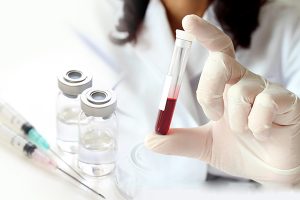
A blood test may be able to detect the most common form of pancreatic cancer while it is still in its early stages while also helping doctors accurately stage a patient’s disease and guide them to the appropriate treatment. A multidisciplinary study found the test—known as a liquid biopsy—was more accurate at detecting disease in a blinded study than any other known biomarker alone, and was also more accurate at staging disease than imaging is capable of alone. The team, which includes researchers from the Perelman School of Medicine, the Abramson Cancer Center, and the School of Engineering and Applied Science, published their findings in Clinical Cancer Research, a journal of the American Association for Cancer Research.
Pancreatic ductal adenocarcinoma (PDAC), the most common form of pancreatic cancer, is the third leading cause of cancer deaths. The overall five-year survival rate is just 9%, and most patients live less than one year following their diagnosis. One of the biggest challenges is catching the disease before it has progressed or spread. If the disease is caught early, patients may be candidates for surgery to remove the cancer, which can be curative. For locally advanced patients—meaning patients whose cancer has not spread beyond the pancreas but who are not candidates for surgery based on the size or location of the tumor—treatment involves three months of systemic therapy like chemo or radiation, then reassessing to see if surgery is an option. For patients whose disease has spread, there are currently no curative treatment options.
“Right now, the majority of patients who are diagnosed already have metastatic disease, so there is a critical need for a test that can not only detect the disease earlier but also accurately tell us who might be at a point where we can direct them to a potentially curative treatment,” says the study’s co-senior author Erica L. Carpenter,director of the Liquid Biopsy Laboratory and a research assistant professor of medicine. The study’s other co-senior author is David Issadore, an associate professor of bioengineering and electrical and systems engineering.
A microscopic view of pancreatic adenocarcinoma. (Image: Emma Furth)
Swept up in a pancreatic cancer diagnosis is inevitably a sense of fear and sadness.
But at Penn, researchers are bringing new hope to this disease. And with patients like Nick Pifani, it’s clear that they’re moving in the right direction.
Pifani, from Delran, New Jersey, first noticed some lingering stomach upset in February 2017. He called his family doctor, concerned—especially given that he was an otherwise healthy marathon runner who was only 42. He was sent to a gastrointestinal specialist. A few weeks later, some crippling stomach pain sent him back to the emergency room and he received an MRI that showed a mass on his pancreas—Stage Three, inoperable, he was told.
He was treated with chemotherapy, along with radiation and, eventually, and after receiving advice from doctors at Penn, his tumor was removed. Thereafter, he realized he had a PALB2 mutation—a cousin of the BRCA gene mutation. At that moment, his long-term needs changed and he found himself seeking specialized care at Penn, where he met Kim Reiss Binder, assistant professor of medicine at the Hospital of the University of Pennsylvania (HUP).
“I’m a planner; I want to understand what [my] potential options are,” Pifani says. “[Reiss Binder] asked why I was there to see her and I explained and quickly I could tell she was—outside of her being remarkably intelligent—a great listener and a compassionate doctor.”
“I have a feeling she worries about me more than I do,” he laughs.
Pifani has now been in remission for two years and four months; he sees Reiss-Binder every three months for checkups. His survival story is inspiring and a sign of momentum, even if a world without pancreatic cancer is still frustratingly out of reach.
Pancreatic cancer at Penn
Pancreatic cancer is the third-leading cause of cancer-related death in the United States, outmatched only by lung cancer (No. 1) and colorectal cancer (No. 2). A person diagnosed with pancreatic cancer is still unlikely to survive past five years—only 9% of survivors do, giving it the highest mortality rate among every major cancer.
In short, pancreatic cancer seldom paves the way for optimistic narratives. Some of the hope that has surfaced, though, is thanks to some talent, dedication to the cause, and hard work at Penn.
A key point of progress in the battle against the disease was made in 2002, when former Assistant Professor of Medicine David Tuveson established a standard model for examining human development of this disease in mice. This model has allowed for a reliable way to study the disease and has influenced progress made here at Penn and elsewhere since.
“There’s been a burst of activity in translational research, from bench to bedside,” explains Ben Stanger, the Hanna Wise Professor in cancer research and director of the Penn Pancreatic Cancer Research Center (PCRC) at the Abramson Cancer Center.
“And there’s a lot of momentum with community building, a dramatic increase in patient volumes, and a dramatic increase in what we know about the cancer,” he says of the status of pancreatic cancer today.
Reiss Binder, meanwhile, explains that one mark of progress at Penn and beyond has been learning about people like Pifani, who have the PALB2 gene, and why they respond differently to treatments than those without it. Platinum-based chemotherapies, for example, are especially effective for people with the PALB2 gene who are battling pancreatic cancer. An ongoing trial at Penn has tested and found some success with using PARP inhibitors—taken orally as an enzyme that fixes single-stranded breaks of DNA—as a maintenance therapy in that same PALB2 demographic after they’ve had chemotherapy. These are less toxic than chemotherapy for patients with the same mutations.
It’s all been slow progress toward better treatments, but there has been progress.
“This is the tip of the iceberg for a disease that we historically have treated with perpetual chemotherapy,” Reiss Binder says. “We owe it to patients to find better options to suppress the cancer but not ruin their quality of life.”
Catching cancer earlier
The consensus on why pancreatic cancer is so deadly? It just can’t be spotted fast enough.
Pancreatic cancer often presents well after it has developed and metastasized, and does so in a way that is not easy to recognize as cancer. Common symptoms include, for example, stomach upset and back pain. And by the time a harder-to-ignore symptom of the cancer surfaces, a sort of yellowing of the skin (a result of a bile duct blockage), it’s likely too late to stop the cancer in its tracks.
One approach to improved detection being tested at Penn, by Research Assistant Professor of Medicine Erica Carpenter, is a liquid biopsy—drawn from a standard blood test. Current means to test for pancreatic cancer—imaging through an endoscopic tube—are invasive and expensive, meaning a common liquid test could transform how many cases are detected early.
Carpenter explains that circulating tumor cells (CTCs) can shed from a tumor that’s adjacent to the wall of a blood vessel; what’s shed then shows up in a blood test. The cells, if detected, can explain more about the nature of the tumor, giving doctors an opportunity to examine characteristics of cancerous cells and decide how to effectively treat a tumor if it can’t be surgically removed. It also allows interpretations of disease burden and the effectiveness of medications—through genome sequencing—that imaging does not.
Ultimately, this gives doctors the potential to track the growth of a tumor before it’s fully developed, all through one tube of blood—detected through an innovative use of technology.
David Issadore, Ph.D.
David Issadore, associate professor of bioengineering and electrical and systems engineering in the School of Engineering and Applied Science, has worked since 2017 to develop a chip that detects cancer in the blood, using machine learning to sort through literally hundreds of billions of vesicles and cells, looking for these CTCs. The chip retrieves data and the machine learning developed interprets that data, attempting to make a diagnosis that not only finds pancreatic cancer but also provides information about its progression—and, importantly, whether a patient might benefit from surgery.
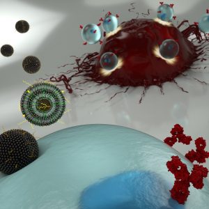
An artist’s illustration of nanoparticles transporting mRNA into a T cell (blue), allowing the latter to express surface receptors that recognize cancer cells (red). (Credit: Ryan Allen, Second Bay Studios)
New cancer immunotherapies involve extracting a patient’s T cells and genetically engineering them so they will recognize and attack tumors. This type of therapy is not without challenges, however. Engineering a patient’s T cells is laborious and expensive. And when successful, the alterations to the immune system immediately make patients very sick for a short period of time, with symptoms including fever, nausea and neurological effects.
Now, Penn researchers have demonstrated a new engineering technique that, because it is less toxic to the T cells, could enable a different mechanism for altering the way they recognize cancer, and could have fewer side effects for patients.
The technique involves ferrying messenger RNA (mRNA) across the T cell’s membrane via a lipid-based nanoparticle, rather than using a modified HIV virus to rewrite the cell’s DNA. Using the former approach would be preferable, as it only confers a temporary change to the patient’s immune system, but the current standard method for getting mRNA past the cell membrane can be too toxic to use on the limited number of T cells that can be extracted from a patient.
Michael Mitchell, Margaret Billingsley, and Carl June
The researchers demonstrated their technique in a study published in the journal Nano Letters. It was led by Michael Mitchell, Skirkanich Assistant Professor of Innovation of bioengineering in the School of Engineering and Applied Science, and Margaret Billingsley, a graduate student in his lab.
They collaborated with one of the pioneers of CAR T therapy: Carl June, the Richard W. Vague Professor in Immunotherapy and director of the Center for Cellular Immunotherapies in the Abramson Cancer Center and the director of the Parker Institute for Cancer Immunotherapy at the Perelman School of Medicine.
University of Washington Researchers Engineer a New Way to Study Circulatory Obstruction
Capillaries are one of the most important forms of vasculature in our body, as they allow our blood to transfer nutrients to other parts of our body. But for how much effect capillary functionality can have on our health, their small size makes them extremely difficult to engineer into models for a variety of diseases. Now, researchers at the University of Washington led by Ying Zheng, Ph. D., engineered a three-dimensional microvessel model with living cells to study the mechanisms of microcirculatory obstruction involved with malaria.
Rather than just achieving a physical model of capillaries, these researchers created a model that allowed them to study typical flow and motion through capillaries, before comparing it to deficiencies in this behavior involved with diseases like malaria. The shape of the engineered model is similar to that of an hourglass, allowing the researchers to study instances where red blood cell transit may encounter bottlenecks between the capillaries and other vessels. Using multiphoton technology, Zheng and her team created 100mm capillary models with etched-in channels and a collagen base, to closely model the typical size and rigidity of the vessels. Tested with malaria-infected blood cells, the model showed similar circulatory obstructive behavior to that which occurs in patients, giving hope that this model can be transferred to other diseases involving such obstruction, like sickle cell anemia, diabetes, and cardiovascular conditions.
Understanding a Cell Membrane Protein Could Be the Key to New Cancer Treatments
Almost every cell in the body has integrins, a form of proteins, on its membrane, allowing cells to sense biological information from beyond their membranes while also using this feedback information to initiate signals within cells themselves. Bioengineers at the Imperial College of London recently looked at the way another membrane protein, called syndecan-4, interacts with integrins as a potential form of future cancer treatment. Referred to as “cellular hands” by lead researcher of the study Armando del Rio Hernandez, Ph.D., syndecan-4 sometimes controls the development of diseases or conditions like cancer and fibrosis. Hernandez and his team specifically studied the ties of syndecan-4 to yes-associated protein (YAP) and enzyme called P13K, both of which are affiliated with qualities of cancer progression like halted apoptosis or cell stiffening. Knowing this, Hernandez and his team hope to continue research into understanding the mechanisms of syndecan-4 throughout the cell, in search of new mechanisms and targets to focus on with future developments of cancer treatments.
A New Medical Device Could Improve Nerve Functionality After Severe Damage
Serious nerve damage remains difficult to repair surgically, often involving the stretching of nerves for localized damage, or the transfer of healthy nerve cells from another part of the body to fill larger gaps in nerve damage. But these imperfect solutions limit the return of full nerve function and movement to the damaged part of the body, and in more serious cases with large areas of nerve damage, can also risk damage in other areas of the body that healthy nerves are borrowed from for treatment. A new study from the University of Pittsburgh published in Science Translational Medicine led by Kacey Marra, Ph. D., has successfully repaired nerve damage in mice and monkeys using a biodegradable tube that releases growth factors called glial-cell-derived neurotrophic factors over time.
Marra and her team showed that this new device restored nerve function up to 80% in nonhuman primates, where current methods of nerve replacement often only achieve 50-60% functionality restoration. The device might have an easier time getting FDA-approval, since it doesn’t involve the use of stem cells in its repair mechanisms. Hoping to start human clinical trials in 2021, Marra and her team hope that the device will help both injured veterans and typical patients with nerve damage, and see potential future applications in facial nerve damage as well.
A New Computational Model Could Improve Treatments for Cancer, HIV, and Autoimmune Diseases
With cancer, HIV, and other autoimmune diseases, the best treatment options for patients are often determined with trial-and-error methods, leading to prolonged instances of ineffective approaches and sometimes unnecessary side effects. A group of researchers led by Wesley Errington, Ph.D., at the University of Minnesota decided to take a computational approach this problem, in an effort to more quickly and efficiently determine the most appropriate treatment for a given patient. Based on parameters controlling interactions between molecules with multiple binding sites, the team’s new model looks primarily at binding strength, linkage rigidity, and size of linkage arrays. Because diseases can often involve issues in molecular binding, the model aimed to model the 78 unique binding configurations for cases of when interacting molecules only have three binding sites, which are often difficult to observe experimentally. This new approach will allow for faster and easier determination of treatments for patients with diseases involving these molecular interactions.
Improved Drug Screening for Glioblastoma Patients
A new microfluidic brain chip from researchers at the University of Houston could help improve treatment evaluations for brain tumors. Glioblastoma patients, who have a five-year survival rate of a little over 5%, are some of the most common patients suffering from malignant brain tumors. This new chip, developed by the lab of Yasemin Akay, Ph.D., can quickly determine cancer drug effectiveness by analyzing a piece of cultured tumor biopsy from a patient by incorporating different chemotherapy treatments through the microfluidic vessels. Overall, Akay and her team found that this new chip holds hope as a future efficient and inexpensive form of drug screening for glioblastoma patients.
People and Places
The brain constructs maps to guide people, not just of physical spaces but also to connect stimuli around them, like conversations and other people. It’s long been known that the brain area responsible for this spatial navigation—the medial temporal lobe—is also involved in recalling memories.
Michael Kahana (left) is principal investigator in the Defense Advanced Research Projects Agency’s RAM program and a professor in the Department of Psychology. Ethan Solomon is an M.D./Ph.D. student in the Department of Bioengineering of the School of Engineering and Applied Science and in the Perelman School of Medicine.
Now, neuroscientists at the University of Pennsylvania have discovered that the signals the brain produces during spatial navigation and episodic memory recall look similar. Low-frequency brain waves called the theta rhythm appear as people jump from one memory to the next, as many prior studies looking only at human navigation have shown. The new findings, which suggest that the brain structures responsible for helping people navigate the world may also “navigate” a mental map of prior experiences, appear in the Proceedings of the National Academy of Sciences.
The Florida Institute of Technology recently announced plans to start construction in spring 2020 on a new Health Sciences Research Center, set to further establish biomedical engineering and pre-medical coursework and research at the institute. With plans to open the new center in 2022, Florida Tech anticipates increased enrollment in the two programs, and hopes that the center will offer more opportunities in a growing professional field.
Anson Ong, Ph.D., the Associate Dean of Administration and Graduate Programs at the University of Texas at San Antonio, was recently elected to the International College of Fellows of Biomaterials Science and Engineering. With a focus on research in biomaterial implants for orthopaedic applications, Ong’s election to the college honors his advancement and contribution to the field of biomaterials research.
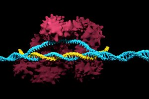
Positive results in first-in-U.S. trial of CRISPR-edited immune cells
3D render of the CRISPR-Cas9 genome editing system
Genetically editing a cancer patient’s immune cells using CRISPR/Cas9 technology, then infusing those cells back into the patient appears safe and feasible based on early data from the first-ever clinical trial to test the approach in humans in the United States. Researchers from the Abramson Cancer Center have infused three participants in the trial thus far—two with multiple myeloma and one with sarcoma—and have observed the edited T cells expand and bind to their tumor target with no serious side effects related to the investigational approach. Penn is conducting the ongoing study in cooperation with the Parker Institute for Cancer Immunotherapy and Tmunity Therapeutics.
“This trial is primarily concerned with three questions: Can we edit T cells in this specific way? Are the resulting T cells functional? And are these cells safe to infuse into a patient? This early data suggests that the answer to all three questions may be yes,” says the study’s principal investigator Edward A. Stadtmauer, section chief of Hematologic Malignancies at Penn. Stadtmauer will present the findings next month at the 61st American Society of Hematology Annual Meeting and Exposition.
Tulane researchers join NIH HEAL initiative for research into opioid crisis
A Tulane University professor and researcher of biomedical engineering will join fellow researchers from over 40 other institutions in the National Institute of Health’s Help to End Addiction Long-Term (HEAL) Initiative. Of the $945 million that make up the project, Michael J. Moore, Ph.D. will receive a share of $1.2 million to advance research in modeling human pain through computer chips, with the help of fellow Tulane researchers Jeffrey Tasker, Ph.D., and James Zadina, Ph.D., each with backgrounds in neuroscience.
Because of the opioid epidemic sweeping the nation, Moore notes that there’s a rapid search going on to develop non-addictive painkiller options. However, he also sees a gap in adequate models to test those new drugs before human clinical trials are allowed to take place. Here is where he hopes to step in and bring some innovation to the field, by integrating living human cells into a computer chip for modeling pain mechanisms. Through his research, Moore wants to better understand not only how some drugs can induce pain, but also how patients can grow tolerant to some drugs over time. If successful, Moore’s work will lead to a more rapid and less expensive screening option for experimental drug advancements.
New machine learning-assisted microscope yields improved diagnostics
Researchers at Duke University recently developed a microscope that uses machine learning to adapt its lighting angles, colors, and patterns for diagnostic tests as needed. Most microscopes have lighting tailored to human vision, with an equal distribution of light that’s optimized for human eyes. But by prioritizing the computer’s vision in this new microscope, researchers enable it to see aspects of samples that humans simply can’t, allowing for a more accurate and efficient diagnostic approach.
Led by Roarke W. Horstmeyer, Ph.D., the computer-assisted microscope will diffuse light through a bowl-shaped source, allowing for a much wider range of illumination angles than traditional microscopes. With the help of convolutional neural networks — a special kind of machine learning algorithm — Horstmeyer and his team were able to tailor the microscope to accurately diagnose malaria in red blood cell samples. Where human physicians typically perform similar diagnostics with a rate of 75 percent accuracy, this new microscope can do the same work with 90 percent accuracy, making the diagnostic process for many diseases much more efficient.
Case Western Reserve University researchers create first-ever holographic map of brain
A Case Western Reserve University team of researchers recently spearheaded a project in creating an interactive holographic mapping system of the human brain. The design, which is believed to be the first of its kind, involves the use of the Microsoft HoloLens mixed reality platform. Lead researcher Cameron McIntyre, Ph.D., sees this mapping system as a better way of creating holographic navigational routes for deep brain stimulation. Recent beta tests with the map by clinicians give McIntyre hope that the holographic representation will help them better understand some of the uncertainties behind targeted brain surgeries.
More than merely providing a useful tool, McIntyre’s project also brings together decades’ worth of neurological data that has not yet been seriously studied together in one system. The three-dimensional atlas, called “HoloDBS” by his lab, provides a way of finally seeing the way all of existing neuro-anatomical data relates to each other, allowing clinicians who use the tool to better understand the brain on both an analytical and visual basis.
Implantable cancer traps reduce biopsy incidence and improve diagnostic
Biopsies are one of the most common procedures used for cancer diagnostics, involving a painful and invasive surgery. Researchers at the University of Michigan are trying to change that. Lonnie Shea, Ph.D., a professor of biomedical engineering at the university, worked with his lab to develop implants with the ability to attract any cancer cells within the body. The implant can be inserted through a scaffold placed under the patient’s skin, making it a more ideal option than biopsy for inaccessible organs like lungs.
The lab’s latest work on the project, published in Cancer Research, details its ability to capture metastatic breast cancer cells in vivo. Instead of needing to take biopsies from areas deeper within the body, the implant allows for a much simpler surgical procedure, as biopsies can be taken from the implant itself. Beyond its initial diagnostic advantages, the implant also has the ability to attract immune cells with tumor cells. By studying both types of cells, the implant can give information about the current state of cancer in a patient’s body and about how it might progress. Finally, by attracting tumor and immune cells, the implant has the ability to draw them away from the area of concern, acting in some ways as a treatment for cancer itself.
People and Places
Cesar de la Fuente-Nunez, PhD
The Philadelphia Inquirer recently published an article detailing the research of Penn’s Presidential Assistant Professor in Psychiatry, Microbiology, and Bioengineering, Cesar de la Fuente, Ph.D. In response to a growing level of worldwide deaths due to antibiotic-resistant bacteria, de la Fuente and his lab use synthetic biology, computation, and artificial intelligence to test hundreds of millions of variations in bacteria-killing proteins in the same experiment. Through his research, de la Fuente opens the door to new ways of finding and testing future antibiotics that might be the only viable options in a world with an increasing level of drug-resistant bacteria
Emily Eastburn, a Ph.D. candidate in Bioengineering at Penn and a member of the Boerckel lab of the McKay Orthopaedic Research Laboratory, recently won the Ashton fellowship. The Ashton fellowship is an award for postdoctoral students in any field of engineering that are under the age of 25, third-generation American citizens, and residents of either Pennsylvania or New Jersey. A new member of the Boerckel lab, having joined earlier this fall, Eastburn will have the opportunity to conduct research throughout her Ph.D. program in the developmental mechanobiology and regeneration that the Boerckel lab focuses on.
The researchers’ experiments involved making synthetic tissues with artificial “cells.” The fibrin network that surrounds these beads pull on them when compressed; by changing the number of beads in their experimental tissues, the researchers could suss out how cell-fiber interplay contributes to the tissue’s overall properties.
Tissue gets stiffer when it’s compressed. That property can become even more pronounced with injury or disease, which is why doctors palpate tissue as part of a diagnosis, such as when they check for lumps in a cancer screening. That stiffening response is a long-standing biomedical paradox, however: tissue consists of cells within a complex network of fibers, and common sense dictates that when you push the ends of a string together, it loosens tension, rather than increasing it.
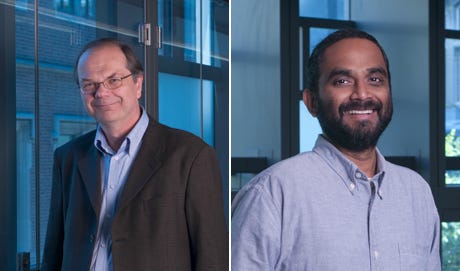
Now, in a study published in Nature, University of Pennsylvania’s School of Engineering and Applied Science researchers have solved this mystery by better understanding the mechanical interplay between that fiber network and the cells it contains.
The researchers found that when tissue is compressed, the cells inside expand laterally, pulling on attached fibers and putting more overall tension on the network. Targeting the proteins that connect cells to the surrounding fiber network might therefore be the optimal way of reducing overall tissue stiffness, a goal in medical treatments for everything from cancer to obesity.
Paul Janmey and Vivek Shenoy
The study was led by Paul Janmey, Professor in the Perelman School of Medicine’s Department of Physiology and in Penn Engineering’s Department of Bioengineering, and Vivek Shenoy, Eduardo D. Glandt President’s Distinguished Professor in Penn Engineering’s Department of Materials Science and Engineering, Mechanical Engineering and Applied Mechanics, and Bioengineering, along with Anne van Oosten and Xingyu Chen, graduate students in Janmey’s and Shenoy’s labs. Van Oosten is now a postdoctoral fellow at Leiden University in The Netherlands.
Shenoy is Director of Penn’s Center for Engineering Mechanobiology, which studies how physical forces influence the behavior of biological systems; Janmey is the co-director of one of the Center’s working groups, organized around the question, “How do cells adapt to and change their mechanical environment?”
Together, they have been interested in solving the paradox surrounding tissue stiffness.
The blue circle is the global symbol for diabetes. Wikimedia Commons.
Diabetes is one of the more common diseases among Americans today, with the American Diabetes Association estimating that approximately 9.5 percent of the population battles the condition today. Though symptoms and causes may vary across types and patients, diabetes generally results from the body’s inability to produce enough insulin to keep blood sugar levels in check. A new experimental treatment from the lab of Sha Jin, Ph.D., a biomedical engineering professor at Binghamton University, aims to use about $1.2 million in recent federal grants to develop a method for pancreatic islet cell transplantation, as those are the cells responsible for producing insulin.
But the catch to this new approach is that relying on healthy donors of these islet cells won’t easily meet the vast need for them in diabetic patients. Sha Jin wants to use her grants to consider the molecular mechanisms that can lead pluripotent stem cells to become islet-like organoids. Because pluripotent stem cells have the capability to evolve into nearly any kind of cell in the human body, the key to Jin’s research is learning how to control their mechanisms and signaling pathways so that they only become islet cells. Jin also wants to improve the eventual culture of these islet cells into three-dimensional scaffolds by finding ways of circulating appropriate levels of oxygen to all parts of the scaffold, particularly those at the center, which are notoriously difficult to accommodate. If successful in her tissue engineering efforts, Jin will not only be able to help diabetic patients, but also open the door to new methods of evolving pluripotent stem cells into mini-organ models for clinical testing of other diseases as well.
A Treatment to Help Others See Better
Permanently crossed eyes, a medical condition called strabismus, affects almost 18 million people in the United States, and is particularly common among children. For a person with strabismus, the eyes don’t line up to look at the same place at the same time, which can cause blurriness, double vision, and eye strain, among other symptoms. Associate professor of bioengineering at George Mason University, Qi Wei, Ph.D., hopes to use almost $2 million in recent funding from the National Institute of Health to treat and diagnose strabismus with a data-driven computer model of the condition. Currently, the most common method of treating strabismus is through surgery on one of the extraocular muscles that contribute to it, but Wei wants her model to eventually offer a noninvasive approach. Using data from patient MRIs, current surgical procedures, and the outcomes of those procedures, Wei hopes to advance and innovate knowledge on treating strabismus.
A Newly Analyzed Brain Mechanism Could be the Key to Stopping Seizures
Among neurological disorders, epilepsy is one of the most common. An umbrella term for a lot of different seizure-inducing conditions, many versions of epilepsy can be treated pharmaceutically. Some, however, are resistant to the drugs used for treatment, and require surgical intervention. Bin He, Ph. D., the Head of the Department of Biomedical Engineering at Carnegie Mellon University, recently published a paper in collaboration with researchers at Mayo Clinic that describes the way that seizures originating at a single point in the brain can be regulated by what he calls “push-pull” dynamics within the brain. This means that the propagation of a seizure through the brain relies on the impact of surrounding tissue. The “pull” he refers to is of the surrounding tissue towards the seizure onset zone, while the “push” is what propagates from the seizure onset zone. Thus, the strength of the “pull” largely dictates whether or not a seizure will spread. He and his lab looked at different speeds of brain rhythms to perform analysis of functional networks for each rhythm band. They found that this “push-pull” mechanism dictated the propagation of seizures in the brain, and suggest future pathways of treatment options for epilepsy focused on this mechanism.
Hyperspectral Imaging Might Provide New Ways of Finding Cancer
A new method of imaging called hyperspectral imaging could help improve the prediction of cancerous cells in tissue specimens. A recent study from a University of Texas Dallas team of researchers led by professor of bioengineering Baowei Fei, Ph.D., found that a combination of hyperspectral imaging and artificial intelligence led to an 80% to 90% level of accuracy in identifying the presence of cancer cells in a sample of 293 tissue specimens from 102 patients. With a $1.6 million grant from the Cancer Prevention and Research Institute of Texas, Fei wants to develop a smart surgical microscope that will help surgeons better detect cancer during surgery.
Fei’s use of hyperspectral imaging allows him to see the unique cellular reflections and absorptions of light across the electromagnetic spectrum, giving each cell its own specific marker and mode of identification. When paired with artificial intelligence algorithms, the microscope Fei has in mind can be trained to specifically recognize cancerous cells based on their hyperspectral imaging patterns. If successful, Fei’s innovations will speed the process of diagnosis, and potentially improve cancer treatments.
People and Places
A group of Penn engineering seniors won the Pioneer Award at the Rothberg Catalyzer Makerthon led be Penn Health-Tech that took place from October 19-20, 2019. SchistoSpot is a senior design project created by students Vishal Tien (BE ‘20), Justin Swirbul (CIS ‘20), Alec Bayliff (BE ‘20), and Bram Bruno (CIS ‘20) in which the group will design a low-cost microscopy dianostic tool that uses computer vision capabilities to automate the diagnosis of schistosomiasis, which is a common parasitic disease. Read about all the winners here.
Virginia Tech University will launch a new Cancer Research Initiative with the hope of creating an intellectual community across engineers, veterinarians, biomedical researchers, and other relevant scientists. The initiative will focus not only on building better connections throughout departments at the university, but also in working with local hospitals like the Carilion Clinic and the Children’s National Hospital in Washington, D.C. Through these new connections, people from all different areas of science and engineering and come together to share ideas.
Associate Professor of Penn Bioengineering Dani Bassett, Ph.D., recently sat down with the Penn Integrates Knowledge University Professor Duncan Watts, Ph.D., for an interview published in Penn Engineering. Bassett discusses the origins of network science, her research in small-world brain networks, academic teamwork, and the pedagogy of science and engineering. You can read the full interview here.
An all-female group of researchers from Northern Illinois University developed a device for use by occupational therapists that can capture three-dimensional images of a patient’s hand, helping to more accurately measure the hand or wrist’s range of motion. The group presented the abstract for their design at this year’s meeting of the Biomedical Engineering Society here in Philadelphia, where Penn students and researchers presented as well.
New 3D Tumor Models Could Improve Cancer Treatment
New ways of testing cancer treatments may now be possible thanks to researchers at the University of Akron who developed three-dimensional tumor models of triple-negative breast cancer. Led by Dr. Hossein Tavana, Ph. D., an associate professor of biomedical engineering at the university, the Tissue Engineering Microtechnologies Lab recently received a $1.13 million grant from the prestigious National Cancer Institute (NCI) of the National Institute of Health (NIH) to continue improving these tumor models. Tumors are difficult to fully replicate in vitro, as they are comprised of cancerous cells, connective tissue, and matrix proteins, among several other components. With this new grant, Tavana sees creating a high-throughput system that uses many identical copies of the tumor model for drug testing and better understanding of the way tumors operate. This high-throughput method would allow Tavana and his lab to isolate and test several different approaches at once, which they hope will help change the way tumors are studied and treated everywhere.
Noise-Induced Hearing Loss Poses Greater Threat to Neural Processing
Even though we all know we probably shouldn’t listen to music at high volumes, most of us typically do it anyway. But researchers at Purdue University recently found that noise-induced hearing loss could cause significant changes in neural processing of more complex sound inputs. Led by Kenneth Henry, Ph.D., an assistant professor of otolaryngology at the University of Rochester Medical Center, and Michael Heinz, Ph.D., a professor of biomedical engineering at Purdue University, the study shows that when compared with age-related hearing loss, noise-induced hearing loss will result in a greater decrease in hearing perception even when the two kinds of hearing loss appear to be of the same degree on an audiogram. This is because noise-induced hearing loss occurs because of physical trauma to the ear, rather than the long-term electrochemical degradation of some components that come happen with age. The evidence of this research is yet another reason why we should be more careful about exposing our ears to louder volumes, as they pose a greater risk of serious damage.
Increasing the Patient Populations for Research in Cartilage Therapy and Regenration
Despite the great progress in research of knee cartilage therapy and regeneration, there are still issues with the patient populations that most studies consider. Researchers often want to test new methods on patients that have the greatest chance of injury recovery without complications – often referred to as “green knees” – but this leaves out those patient populations who suffer from conditions or defects that have the potential to cause complications – often referred to as “red knees.” In a new paper published in Regenerative Medicine, the Mary Black Ralston Professor for Education and Research in Orthopaedic Surgery and secondary faculty in the Department of Bioengineering at Penn, Robert Mauck, Ph.D., discusses some cartilage therapies that may be suitable for red knee populations.
Working with James Carey, M.D., the Director of the Penn Center for Cartilage Repair and Osteochondritis, Mauck and his research team realized that even those with common knee cartilage conditions such as the presence of lesions or osteoarthritis were liable to be excluded from most regeneration studies. In discussing alternatives methods and structures of studying cartilage repair and regeneration, Mauck and Carey hope that future therapies will be applicable to a wider range of patient populations, and that there will soon be more options beyond full joint replacement for those with red knee conditions.
Plant-Like Superhydrophobicity Has Applications in Biomedical Engineering
Researchers in the Department of Biomedical Engineering at Texas A&M University recently found ways of incorporating the superhydrophobic properties of some plant leaves into biomedical applications through what they’re calling a “lotus effect.” The Gaharwar Lab, led by principal investigator and assistant professor of biomedical engineering Akhilesh Gaharwar, Ph.D., developed an assembly of two-dimensional atomic layers that they describe as a “nanoflower” to help control surface wetting in a biomedical setting. A recent paper published in Chemical Communications describes Gaharwar and his team’s work as expanding the use of superhydrophobic surface properties in biomedical devices by demonstrating the important role that atomic vacancies play in the wetting characteristic. While Gaharwar hopes to research the impact that controlling superhydrophobicity could have in stem cell applications, his work already allows for innovations in self-cleaning and surface properties of devices involving labs-on-a-chip and biosensing.
People and Places
Nader Engheta, H. Nedwill Ramsey Professor in Electrical and Systems Engineering, Bioengineering and Materials Science and Engineering, has been inducted into the Canadian Academy of Engineering (CAE) as an International Fellow. The CAE comprises many of Canada’s most accomplished engineers and Engheta was among the five international fellows that were inducted this year.
The Academy’s President Eddy Isaacs remarked: “Over our past 32 years, Fellows of Academy have provided insights in the fields of education, infrastructure, and innovation, and we are expecting the new Fellows to expand upon these contributions to public policy considerably.”
We would like to congratulate Anthony Lowman, Ph.D., on his appointment as the Provost and Senior Vice President for Academic Affairs at Rowan University. Formerly the Dean of Rowan’s College of Engineering, Lowman helped the college double in size, and helped foster a stronger research community. Lowman also helped to launch a Ph.D. program for the school, and added two new departments of Biomedical Engineering and Experiential Engineering Education in his tenure as the dean. Widely recognized for his research on hydrogels and drug delivery, Lowman was also formerly a professor of bioengineering at Temple University and Drexel University.
Lastly, we would like to congratulate Daniel Lemons, Ph.D., on his appointment as the Interim President of Lehman College of the City University of New York. Lemons, a professor in the Department of Biology at City College, specializes in cardiovascular and comparative physiology, and was also one of the original faculty members of the New York Center for Biomedical Engineering. With prior research funded by both the National Institute of Health (NIH) and the National Science Foundation (NSF), Lemons also holds patents in biomechanics teaching models and mechanical heart simulators.

 A blood test may be able to detect the most common form of pancreatic cancer while it is still in its early stages while also helping doctors accurately stage a patient’s disease and guide them to the appropriate treatment. A multidisciplinary study found the test—known as a liquid biopsy—was more accurate at detecting disease in a blinded study than any other known biomarker alone, and was also more accurate at staging disease than imaging is capable of alone. The team, which includes researchers from the
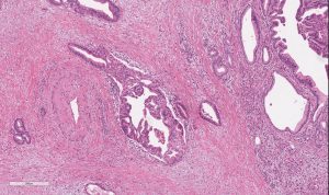

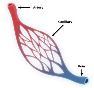 Capillaries are one of the most important forms of vasculature in our body, as they allow our blood to transfer nutrients to other parts of our body. But for how much effect capillary functionality can have on our health, their small size makes them extremely difficult to engineer into models for a variety of diseases. Now, researchers at the University of Washington led by


 New ways of testing cancer treatments may now be possible thanks to researchers at the University of Akron who developed three-dimensional tumor models of triple-negative breast cancer. Led by