While modern cancer treatments can have tremendous therapeutic impact, formidable obstacles remain. Foremost among these is drug resistance, the ability of cancers to withstand and ultimately progress despite the presence of an anti-cancer drug. However, ongoing research provides hope that these challenges can be overcome, including recent work performed by Penn Engineers.
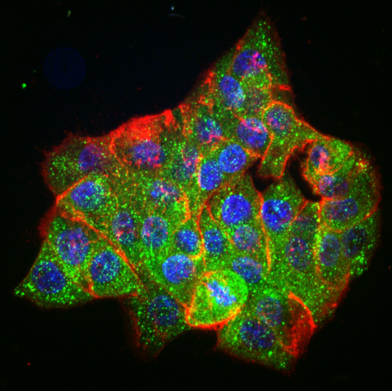
The lab of Lukasz J. Bugaj, Assistant Professor in the Department of Bioengineering, recently published an article that uncovers new mechanisms of how oncogenes interact with important pathways of cellular signaling that are associated with resistance. This work, titled “Oncogenic EML4-ALK Assemblies Suppress Growth Factor Perception and Modulate Drug Tolerance,” applied a new technique called ‘optogenetic functional profiling’ that allowed measurement of how important molecular signaling pathways respond to precise perturbations applied by the researchers. By applying this technique to many different cell types, the group found important differences in resistance-associated signaling between cancer cells and healthy cells
Specifically, the research showed that an oncogene called EML4-ALK, which activates oncogenic signaling, simultaneously inactivates adjacent pathways that can cause resistance. As a consequence, once an oncogene-blocking drug is applied, the inactivation is relieved, thus boosting activity through these adjacent, resistance-associated pathways. The study also showed that these pathways were not only de-repressed, but were actively stimulated by neighboring cancer cells, further enhancing cell survival in the presence of the drug.
“Our work shows that oncogenes, while driving cell division in cancer cells, simultaneously suppress the cells’ regulation by their environment,” said Dr. Bugaj. “While the work reveals mechanisms of paradoxical responses to drug treatment related to resistance, they may also inspire new ideas for therapies that can more efficiently kill cancer cells while maintaining suppression of resistance signaling. This work was co-led by PhD student David Gonzalez-Martinez and by Lee Roth, PhD, a postdoctoral fellow, and was supported by a grant from the American Cancer Society.
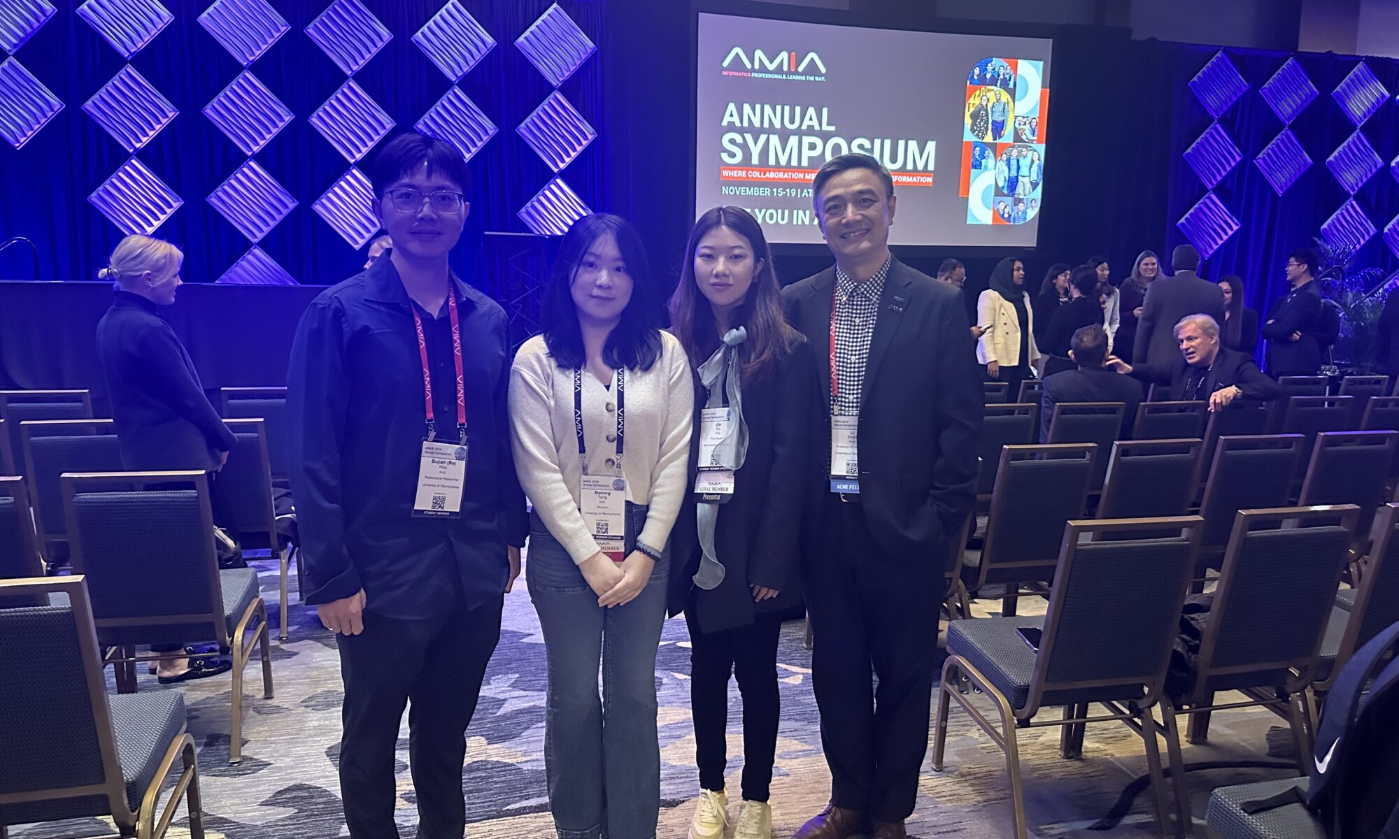
Boning Tong, a student in the Department of Bioengineering, has been awarded the Distinguished Paper Award from the AMIA 2024 Annual Symposium. The Awards Committee recognizes five notable papers that best encapsulate the potential of tremendous breakthroughs in the medical community. Ms. Tong works in the laboratory of Dr. Li Shen, who acts as her doctoral advisor and is a professor of Informatics in Biostatistics and Epidemiology.
“Our research tackles challenges in early Alzheimer’s Disease detection by addressing diagnosis label imbalances and fairness issues simultaneously in machine learning models,” said Ms. Tong. “Unlike traditional models, our approach achieves better prediction performance while minimizing bias related to sensitive factors like race, sex, and age. This advancement holds promise for improving the reliability and fairness of early AD detection, ultimately aiding better patient outcomes and equitable care.”
In the future, Ms. Tong plans to take the research they have gained and use them to obtain greater amounts of data. “Our plan is to apply our proposed model to other datasets with larger sample size and more detailed attribute subgroup information to explore the bias issue in AD further,” said Ms. Tong.
Ms. Tong’s work was supported by NIH grants and the ADNI data sets were obtained from the Alzheimer’s Disease Neuroimaging Initiative Database.
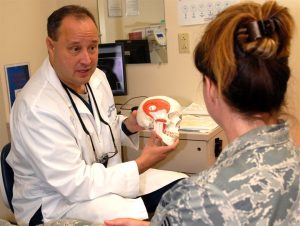
TMD is a common condition affecting movement of the jaw
Medical researchers have long been baffled by the need to find safe and effective treatment for a common condition called temporomandibular joint dysfunction (TMD). Affecting around twenty-five percent of the adult population worldwide, TMD appears overwhelmingly in adolescent, premenopausal women. Many different factors such as injury, arthritis, or grinding of the teeth can lead to the disintegration of or damage to the temporomandibular joint (TMJ), which leads to TMD, although the root cause is not always clear. A type of temporomandibular disorder, TMD can result in chronic pain in the jaw and ears, create difficulty eating and talking, and even cause occasional locking of the joint, making it difficult to open or close one’s mouth. Surgery is often considered a last resort because the results are often short-lasting or even dangerous.
The state of TMD treatment may change with the publication of a study in Science Translational Medicine. With contributions from researchers at the University of California, Irvine (UCI), UC Davis, and the University of Texas School of Dentistry at Houston, this new study has successfully implanted engineered discs made from rib cartilage cells into a TMJ model. The biological properties of the discs are similar enough to native TMJ cells to more fully reduce further degeneration of the joint as well as potentially pave the way for regeneration of joints with TMD.
Senior author Kyriacos Athanasiou, PhD, Distinguished Professor of Biomedical Engineering at UCI, states the next steps for the team of researchers include a long-term study to ensure ongoing effectiveness and safety of the implants followed by eventual clinical trials. In the long run, this technique may also prove useful and relevant to the treatment of other types of arthritis and joint dysfunction.
Advances in Autism Research
Currently, diagnosis of autism spectrum disorders (ASD) has been limited entirely to clinical observation and examination by medical professionals. This makes the early identification and treatment of ASD difficult as most children cannot be accurately diagnosed until around the age of four, delaying the treatment they might receive. A recent study published in the journal of Bioengineering & Translational Medicine, however, suggests that new blood tests may be able to identify ASD with a high level of accuracy, increasing the early identification that is key to helping autistic children and their families. The researchers, led by Juergen Hahn, PhD, Professor and Department Head of Biomedical Engineering at the Rensselaer Polytechnic Institute, hope that after clinical trials this blood test will become commercially available.
In addition to work that shows methods to detect autism earlier, the most recent issue of Nature Biomedical Engineering includes a study to understand the possible causes of autism and, in turn, develop treatments for the disease. The breakthrough technology of Cas9 enzymes allowed researchers to edit the genome, correcting for symptoms that appeared in mice which resembled autism, including exaggerated and repetitive behaviors. This advance comes from a team at the University of California, Berkeley, which developed the gene-editing technique known as CRISPR-Gold to treat symptoms of ASD by injecting the Cas9 enzyme into the brain without the need for viral delivery. The UC Berkeley researchers suggest in the article’s abstract that these safe gene-editing technologies “may revolutionize the treatment of neurological diseases and the understanding of brain function.” These treatments may have practical benefits for the understanding and treatment of such diverse conditions as addiction and epilepsy as well as ASD.
Penn Professor’s Groundbreaking Bioengineering Technology
Our own D. Kacy Cullen, PhD, was recently featured in Penn Today for his groundbreaking research which has led to the first implantable tissue-engineered brain pathways. This technology could lead to the reversal of certain neurodegenerative disorders, such as Parkinson’s disease.
With three patents, at least eight published papers, $3.3 million in funding, and a productive go with the Penn Center for Innovation’s I-Corps program this past fall, Dr. Cullen is ready to take this project’s findings to the next level with the creation of a brand new startup company: Innervace. “It’s really surreal to think that I’ve been working on this project, this approach, for 10 years now,” he says. “It really was doggedness to just keep pushing in the lab, despite the challenges in getting extramural funding, despite the skepticism of peer reviewers. But we’ve shown that we’re able to do it, and that this is a viable technology.” Several Penn bioengineering students are involved in the research conducted in Dr. Cullen’s lab, including doctoral candidate Laura Struzyna and recent graduate Kate Panzer, who worked in the lab all four years of her undergraduate career.
In addition to his appointment as a Research Associate Professor of Neurosurgery at the Perelman School of Medicine at the University of Pennsylvania, Dr. Cullen also serves as a member of Penn’s Department of Bioengineering Graduate Group Faculty, and will teach the graduate course BE 502 (From Lab to Market Place) for the BE Department this fall 2018 semester. He also serves as the director for the Center of Neurotrauma, Neurodegeneration, and Restoration at the VA Medical Center.
New Prosthetics Will Have the Ability to Feel Pain
New research from the Department of Biomedical Engineering at Johns Hopkins University (JHU) has found a way to address one of the difficult aspects of amputation: the inability for prosthetic limbs to feel. This innovative electronic dermis is worn over the prosthetic, and can detect sensations (such as pain or even a light touch), which are conveyed to the user’s nervous system, closing mimicking skin. The findings of this study were recently published in the journal ScienceRobotics.
While one might wonder at the value of feeling pain, both researchers and amputees verify that physical sensory reception is important both for the desired realism of the prosthetic or bionic limb, and also to alert the wearer of any potential harm or damage, the same way that heat can remind a person to remove her hand from a hot surface, preventing a potential burn. Professor Nitish Thakor, PhD, and his team hope to make this exciting new technology readily available to amputees.
People and Places
Women are still vastly outnumbered in STEM, making up only twenty percent of the field, and given the need for diversification, researchers, educators, and companies are brainstorming ways to proactively solve this problem by promoting STEM subjects to young women. One current initiative has been spearheaded by GE Healthcare and Milwaukee School of Engineering University (MSOE) who are partnering to give middle school girls access to programs in engineering during their summer break at the MSOE Summer STEM Camp, hoping to reduce the stigma of these subjects for young women. GE Girls also hosts STEM programs with a number of institutions across the U.S.
The National Science Policy Network (NSPN) “works to provide a collaborative resource portal for early-career scientists and engineers involved in science policy, diplomacy, and advocacy.” The NSPN offers platforms and support including grant funding, internships, and competitions. Chaired and led by emerging researchers and professors from around the country, including biomedical engineering PhD student Michaela Rikard of the University of Virginia, the NSPN seeks to provide a network for young scientists in the current political climate in which scientific issues and the very importance of the sciences as a whole are hotly contested and debated by politicians and the public. The NSPN looks to provide a way for scientists to have a voice in policy-making. This new initiative was recently featured in the Scientific American.
Upon its original founding in 2000, the Bill and Melinda Gates Foundation has included the eradication of malaria as part of its mission, pledging around $2 billion to the cause in the years since. One of its most recent initiatives is the funding of a bioengineering project which targets the type of mosquitoes which carry the deadly disease. Engineered mosquitoes (so-called “Friendly Mosquitoes”) would mate in the wild, passing on a mosquito-killing gene to their female offspring (only females bite humans) before they reach maturity. While previous versions of “Friendly Mosquitoes” have been met with success, concerns have been raised about the potential long-term ecological effects to the mosquito population. UK-based partner Oxitec expects to have the new group ready for trials in two years.
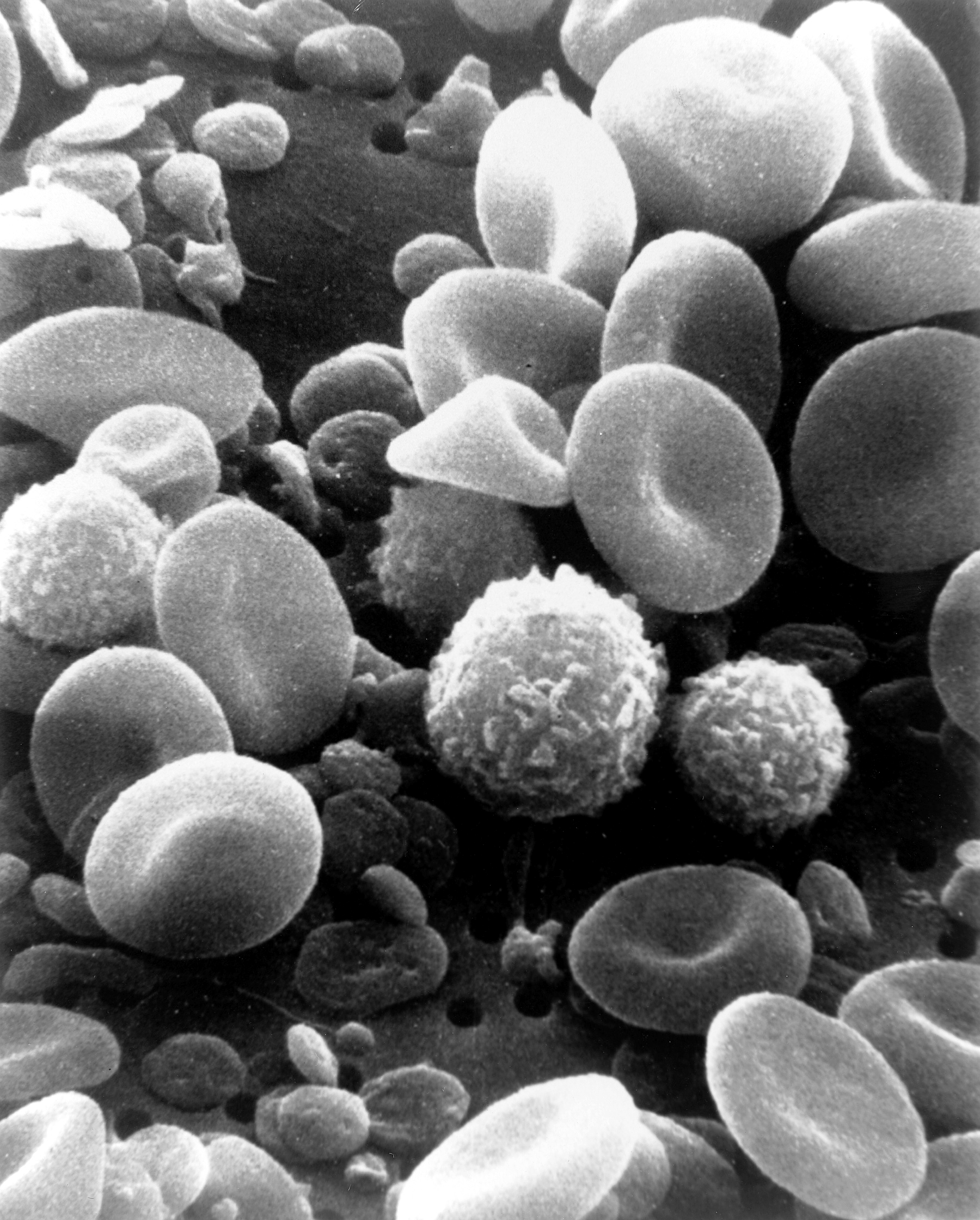
Several types of blood cells under scanning electron microscope.
We cover many highly complex innovations here, and many of these innovations solve vexing problems in the healthcare field. However, sometimes the problems addressed by these innovations are not particularly complex, even if the impact is economically significant. For instance, venipuncture — commonly referred to as a blood draw — is one of the most basic medical procedures and is mainly performed by medical technicians. There are two problems with venipuncture, however. First, either the health specialist might not be very good at drawing blood or the patient might be non-compliant. Second, the sample must be sent to a lab for analysis by someone else days later, making the costs for blood draws high. A device that could combine these procedures has been considered the “holy grail” of blood testing.
Engineers at Rutgers University might have found this holy grail. Reporting in the journal Technology, the engineers, led by Martin L. Yarmush, PhD, Paul & Mary Monroe Chair and Professor in the Department of Biomedical Engineering at Rutgers, describe how they combined robotics and lab-on-chip technology to create a point-of-care blood testing device. In the article, the authors report the testing of their device with a blood-like fluid loaded with microbeads and with model veins. The automated blood draw technique is the same across all patients, and the analysis of blood can occur immediately after isolating the blood, making it safer, faster, and more cost-effective. Animal testing should follow soon, and a longer-term view envisions expansion of the initial model to accommodate different types of blood testing.
Preventing a Water Crisis
One of the looming crises humankind faces is access to clean water. Nearly one third of the human population either lacks access or has threatened access to potable water. The rapid population growth in the southern hemisphere means that this proportion will increase, even as preventable water-borne diseases like cholera take their toll. Solutions such as desalinization or mass purification could provide solutions, but they are currently prohibitively expensive and create environmental problems of their own. Less expensive and less burdensome solutions continue to be sought.
Now, engineering professors from Carnegie Mellon University (CMU) might have identified a solution. Robert Tilton, PhD, and Todd Przybycien, PhD, both Professors in the Departments of Biomedical Engineering and Chemical Engineering at CMU, are lead authors on a new study in ACS Langmuirdescribing how proteins produced by the drumstick tree — a very hearty tree native to India that is conducive to a broad range of climate and is already widely cultivated for its fruit and oils — could be used to address water scarcity. The authors exploited the knowledge that cationic protein-modified sand can be used to filter water and showed how to engineer drumstick tree proteins to optimize the filtration process. Testing showed that the authors’ engineered filtration system was more effective and might even lend itself to repeated use.
Innovating for Pediatric Care
Penn Health-Tech is one of the newer initiatives here at the University of Pennsylvania dedicated to catalyzing medical device innovation. However, Penn isn’t the only Philadelphia institution dedicating resources to innovation and invention in medical devices. In the June issue of DOTmed HealthCare Business News magazine, Andrew Rich, who is Senior Director of Biomedical Engineering at the Children’s Hospital of Philadelphia (CHOP), discusses the initiatives being undertaken at CHOP to integrate data from medical devices with electronic health records, as well as other projects.
Improving Limb Prosthetics
Prosthetics have provided a solution for amputees for more than a century, and engineering has been the source of many improvements over that time. A significant goal of scientists studying prosthetics has been the ability of patients to control their artificial limbs with their own neuromuscular signals. While progress has been made in this direction with machine learning, many patients have to spend a lot of time “training” their prostheses to react properly to these signals, which can be deeply discouraging to patients who have already experienced trauma.
A possible solution has been suggested by He (Helen) Huang, PhD, Professor of Biomedical Engineering in the joint department of the University of North Carolina and North Carolina State University, and Stephanie Huang, PhD, Research Assistant Professor in the joint department. In a paper newly published in IEEE Transactions on Neural Systems and Rehabilitation Engineering, the professors used the muscle activation patterns of the residual muscles remaining in patients after amputation. They tested their approach in 10 patients and found highly statistically significant improvement in movement accuracy. Testing in more subjects and allowing users longer training periods during testing could both yield even more impressive outcomes.
People and Places
Synthetic biologists at Colorado State University received a $1.7 million grant from the Defense Advanced Research Projects Agency (DARPA) to genetically engineer sporopollenin — a naturally occurring, chemically inert polymer found in pollen grains — to create what they hope will be the world’s strongest material. Early success could lead to another $2 million in DARPA funding in a couple of years. Matt Kipper, PhD, Associate Professor of Chemical and Biological Engineering, is a co-investigator on the grant.
Rose-Hulman Institute of Technology in Terre Haute, Indiana, has announced that it will be adding a new major in engineering design to its curriculum. Patsy Brackin, PhD, Professor of Mechanical Engineering, will lead the new program as director.
Dolphins are among the most intelligent creatures on earth, showing behaviors such as teaching, learning, cooperation, delayed gratification, and other markers of high intelligence. Dolphins communicate vocally with one another, although we aren’t sure exactly what they communicate. While this communication isn’t “language” as humans define it, it uses echolocation — finding objects and orienteering on the basis of reflected sound — which humans don’t use in their communications.
Now, we have new information about dolphin echolocation thanks to an article recently published in the Journal of the Acoustical Society of America by mathematicians and biomedical engineers in Sweden. On the basis of earlier research finding that dolphin echolocation signals consist of two tones, rather than one, the new study finds that these two tones are emitted at slightly different times and that the sound waves have a Gaussian shape, similar to a bell curve. Using a mathematical algorithm, the authors successfully simulated echolocation signals in the lab.
The findings explain how dolphins use echolocation effectively but could also contribute to more accurate sound-based diagnostic techniques — particularly ultrasound, which relies heavily on methods similar to echolocation to provide images of moving tissues within the body, e.g., prenatal imaging and heart contraction.
Modeling Diseased Blood Vessels for Drug and Device Testing
Drugs and devices require extensive testing before they are approved by regulatory agencies and used to treat human patients. Tissue engineering has helped bridge the gap between a promising idea and its use in a patient by creating technologies that mimic the complex structure of human tissue. Most of these technologies focus on the engineering of healthy tissues and much less on constructing models of diseased tissue. These models of diseased tissue may be useful for designing treatments for diseases and understanding how diseases are caused.
In this light, Marsha W. Rolle, PhD, Associate Professor of Biomedical Engineering at Worcester Polytechnic Institute (WPI), is working to create engineered blood vessels that are already diseased as a way to test possible treatments. With three years of funding from the National Institutes of Health’s National Heart Lung and Blood Institute amounting to nearly $500,000, Dr. Rolle and her research team create these damaged vessels by engineering smooth muscle cells to form tubes 2 mm in diameter. These synthetic vessels are then modified to resemble features of diseases. For example, growth factors attached to microspheres can encourage the growth of tissue in small parts of the vessel wall, eventually becoming areas of narrowing in the vessel. Similarly, other factors could lead to changes in the vessel that resemble aneurysms. In both cases, the function of the microengineered vessel could be measured as the change happens, providing insight into either vascular stenosis or aneurysms, neither of which is possible in humans.
Dr. Rolle’s first step will be to test the damaged engineered vessels with existing medications. If successful, this new technique could be used for testing of new drugs and devices prior to testing in animals.
New Heart Implant Can Deliver Drug
Speaking of damage to the circulatory system, a new article in Nature Biomedical Engineering details how engineers at MIT, Harvard, and Trinity College, Dublin, created a heart implant that can deliver targeted therapy to damaged heart tissue. The authors, led in part by Conor J. Walsh, PhD, and David J. Mooney, PhD, of Harvard, created a device called Therepi, approximately 4 mm in size, which is deployed with a hypodermic. Once placed, a reservoir of medicine within the Therepi treats the damaged heart muscle. In addition, it can be refilled without needing to remove the implant. The Nature Biomedical Engineering study is limited to testing in rats, but the authors see testing in humans in the near future.
Erdős-Rényi Prize for Penn Professor
Danielle S. Bassett, PhD, Eduardo D. Glandt Faculty Fellow and Associate Professor of Bioengineering at the University of Pennsylvania, has been named the 2018 recipient of the Erdős-Rényi Prize in Network Science by the Network Science Society (NetSci). NetSci has recognized Dr. Bassett for “fundamental contributions to our understanding of the network architecture of the human brain, its evolution over learning and development, and its alteration in neurological disease.” Dr. Bassett will receive the award and deliver a lecture on June 14 at the International Conference on Network Science in Paris. She is the seventh scientist and fourth American to receive the prize.
The Erdős-Rényi Prize is awarded annually to a scientist younger than 40 years old for his/her achievements in the field of network science. It is named for the Hungarian mathematicians Paul Erdős, whose surname provided a measurement for research collaboration by academic mathematicians, and Alfréd Rényi, whose work focused on probability and graph theory. In network science, an Erdős-Rényi model is a model for generating random graphs. Dr. Bassett’s research applies the principles of network science in neuroscience, with the intention of understanding the brain better by modeling the networks and circuits of our most complex organ.
People and Places
Two new centers dedicated to health sciences are opening. Western New England University opened its new Center for Global Health Engineering in April, with Michael J. Rust, PhD, Associate Professor of Biomedical Engineering, as the codirector under director Christian Salmon, PhD. Elsewhere, Northwestern University launched a new center — the Center for Advanced Regenerative Engineering — with Guillermo Ameer, PhD, Daniel Hale Williams Professor of Biomedical Engineering and Surgery at Northwestern, as founding director.
Finally, Joseph J. Pancrazio, Ph.D., Professor of Bioengineering at the University of Texas at Dallas and Associate Provost, has been named Vice President for Research. Before moving to UT Dallas in 2015, Dr. Pancrazio was the founding chair of Bioengineering at George Mason University in Virginia.
Danielle S. Bassett, PhD, Eduardo D. Glandt Faculty Fellow and Associate Professor of Bioengineering at the University of Pennsylvania, has been named the 2018 recipient of the Erdős-Rényi Prize in Network Science by the Network Science Society (NetSci). NetSci has recognized Dr. Bassett for “fundamental contributions to our understanding of the network architecture of the human brain, its evolution over learning and development, and its alteration in neurological disease.” Dr. Bassett will receive the award and deliver a lecture on June 14 at the International Conference on Network Science in Paris. She is the seventh scientist and fourth American to receive the prize.
“Receiving the Erdos prize is a clear recognition from her colleagues that Dani is a true pioneer with many significant accomplishments to date and even more ahead of her,” says Bioengineering Chair Dave Meaney. “She is an amazing role model for all of us.”
The Erdős-Rényi Prize is awarded annually to a scientist younger than 40 years old for his/her achievements in the field of network science. It is named for the Hungarian mathematicians Paul Erdős, whose surname provided a measurement for research collaboration by academic mathematicians, and Alfréd Rényi, whose work focused on probability and graph theory. In network science, an Erdős-Rényi model is a model for generating random graphs. Dr. Bassett’s research applies the principles of network science in neuroscience, with the intention of understanding the brain better by modeling the networks and circuits of our most complex organ.
“I am thrilled and honored to receive this prestigious award,” Dr. Bassett says. “Network science is a true passion for me, and it is heartwarming to see my work, and that of my fantastic collaborators and brilliant students, acknowledged in this way.”
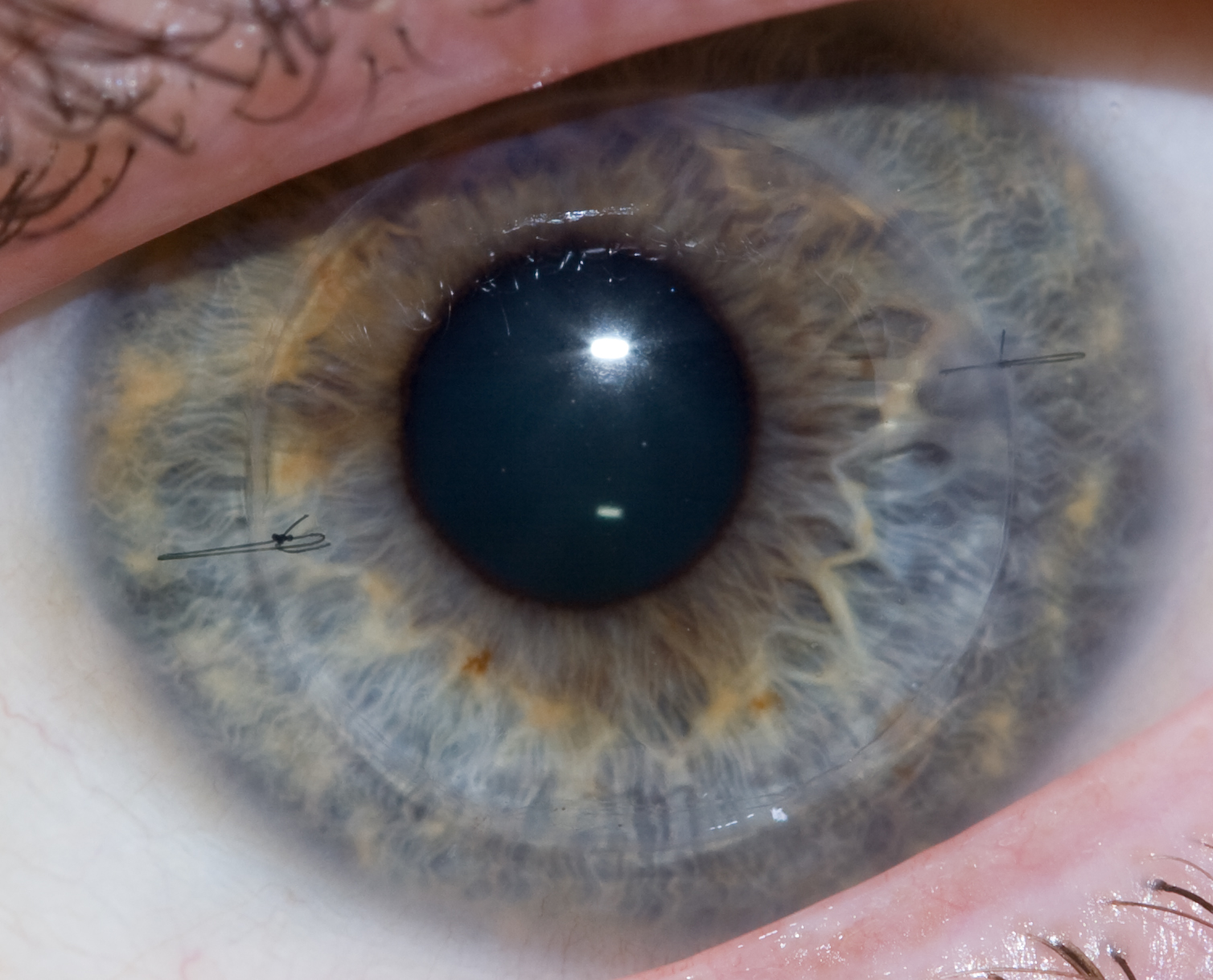
A human eye that received a cornea transplant one year postoperatively.
Disorders of or damage to the cornea — the clear covering over the lens of the eye — can be threatening to vision, and for the last century, corneal transplantation has been a cornerstone of treatment for these conditions. However, corneal transplants are complicated by two key facts: first, as with virtually all transplant procedures, donor organs are in short supply; and second, rejection is common, and recipients of transplants face repeated procedures or a lifetime of steroid eyedrops to prevent rejection.
One way of obviating these issues is the use of synthetic materials, which can now be manufactured with three-dimensional printing. In a new study from scientists at the Institute of Genetic Medicine at Newcastle University in the UK, to be published this summer in Experimental Eye Research, synthetic corneal tissue was 3D printed using a bioink loaded with encapsulated keratocytes (corneal cells), in combination with computer modeling based on actual corneas. The study is only proof to show that printing a biological replicate of the cornea is possible, but it lays the groundwork for future studies in animals.
Engineering Brain Recovery
One of the reasons why stroke is such a damaging event is the inability of damaged brain tissue to regenerate. Angiogenesis, the growth of new blood vessels, can help to regenerate brain tissue but properly guiding the process of angiogenesis is rather difficult.
However, a new report in Nature Materials indicates success using an injectable biogel for this purpose. In the report, a team led by Tatiana Segura, PhD, Professor of Biomedical Engineering at Duke with colleagues at UCLA, details its engineering of an injectable gel using nanoparticles consisting of heparin (a blood-thinning agent to prevent unwanted blood clotting) and vascular endothelial growth factor (VEGF) to stimulate brain regeneration. After injecting the gel in a mouse model of stroke, the mice showed a significant improvement in recovery compared to animals not receiving the engineered nanomaterial.
Here at Penn, D. Kacy Cullen, PhD, Research Associate Professor of Neurosurgery in the Perelman School of Medicine, has been investigating the use of implantable tissue-engineered brain pathways to treat and perhaps reverse the effects of neurodegnerative diseases like Parkinson’s disease. Penn Today has the story, with video of Dr. Cullen and photos and quotes from several of our own Bioengineering students.
Streamlining Environmental Bioengineering
Outside of the health sciences, bioengineering has applications in diverse fields, including energy development and environmental protection. Biofuels are one application for bioengineering that received a major boost recently. In an article published in NPJ Systems Biology and Applications, engineers from the US Department of Energy’s Lawrence Berkeley National Laboratory describe how they used machine learning to better predict the ability of engineered microbes to produce biofuel. With this information, they can then better adjust fuel-producing microbial pathways to maximize production. The machine learning model is a significant improvement over earlier, traditionally algorithmic approaches requiring complex differential equations. The time saved could, over generations of adjustments, result in a significant increase in output.
More on Pilots
Last week, we discussed how the cognitive load borne by airline pilots differs between simulated and real flight. Other scientists, it turns out, are looking at ways that pilots — in particular, fighter pilots — can overcome fatigue. With more than $1 million in grants from the US Department of Defense, Merhavan Singh, PhD, Dean of the Graduate School of Biomedical Sciences at the University of North Texas Health Science Center, and Kai Shen, PhD, Associate Professor in the Department of Chemistry and Forensic Science at Savannah State University in Georgia, are investigating compounds targeting the sigma 1 receptor, which the scientists believe could combat fatigue and also have neuroprotective effects if activated. This is particularly important among fighter pilots serving in conflict, who are often sleep deprived but must remain alert during missions.
People and Places
Having achieved success in its mission, the University of Alabama at Birmingham’s PREP Scholars Program, which supports underrepresented minority students in pursuing graduate study in bioengineering and biomedical engineering, has received an additional $1.8 million in support from the National Institutes of Health. The money will enable the funding of 40 students over the next five years.
Jeffrey Collins Wolchok, PhD, and Kartik Balachandran, PhD, both associate professors in the Department of Biomedical Engineering at the University of Arkansas, have received a $375,000 grant from the National Science Foundation to study the long-term effects of multiple concussions on the brain. With the increased emphasis in the scientific community and media on traumatic brain injury and chronic traumatic encephalopathy, including among former athletes, the two scientists will develop brain on a chip technology to examine the issue.
Finally, this week, the Best College Reviews website published its Top 10 list of online Master’s programs in biomedical engineering. Purdue University’s program finished in first place, with appearances on the list by Colorado State, UC Riverside, Stevens Tech, and Worcester Tech.
By Summer Kollie, Health & Societies ’19; Amber Figueroa, Biology ’21; and Bosede Ajiboye, Psychology ‘19
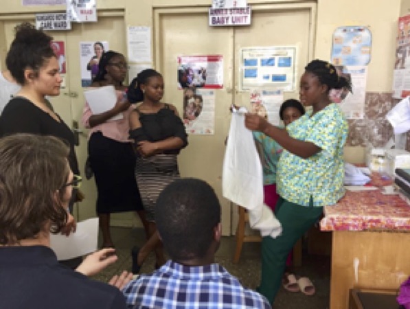
(left to right) Penn students Amber Figueroa, Bosede Ajiboye, and Summer Kollie watch a nurse at KATH demonstrate how she teases Kangaroo Mother Care to new mothers.
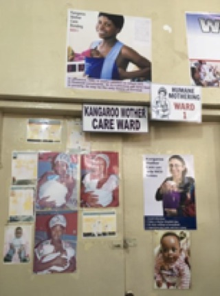
A photo of the Kangaroo Mother Care ward as well as public service posters explaining their benefits.
Today, we made a visit to the premature infants and Kangaroo Mother Care (KMC) wing of Komfo Anokye Teaching Hospital (KATH). Our visit was very informative. The overall goal for premature babies was for mothers to engage in KMC. When talking to the doctor in the premature clinic, she mentioned that, on average, a baby would stay in the clinic, where there are incubators and CPAP machines, for about one week before being referred to the KMC ward. In some cases, babies who have more severe cases might stay for a month or longer. The goal of the premature clinic was to stabilize the baby. An infant was stable when they no longer needed supplied oxygen and IV fluids, they had no more difficulties in breathing, and they were able to take food, as in breastmilk, through their mouths. After the baby was stable, then they were moved to the maternal ward, where the mother could administer KMC.
Kangaroo Mother Care (KMC) is an efficient way to take care of premature infants without using an incubator. With skin-to-skin contact, the baby is placed on the mother’s chest between her breast. Then, two to three blankets are wrapped securely around the baby to keep them warm.
At the KMC ward, a nurse demonstrated on herself how a mother would tie her infant onto her chest. Th nursey emphasize for mothers to be able to perform the process of tying the blanket on their own. In this ward, KMC is administered 24/7. The only time a mother gets a break is if she needs to use the bathroom or to buy food. Fathers and other relatives only assist in this process for short periods when the mother needs a break or when the mother has more than one baby, as in twins or triplets, and cannot physically carry more than one on her chest.
By Shihan Dong, Biotechnology MS ’19; Xuanjie (Lucas) Gong, Biotechnology MS ’19; and Princess Aghayere, Health & Societies ‘19
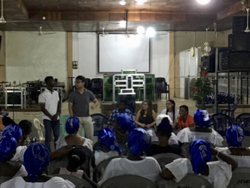
From left to right: KNUST medical student Kwabena Asoka Sarpong, Penn students Ethan Zhao, Sheldon Amoo-Mitchual, Rebecca Zappala, Yasmina Al Ghadban, and KNUST medical student Muti Agyekum present to Christ Apostolic church on the causes and prevention of cancer.
As usual, today we left for to visit the psychiatric ward of the Komfo Anokye Teaching Hospital (KATH), the biggest hospital in Kumasi and the second biggest in Ghana. However, when we arrived at the KATH it turned out that the clinic wasn’t ready for our visit, because there was a mix-up with the letter sent to inform them of our visit. So, we went back to School of Public Health to access to internet to let everyone research and work on their projects.
In the evening we returned to Christ Apostolic Church, a church for women who are poorly educated and mostly petty traders. Last Tuesday, we spoke to inform them about nutrition. This time, presentation was delivered by Ethan Zhao, Sheldon Amoo-Mitchual, Rebecca Zappala and Yasmina Al Ghadban, with KNUST medical students Kwabena Asoka Sarpong and Muti Agyekum as translators.
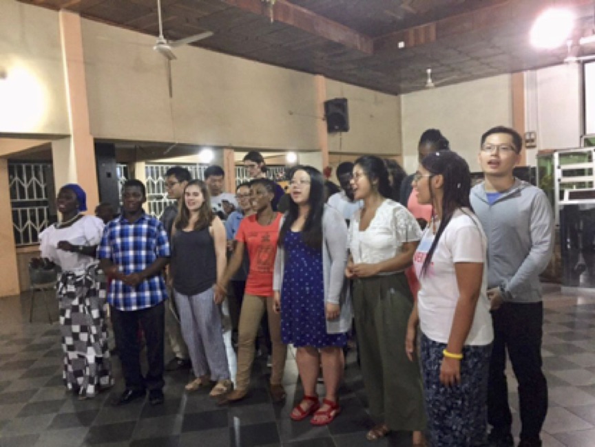
The APOC team sings a praise song in Twi for the congregation at Christ Apostolic church
Since last time there was a woman asked us to talk about cervical cancer, we decided talk about that as well as about cancer in general this time. At first, Ethan introduced the cancer was the result of cellular instructions being modified, and that there are ways to either protect those instructions or increase the risk of modification. Next, Sheldon talked about the importance of nutrition in cancer prevention, as introduced in last week, as well as the importance of good habits like exercising regularly and not smoking. Then, Rebecca introduced prevent cervical cancer and how to help prevent it through things like HPV vaccines and diagnosing it at early stages through Pap smear test. Finally, Yasmina covered the breast cancer, another cancer prevalent among women. She introduced that everyone is at risk of it and some certain factors would increase the risk like family breast cancer history. She taught them the self-exam, and recommended that women over 40 get a mammogram annually. During the question section, the audience was really active to ask questions, so much so that the other group that was supposed to present on pregnancy, Amber Figueroa, Bosede Ajiboye, and Summer Kollie had to be rescheduled to present next week. The churchgoers were curious about things like how and how often they should wash genitals area, as well as how to take care of their pre-pubescent daughters. They also wanted to clarify some rumors they were told about the causes of cancer, such as if things like phone vibration, or putting a phone to the left ear could cause cancer.
After the presentation, we sang a simple church song in Twi, Asem papa bia mate ne s3 oye oye, which we had prepared before coming. The churchgoers were delighted and they joined in, creating beautiful harmonies as our voices combined.
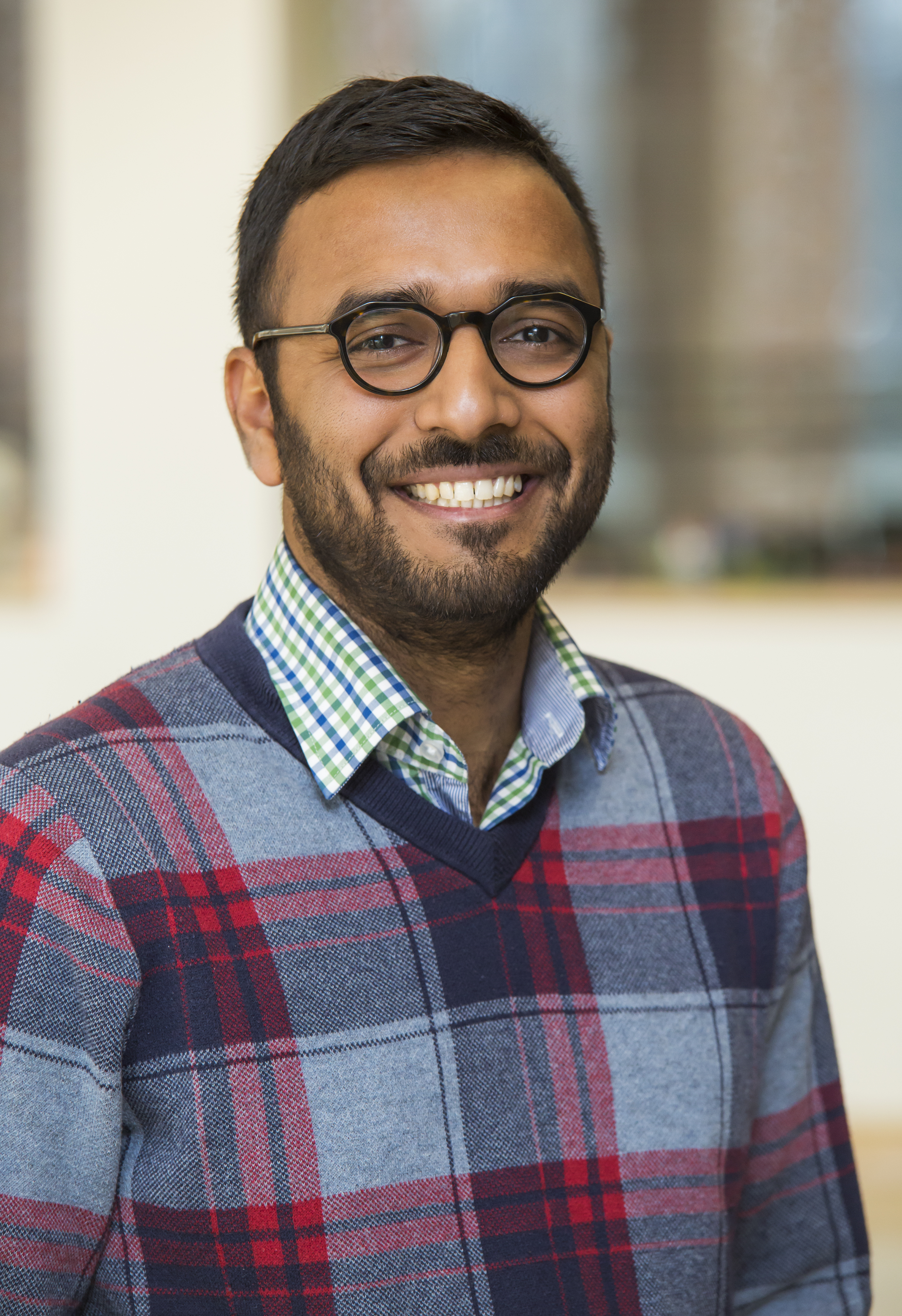
The University of Pennsylvania Department of Bioengineering is proud to announce that Yogesh Goyal, a postdoctoral fellow in the laboratory of Professor Arjun Raj, PhD, has received two pretigious awards. First, has received the Jane Coffin Childs (JCC) Memorial Fund Fellowship, which is a premier fellowship for biomedical studies. The JCC fellowship provides three years of funding at approximately $50,000 per year to top scholars having received the PhD in the previous 18 months. In addition, along with recently minted Bioengineering PhD Jina Ko, Yogesh has been named one of the 14 inaugural Schmidt Science Fellows, each of whom receives $100,000 to cover living expenses while working as a postdoctoral fellow under the auspices of the Rhodes Trust.
“I am thrilled to have the opportunity to work with such a talented scientist in the coming years on quantitative problems related to development and cancer,” Dr. Raj said. “These fellowships are a well-deserved recognition of Yogesh’s scientific vision and dedication.”
Yogesh, a native of Jammu and Kashmir, India, received his undergraduate in chemical engineering at the Indian Institute of Technology. He then came to Princeton University and studied for the PhD in the Department of Chemical and Biological Engineering and the Lewis-Sigler Institute for Integrative Genomics, under the advisement of Stanislav Shvartsman, PhD, and Trudi Schüpbach, PhD. He came to Penn Bioengineering after finishing his doctorate.
“I am very excited to be selected for two prestigious fellowships,” Yoghes says. “I am looking forward to working with Arjun on learning experimental and computational single-cell techniques to understand developmental and invasive systems.”


 Danielle Bassett, PhD