Penn Engineering’s Latest ‘Organ-On-a-Chip’ is a New Way to Study Cancer-related Muscle Wasting

by Melissa Pappas

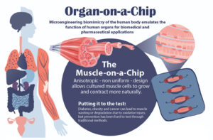
Bioengineering’s Dan Huh and colleagues have developed a number of organ-on-a-chip devices to simulate how human cells grow and perform in their natural environments. Their latest is a muscle-on-a-chip, which carefully captures the directionality of muscle cells as they anchor themselves within the body. See the full infographic at the bottom of this story. (Illustration by Melissa Pappas).

Studying drug effects on human muscles just got easier thanks to a new “muscle-on-a-chip,” developed by a team of researchers from Penn’s School of Engineering and Applied Science and Inha University in Incheon, Korea.

Muscle tissue is essential to almost all of the body’s organs, however, diseases such as cancer and diabetes can cause muscle tissue degradation or “wasting,” severely decreasing organ function and quality of life. Traditional drug testing for treatment and prevention of muscle wasting is limited through animal studies, which do not capture the complexity of the human physiology, and human clinical trials, which are too time consuming to help current patients.

An “organ-on-a-chip” approach can solve these problems. By growing real human cells within microfabricated devices, an organ-on-a-chip provides a way for scientists to study replicas of human organs outside of the body.

Using their new muscle-on-a-chip, the researchers can safely run muscle injury experiments on human tissue, test targeted cancer drugs and supplements, and determine the best preventative treatment for muscle wasting.

organ-on-a-chip
Dan Huh, Ph.D.

This research was published in Science Advances and was led by Dan Huh, Associate Professor in the Department of Bioengineering, and Mark Mondrinos, then a postdoctoral researcher in Huh’s lab and currently an Assistant Professor of Biomedical Engineering at Tulane University. Their co-authors included Cassidy Blundell and Jeongyun Seo, former Ph.D. students in the Huh lab, Alex Yi and Matthew Osborn, then research technicians in the Huh lab, and Vivek Shenoy, Eduardo D. Glandt President’s Distinguished Professor in the Department of Materials Science and Engineering. Lab members Farid Alisafaei and Hossein Ahmadzadeh also contributed to the research. The team collaborated with Insu Lee and professors Sun Min Kim and Tae-Joon Jeon of Inha University.

In order to conduct meaningful drug testing with their devices, the research team needed to ensure that cultured structures within the muscle-on-a-chip were as close to the real human tissue as possible. Critically, they needed to capture muscle’s “anisotropic,” or directionally aligned, shape.

“In the human body, muscle cells adhere to specific anchor points due to their location next to ligament tissue, bones or other muscle tissue,” Huh says. “What’s interesting is that this physical constraint at the boundary of the tissue is what sculpts the shape of muscle. During embryonic development, muscle cells pull at these anchors and stretch in the spaces in between, similar to a tent being held up by its poles and anchored down by the stakes. As a result, the muscle tissue extends linearly and aligns between the anchoring points, acquiring its characteristic shape.”

The team mimicked this design using a microfabricated chip that enabled similar anchoring of human muscle cells, sculpting three-dimensional tissue constructs that resembled real human skeletal muscle.

The the full story in Penn Engineering Today.

“’Electronic Nose’ Accurately Sniffs Out Hard-to-Detect Cancers”

A.T. Charlie Johnson, Ph.D.

A.T. Charlie Johnson, Rebecca W. Bushnell Professor of Physics and Astronomy at the Penn School of Arts & Sciences, and member of the Penn Bioengineering Graduate Group has been working with a team of researchers on a new “electronic nose” that could help track the spread of COVID-19 based on the disease’s unique odor profile. Now, similar research shows that vapors emanating from blood samples can be tested to distinguish between benign and cancerous pancreatic and ovarian cells. Johnson presented the results at the annual American Society of Clinical Oncology meeting on June 4 (Abstract # 5544):

“It’s an early study but the results are very promising,” Johnson said. “The data shows we can identify these tumors at both advanced and the earliest stages, which is exciting. If developed appropriately for the clinical setting, this could potentially be a test that’s done on a standard blood draw that may be part of your annual physical.”

Read the full story in Penn Medicine News.

2021 CAREER Award recipient: Alex Hughes, Assistant Professor in Bioengineering

by Melissa Pappas

Alex Hughes (illustration by Melissa Pappas)

The National Science Foundation’s CAREER Award is given to early-career researchers in order to kickstart their careers in innovative and pivotal research while giving back to the community in the form of outreach and education. Alex Hughes, Assistant Professor in Bioengineering and in Cell and Developmental Biology, is among the Penn Engineering faculty members who have received the CAREER Award this year.

Hughes plans to use the funds to develop a human kidney model to better understand how the development of cells and tissues influences congenital diseases of the kidney and urinary tract.

The model, known as an “organoid,” is a lab-grown piece of human kidney tissue on the scale of millimeters to centimeters, grown from cultured human cells.

“We want to create a human organoid structure that has nephrons, the filters of the kidney, that are properly ‘plumbed’ or connected to the ureteric epithelium, the tubules that direct urine towards the bladder,” says Hughes. “To achieve that, we have to first understand how to guide the formation of the ureteric tubule networks, and then stimulate early nephrons to fuse with those networks. In the end, the structures will look like ‘kidney subunits’ that could potentially be injected and fused to existing kidneys.”

The field of bioengineering has touched on questions similar to those posed by Hughes, focusing on drug testing and disease treatment. Some of these questions can be answered with the “organ-on-a-chip” approach, while others need an even more realistic model of the organ. The fundamentals of kidney development and questions such as “how does the development of nephrons affect congenital kidney and urinary tract anomalies?” require an organoid in an environment as similar to the human body as possible.

“We decided to start with the kidney for a few reasons,” says Hughes. “First, because its development is a beautiful process; the tubule growth is similar to that of a tree, splitting into branches. It’s a complex yet understudied organ that hosts incredibly common developmental defects.

“Second,” he says, “the question of how things form and develop in the kidney has major medical implications, and we cannot answer that with the ‘organ-on-a-chip’ approach. We need to create a model that mimics the chemical and mechanical properties of the kidney to watch these tissues develop.”

The fundamental development of the kidney can also answer other questions related to efficiency and the evolution of this biological filtration system.

“We have the tendency to believe that systems in the human body are the most evolved and thus the most efficient, but that is not necessarily true,” says Hughes. “If we can better understand the development of a system, such as the kidney, then we may be able to make the system better.”

Hughes’ kidney research will lay the foundation for broader goals within regenerative medicine and organ transplantation.

Read the full story in Penn Engineering Today.

How HIV Infection Shrinks the Brain’s White Matter

by Katherine Unger Baillie

Researchers from Penn and CHOP detail the mechanism by which HIV infection blocks the maturation process of brain cells that produce myelin, a fatty substance that insulates neurons.

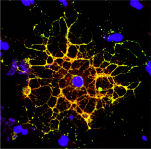
A confocal microscope image shows an oligodendrocyte in cell culture, labeled to show the cell nucleus in blue and myelin proteins in red, green, and yellow. Researchers from Penn and CHOP have shown that HIV infection prevents oligodendrocytes from maturing, leading to a reduction in white matter in the brain. (Image: Raj Putatunda)

It’s long been known that people living with HIV experience a loss of white matter in their brains. As opposed to gray matter, which is composed of the cell bodies of neurons, white matter is made up of a fatty substance called myelin that coats neurons, offering protection and helping them transmit signals quickly and efficiently. A reduction in white matter is associated with motor and cognitive impairment.

Earlier work by a team from the University of Pennsylvania and Children’s Hospital of Philadelphia (CHOP) found that antiretroviral therapy (ART)—the lifesaving suite of drugs that many people with HIV use daily—can reduce white matter, but it wasn’t clear how the virus itself contributed to this loss.

In a new study using both human and rodent cells, the team has hammered out a detailed mechanism, revealing how HIV prevents the myelin-making brain cells called oligodendrocytes from maturing, thus putting a wrench in white matter production. When the researchers applied a compound blocking this process, the cells were once again able to mature.

The work is published in the journal Glia.

“Even when people with HIV have their disease well-controlled by antiretrovirals, they still have the virus present in their bodies, so this study came out of our interest in understanding how HIV infection itself affects white matter,” says Kelly Jordan-Sciutto, a professor in Penn’s School of Dental Medicine and co-senior author on the study. “By understanding those mechanisms, we can take the next step to protect people with HIV infection from these impacts.”

“When people think about the brain, they think of neurons, but they often don’t think about white matter, as important as it is,” says Judith Grinspan, a research scientist at CHOP and the study’s other co-senior author. “But it’s clear that myelination is playing key roles in various stages of life: in infancy, in adolescence, and likely during learning in adulthood too. The more we find out about this biology, the more we can do to prevent white matter loss and the harms that can cause.”

Jordan-Sciutto and Grinspan have been collaborating for several years to elucidate how ART and HIV affect the brain, and specifically oligodendrocytes, a focus of Grinspan’s research. Their previous work on antiretrovirals had shown that commonly used drugs disrupted the function of oligodendrocytes, reducing myelin formation.

In the current study, they aimed to isolate the effect of HIV on this process. Led by Lindsay Roth, who recently earned her doctoral degree within the Biomedical Graduate Studies group at Penn and completed a postdoctoral fellowship working with Jordan-Sciutto and Grinspan, the investigation began by looking at human macrophages, one of the major cell types that HIV infects.

Read the full story in Penn Today.

Kelly Jordan-Sciutto is vice chair and professor in the University of Pennsylvania School of Dental Medicine’s Department of Basic & Translational Sciences and is director of Biomedical Graduate Studies. She is a member of the Penn Bioengineering Graduate Group.

Katherine Reuther Appointed Practice Associate Professor in Bioengineering

Katie Reuther, PhD, MBA

Katherine (Katie) Reuther, Ph.D., M.B.A. will return to Penn Engineering in July 2021 as the new Executive Director of Penn Health-Tech (PHT) and as Practice Associate Professor in Bioengineering. Reuther is an alumna of Penn Bioengineering, having obtained her Ph.D. at Penn in the laboratory of Louis Soslowsky, Fairhill Professor in Bioengineering and Orthopaedic Surgery.

“Dr. Reuther is a role model for biomedical innovation, linking formal training in engineering and entrepreneurship with deep practical experience in leading technologies through the commercialization pipeline. Dr. Reuther graduated with her Bachelor of Science in Biomedical Engineering, Magna cum Laude, from the College of New Jersey; she obtained her Ph.D. in Bioengineering at Penn in the laboratory of Dr. Louis Soslowsky and completed her MBA at Columbia, where she currently is a Senior Lecturer in Design, Innovation, and Entrepreneurship in the Department of Biomedical Engineering. During her tenure at Columbia, Dr. Reuther helped create and led Columbia’s Biomedical Engineering Technology Accelerator (BiomedX), overseeing more than 60 technologies leading to $80M in follow-on funding and 18 licenses to start-ups or start-ups industry.  Introducing both new courses and a new curriculum in biomedical innovation, Dr. Reuther was recently awarded Columbia’s highest teaching honor, the ‘2021 Presidential Award for Outstanding Teaching,’ this Spring as a recognition of her excellence in teaching and dedication to students.

Katie has extensive experience in developing and translating early-stage medical technologies and discoveries and providing formal educational training for aspiring medical entrepreneurs.  Dr. Reuther served as Director of Masters’ Studies for the Department of Biomedical Engineering and spearheaded the development of a graduate-level medical innovation program, including an interdisciplinary course available to scientists, engineers, and clinicians. Dr. Reuther provided advising and educational support to more than 100 student/faculty teams and start-ups, as they worked to develop and commercialize medical technologies. She will bring these extensive skills to PHT and Penn Bioengineering in two new, hands-on graduate courses in medical innovation centered around Penn Health-Tech ventures.”

Read the full announcement in OVPR news.

César de la Fuente Featured in “40 Under 40” List

César de la Fuente, Ph.D.

César de la Fuente, PhD, Presidential Assistant Professor in Bioengineering, Chemical and Biomolecular Engineering, Psychiatry, and Microbiology, was featured in the Philadelphia Business Journal’s Class of 2021 “40 Under 40” list. Currently focused on antibiotic discovery, creating tools for microbiome engineering, and low-cost diagnostics, de le Fuente pioneered the world’s first computer-designed antibiotic with efficacy in animal models.

De la Fuente was previously included in the AIChE’s “35 Under 35” list in 2020 and most recently published his work demonstrating a rapid COVID-19 diagnostic test which delivers highly accurate results within four minutes.

Read “40 Under 40: Philadelphia Business Journal’s complete Class of 2021” here.

Read other BE blog posts featuring Dr. de la Fuente here.

Penn Health-Tech Q&A with César de la Fuente

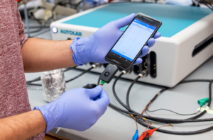
Created in the lab of César de la Fuente, this miniaturized, portable version of rapid COVID-19 test, which is compatible with smart devices, can detect SARS-CoV-2 within four minutes with nearly 100% accuracy. (Image: Courtesy of César de la Fuente)

César de la Fuente, Presidential Assistant Professor in Bioengineering, Chemical and Biomolecular Engineering, Microbiology, and Psychiatry, was the inaugural recipient of the Nemirovsky Engineering and Medicine Opportunity (NEMO) Prize from Penn Health-Tech in 2020 for his low-cost, rapid COVID test. Now with promising results recently published in the journal Matter (showing 90 percent accuracy in as little as four minutes), Penn Health-Tech caught up with de la Fuente to discuss his experience over the past year:

“How did [your project] evolve in the past year?

‘We started with one prototype and now have three entirely different prototypes for the test. Two use electrochemistry, and we are now working on a new technology that uses calorimetry. With calorimetry, when the cotton swabs are exposed to the virus, they change color. This means users are able to see if they’re affected by a virus through a simple color change, making it more of a visual detection method.'”

Read the full Q&A in the Penn Health-Tech blog.

Rapid COVID-19 Diagnostic Test Delivers Results Within 4 Minutes With 90 Percent Accuracy

RAPID, a low-cost COVID-19 diagnostic test, can detect SARS-CoV-2 within four minutes with 90 percent accuracy

Even as COVID-19 vaccinations are being rolled out, testing for active infections remains a critical tool in fighting the pandemic. Existing rapid tests that can directly detect the virus rely on reverse transcription polymerase chain reaction (RT-PCR), a common genetic assay that nevertheless requires trained technicians and lab space to conduct.

Alternative testing methods that can be scaled up and deployed in places where those are in short supply are therefore in high demand.

Penn researchers have now demonstrated such a method, which senses the virus by measuring the change in an electrical signal when a piece of the SARS-CoV-2 virus binds to a biosensor in their device, which they call RAPID 1.0.

The work, published in the journal Matter, was led by César de la Fuente, a Presidential Assistant Professor who has appointments in Engineering’s departments of Chemical and Biomolecular Engineering, and Bioengineering, as well as in Psychiatry and Microbiology in the Perelman School of Medicine.

“Prior to the pandemic, our lab was working on diagnostics for bacterial infections. But then, COVID-19 hit. We felt a responsibility to use our expertise to help—and the diagnostic space was ripe for improvements,” de la Fuente said. “We feel strongly about the health inequities witnessed during the pandemic, with testing access and the vaccine rollout, for example. We believe inexpensive diagnostic tests like RAPID could help bridge some of those gaps.”

The RAPID technology uses electrochemical impedance spectroscopy (EIS), which transforms the binding event between the SARS-CoV-2 viral spike protein and its receptor in the human body, the protein ACE2 (which provides the entry point for the coronavirus to hook into and infect human cells), into an electrical signal that clinicians and technicians can detect. That signal allows the test to discriminate between infected and healthy human samples. The signal can be read through a desktop instrument or a smartphone.

Read more about RAPID at Penn Medicine News.

Originally posted on Penn Engineering Today.

Bioengineering Graduate Students Take the Annual BETA Day Online

By GABE Outreach Chairs and Ph.D. students David Gonzalez-Martinez and David Mai

BETA Day Biomaterials workshop

Every spring, the Graduate Association of Bioengineers (GABE) at Penn partners up with iPraxis, an educational non-profit organization based in Philadelphia, to organize BETA Day, an event that brings together Bioengineering graduate students and local Philadelphia grade school students to introduce them to the field of bioengineering, the life of graduate students, and hands-on scientific demonstrations. Due to COVID-19 restrictions, we adapted the traditional in-person BETA Day into a virtual event on Zoom. This year, we assembled kits containing the necessary materials for our chosen demonstrations and worked with iPraxis to coordinate their delivery to partner schools and their students. This enabled students to perform their demonstrations in a hands-on manner from their own homes; over 40 students were able to participate in extracting their own DNA and making biomaterials with safe household materials.

Michelle Johnson presents on her work in robotics

The day began with a fantastic lecture by Michelle Johnson, Associate Professor in Bioengineering and Physical Medicine and Rehabilitation, who introduced students to the field of rehabilitation robotics and shared her experience as a scientist. Students then learned about DNA and biomaterials through lectures mediated by the graduate students Dayo Adetu and Puneeth Guruprasad. After each lecture, students broke into breakout rooms with graduate student facilitators where they were able to get some hands-on scientific experience as they extracted DNA from their cheek cells and fabricated alginate hydrogels. Michael Sobrepera, a graduate student in Dr. Johnson’s lab, concluded the event by giving a lecture on the process of robotics development and discussed where the field is heading and some important considerations for the field.

Dayo Adetu, Bioengineering Master’s student and GABE President, teaches the students about Genetic Engineering

While yet another online event may seem unexciting, throughout the lectures students remained exceptionally engaged and raised fantastic questions ranging from the accessibility of low income communities to novel robotic therapeutic technologies to the bioethical questions robotic engineers will face as technologies advance. The impact of BETA day was evident as the high school students began to discuss the possible majors they would like to pursue for their bachelor’s degrees. Events like BETA Day give a glimpse into possible STEM fields and careers students can pursue.

“Educating the Next Generation of Civically Engaged Technologists”

Brit Shields, Ph.D.

Brit Shields, Senior Lecturer in Bioengineering, has brought her expertise in the history and sociology of science to her leading role in developing and improving the ethics curriculum for all students in the School of Engineering and Applied Science. Most recently, this includes adapting the core ethics engineering ethics course “Technological Innovation and Civil Discourse in a Dynamic World” (EAS 204) for the Stavros Niarchos Foundation (SNF) Paideia Program. SNF Paideia courses, open to all Penn undergraduates, “integrate students’ personal, professional, and civic development […] focus[ing] on dialogue, wellness, service, and citizenship from different disciplinary and interdisciplinary perspectives.” A recent SNF Paideia blog post goes into detail about the changes made by Shields and co-instructor Christopher Yoo, John H. Chestnut Professor of Law, Communication, and Computer and Information Science, to suit the SNF Paideia Program, including its “explicit focus on civil discourse and technology.” According to Shields:

“I really wanted to break down the false dichotomy between technological expertise or humanities training for the students and open up the opportunity for Engineering students to consider themselves to have an important role, not just creating technological systems but also being important participants in civil discourse.”

Michelle Johnson, Ph.D.

The course also includes guest lectures by Penn faculty, including Michelle Johnson, Associate Professor in Bioengineering and Physical Medicine and Rehabilitation, and students learn to analyze how guest lecturers communicate their research to the public, for example, in the case of Johnson, in the form of a TED Talk and scholarly articles: “Through her TedTalk, journal articles and visit to the class, Michelle Johnson demonstrates how researchers are attuned to the specific preferences of the rehabilitative robots they are creating for patients…engaged scholarship at its finest.”

Read “Educating the Next Generation of Civically Engaged Technologists” in SNF Paideia Perspectives.