Last month, the National Toxicology Program (NTP), a division of the U.S. Department of Health and Human Services, announced the findings of a draft study in which it was shown that high exposure to radiofrequency radiation, similar to that caused by persistent use of cell phones, resulted in the formation of tumors in nerves surrounding the hearts of male rats — but not female rats or mice. These are the final results of the study, the preliminary results of which were released in 2016. The study must still undergo peer review later this month.
Among the experts studying this question for the last decade is Kenneth R. Foster, Ph.D., Professor Emeritus of Bioengineering at the University of Pennsylvania. He’s not so sure that there’s really any link between the radiation emitted by cell phones and cancers. “People have been using cell phones for decades and so far there has been no noticeable increase in brain cancer,” Dr. Foster said. “This means that the risk, if it occurs at all, is too low to detect with any reliability.”
Kenneth R. Foster, Ph.D.
Asked whether the findings of the NTP study could be generalized to people, Dr. Foster responded, “If you look at enough tissues and compare enough endpoints, you will pick up things that may just be one-off findings. Health agencies will have to assess the findings carefully in the light of the considerable previous literature of related studies. The results of the study are unlikely to change their previous assessments, which is that there is no clear evidence of health hazards from using cell phones.” The exposed rats in the NTP study consistently lived longer than the exposed rats, he added.
Dr. Foster noted that the exposure levels for the rats that developed tumors was far higher than safety limits for humans in terms of whole body exposure and not at all similar to the exposures that people receive from using cell phones. “Also,” he said, “people don’t use cell phones for nine hours a day for two years at a time, which were the exposures in the NTP study. After all of this research on the issue, my own view is that cell phones don’t cause brain cancer. But they do contribute to traffic accidents!”
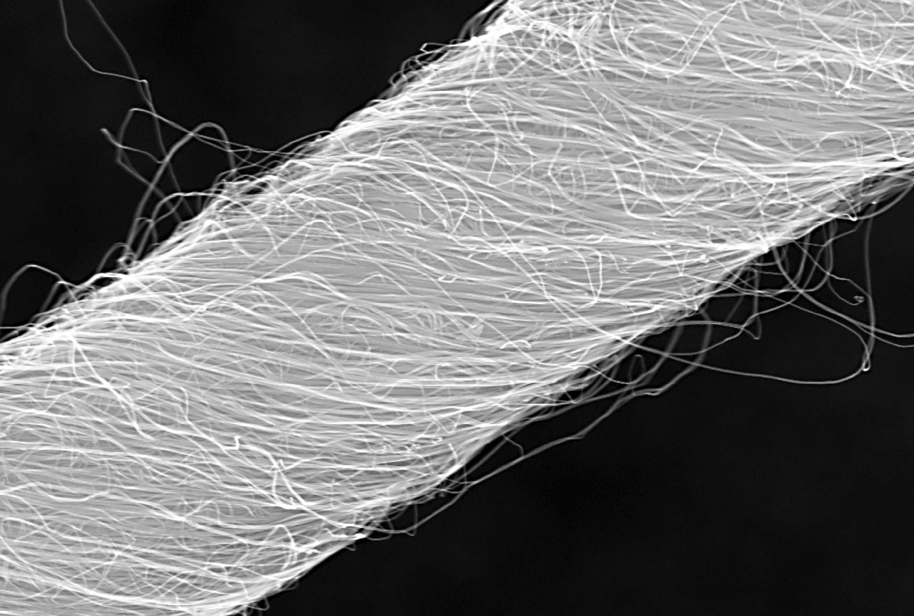
Tapping into the autonomous nervous system – the control center for things like heartbeat and breathing – is a relatively new part of neurostimulation technologies to both record and direct organ function. Implants designed for stimulating peripheral nerves often fail because the protective tissue surrounding nerve bundles (the perineurium) is difficult to penetrate, and the body’s immune response often builds a scar around the implanted device.
Now, a team of scientists from Case Western Reserve University (CWRUL) has used carbon nanotubes to overcome these obstacles, reporting their findings in Scientific Reports. The authors, led by Dominique M. Durand, Ph.D., Director of the Neural Engineering Center and El Lindseth Professor of Biomedical Engineering, Neurosciences, Physiology and Biophysics at CWRU, fabricated yarn made of carbon nanotubes that was 10 to 20 µm in diameter. The yarn was then used to create electrodes, which were implanted into rats to monitor activity of the glossopharyngeal and vagus nerves.
The authors found that they could use the implants to monitor nerve activity under conditions of hypoxia and stomach distention. They report that the success of their experiments likely derives from the similarity of the nanotube yarn to the actual neural tissue surrounding the implant. The implants are a long way from being tried in humans, but the large number of functions controlled by just these two nerves indicates that such implants could find use in an enormous number of diseases.
Better Screening of Nanoparticle Delivery
As we discussed last week, the development of gene-based therapies is hindered by the sheer size of the human genome. The immense volume of information involved can quickly become difficult to manage, so one way in which scientists “keep track” of genetic information during the process of introducing new genetic material into an organism is DNA barcoding. This process attaches a small piece of DNA to the gene being studied; if and when the gene causes cells to replicate, these cells will bear the barcode, thus allowing the observer to be certain of the gene identities the whole time.
Seeking to determine whether DNA barcoding of lipid nanoparticles for injection into living models would outperform in vitro testing, a team of investigators at Georgia Tech and Emory University conducted a comparison of the two techniques, reporting their findings in Nano Letters. The authors, led by James Dahlman, Ph.D., Assistant Professor of Biomedical Engineering at GT/Emory, found that in vitro testing did not predict in vivo delivery. Further, they were able to track several dozen barcodes delivered by nanoparticles to eight different cell lines.
The authors believe that their technique, which they call JOint Rapid DNA Analysis of Nanoparticles (JORDAN), is superior to in vitro screening of nanoparticles to predict successful transplantation. They are offering JORDAN online as open source software so other scientists can use the technology to more accurately screen nanomaterials.
Synthetic Biologists Create Gene Circuits
Among the many types of molecules that regulate genetic expression in the body are microRNAs, non-coding strands of RNA that are responsible for gene silencing and other forms of gene expression regulation. The ability to harness and control the functions of microRNAs could have important implications for disease prevention and treatment.
In a recent article in Systems Biology and Applications, researchers at the University of Texas, Dallas, report on their engineering of a microRNA-based genetic circuit and its deployment in living cells. They created the circuit using strands of RNA from a variety of organisms, including viruses and jellyfish. The authors, led by Leonidas Bleris, Ph.D., Associate Professor of Bioengineering at UT Dallas, used the circuits to better understand how microRNAs change gene expression under different conditions.
More importantly, the authors found that their circuit had the ability to outproduce types of gene expression, which decreased as the number of gene replications increased. The authors believe that their discoveries could have applications in a number of genetic disorders.
Discouraging Smoking at the Level of the Brain
Cigarette smoking is the single greatest contributor to negative health outcomes in the population. Nicotine addiction often appears during the teenage years, and aggressive advertising has been used for the last couple of decades to encourage people to quit smoking and younger people not to start. Despite the widespread use of advertising to change human behavior, remarkably little is known on how the brain responds to advertising messages.
Danielle S. Bassett, Ph.D., Eduardo D. Glandt Faculty Fellow and Associate Professor of Bioengineering at Penn, recently collaborated with faculty from Penn’s Annenberg School of Communication to determine the neuroscience underlying this outcome. The collaborators showed graphic warning labels to a cohort of smokers while they were subjected to functional magnetic resonance imaging, which images brain activity during specific tasks. They found that smokers whose brains showed greater coherence between regions in the valuation network were more likely to quit smoking. Determining why these brain regions acted as they did could yield even more effective smoking-cessation messaging.
Purdue Startup Working to Expand MRI
Engineers at Purdue, including Zhongming Liu, Ph.D., Assistant Professor of Biomedical Engineering and Electrical and Computer Engineering, have cofounded at startup company, called MR-Link, to develop and produce a coin-sized device that can be inserted into MRI machines, allowing them to perform multiple scans simultaneously.
The device could be useful in reducing the amount of electromagnetic force to which patients are exposed during an MRI scan. In addition, Dr. Liu and his colleagues believe the device will cost perhaps less than a tenth of what similar devices currently cost. Given the widespread use of MRI, the device could ultimately impact how a number of diseases and disorders are diagnosed and tracked.
Konrad Kording, professor in the Department of Bioengineering, and colleagues have a new technique for identifying fraudulent scientific papers by spotting reused images. Rather than scrap a failed study, for example, a researcher might attempt to pass off images from a different experiment to give the false impression that their own was a success.
Kording, a Penn Integrates Knowledge (PIK) Professor who also has an appointment in the Department of Neuroscience in Penn’s Perelman School of Medicine, and his collaborators developed an algorithm that can compare images across journal articles and detect such replicas, even if the image has been resized, rotated, or cropped.
They describe their technique in a paper recently published on the BioRxiv preprint server.
“Any fraudulent paper damages science,” Kording says. “In biology, many times fraud is detected when someone looks at a few papers and says ‘hey, these images look a little similar.’ We reckoned we could make an algorithm that does the same thing.”
“Science depends on building upon other people’s work,” adds Daniel Acuna, lead author on the paper, and a student in Kording’s lab at Northwestern University at the time the study was conducted. “If you cannot trust other people’s work, the scientific process collapses and, worse, the general public loses trust in us. Some websites were doing this, anonymously, but at a painstakingly slow rate.” Acuna is now an assistant professor in the School of Information Studies at Syracuse University.
While much of Kording’s work focuses on using data science to understand the brain, he is also curious about the process of research itself, or, as he puts it, “the science of science.” One of the Kording lab’s previous projects closely analyzed common methods of neuroscience research, and another turned a mirror on itself, describing how to structure a scientific paper.
Nicholas Stiansen, a senior Bioengineering major at Penn, is one of eight students and alumni receiving a Thouron Award. Nick will receive a full scholarship to cover tuition and fees, plus a stipend of £19,500 (approximately $27,000). He is still awaiting decisions from graduate programs, but his first choice is to study at Imperial College London (ICL) in the United Kingdom.
Named for Sir John Thouron, a British aristocrat and husband of Esther Driver du Pont, great-granddaughter of Alfred V. du Pont, founder of the chemical company, the Thouron Award is given to graduates of Penn and of universities in the U.K. Each year, a small number of Penn students receives awards, as well as a similar number of British students. Previous awardees include: the current nominee to head the SEC, Jay Clayton; Pulitzer-prize winning novelist Jennifer Egan; and John J. Leonard, Professor of Mechanical and Ocean Engineering and Samuel C. Collins Professor at MIT.
In addition to majoring in Bioengineering, Nick works as an undergraduate research assistant in the Spine Pain Research Laboratory of Beth Winkelstein, Vice Provost for Education and Professor, and a teaching assistant for BE 310, the second half of the junior year bioengineering lab series. Plus, he holds or has held positions with the Engineering Deans’ Advisory Board and the Biomedical Engineering Society, and he is involved in the Theta Tau Professional Engineering Fraternity and Tau Beta Pi Engineering Honor Society. If he enrolls at ICL, Nick intends to study in the university’s Master’s program in Medical Device Design and Entrepreneurship.
“I am honored to be named a Thouron Scholar,” Nick says, “and I am extremely excited to continue my graduate studies in the U.K. I am eager to immerse myself in a new, vibrant culture and learn about medical technology from an entirely new perspective. This experience will be integral towards achieving my long-term goal of developing the next wave of innovative and accessible medical devices.”
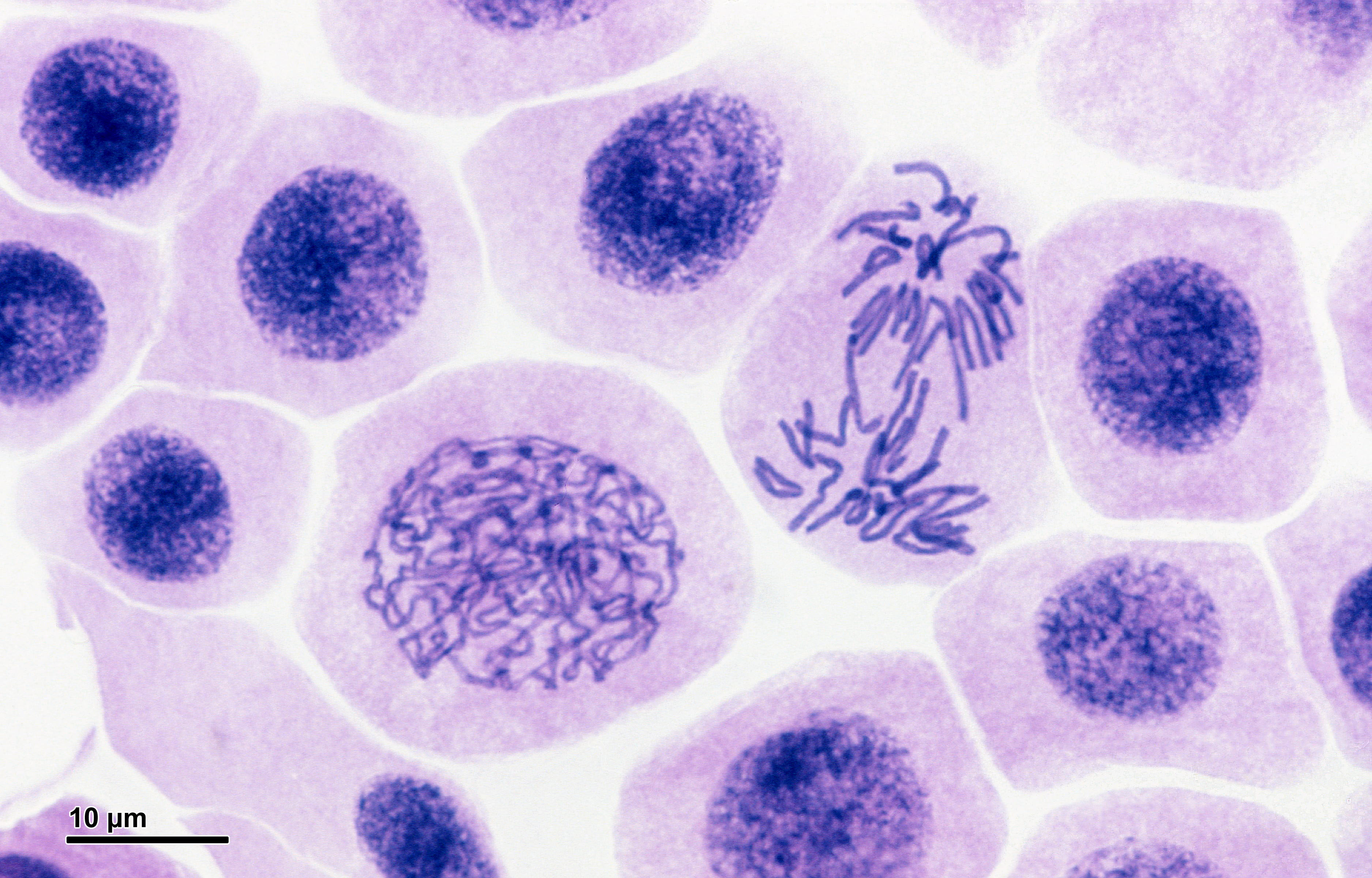
Online Tool for 3D Visualization of Gene Mutations
DNA inside cell nuclei undergoing the process of mitosis.
Fifteen years ago marked a major milestone in the Human Genome Project: scientists successfully sequenced all of the base pairs in our 23 sets of chromosomes. Following this accomplishment, researchers assembled generations of mathematical models to understand how gene mutations result in disease. A key barrier in developing these models is the size of genome itself: a single human genome requires approximately 2 GB of storage, and many studies examine thousands of genomes to detect changes in a small number of patients. Both processing these large datasets and efficiently storing them create challenges. Making these model predictions accurate and complete is another challenge.
Scientists collaborating among several universities on three continents developed an online computational tool to help overcome these barriers. The scientists, who include Bernhard Palsson, Ph.D., Galletti Professor of Bioengineering at University of California, San Diego, as one of the lead authors, report on the resource in a recent issue of Nature Biotechnology.
Called Recon3D, the new resource provides a metabolic network model using approximately 17% of known human genes. The model combines data on the genes, metabolites, proteins, and metabolic reactions for human metabolism. In addition, as the model’s name implies, Recon3D accounts for the physical structure of model components, imporving significantly on past models that relied on linear, two-dimensional models. Although the model still has 83% of genes left to incorporate, it could ultimately unravel some of the mysteries underlying virtually any disease with a genetic cause, from inborn errors of metabolism to cancer.
Bioengineering for Refugees
As the war in Syria enters its seventh year, at least five million refugees have left the country to seek asylum elsewhere. Roughly 20% of the refugees are now in Lebanon, where many reside in refugee camps. Although these refugees are now much safer than before, even in the best of circumstances, the conditions in refugee camps can compromise health and wellness.
Engineering can offer relief for some of these conditions. A three-week course offered in January at the American University of Beirut, co-designed and taught by Muhammad Zaman, Ph.D., Professor of Biomedical Engineering at Boston University, and entitled “Humanitarian Engineering: Designing Solutions for Health Challenges in Crises,” had students devising solutions to the issues facing these refugees.
Among the ideas generated by the students was “3D Safe Water” – a device designed to detect the contamination of water, decontaminate it, and deploy the technology in low resource settings. The device uses sensors to detect contamination and chlorine to decontaminate. With water-borne diseases taking an especially hard toll on camps like these, the device could significantly improve living conditions for refugees.
Placenta on a Chip
Organ-on-chip technologies use microfluidics to model organs or organ systems. So far, engineers have developed chip-based models of the lungs, heart, and kidneys, as well as the circulatory system.
The most recent addition to the organ-on-chip family is the placenta-on-a-chip, developed by Dan Huh, Ph.D., Wilf Family Term Assistant Professor of Bioengineering at the University of Pennsylvania. Modeling the organ that mediates and communicates between a pregnant woman and the fetus, Dr. Huh created a chip to study how drugs move from the bloodstream of the mother to the fetus. With this knowledge, one could determine more safely and more accurately how drugs taken by the mother can affect a pregnancy.
People and Places
Two colleges have announced new biomedical engineering programs. George Fox University, a Christian college in Oregon, will offer a BME concentration for engineering majors starting this fall. On the other side of the country, Springfield Technical Community College in western Massachusetts will offer a two-year associate’s degree in BME technology.
The University of Arizona, in cooperation with the City of Phoenix, will launch a new medical technology accelerator program, to be called InnoVention. It will be located on UofA’s Phoenix Biomedical Campus. Frederic Zenhausern, PhD, MBA, Professor of Basic Medical Sciences and Director of the Center for Applied NanoBioscience and Medicine at Arizona, is among the people leading the effort.
Finally, Distinguished Professor Craig Simmons of the University of Toronto’s Institute of Biomaterials and Biomedical Engineering is among 10 awardees sharing a $3.5 million grant (approximately $2.7 million in U.S. currency) for the development of medical devices and technologies. Dr. Simmons, a former postdoc at Penn, will use his funding to investigate the use of stem cells to repair congenital heart defects in infants.
Penn Bioengineering PhD students Meagan Ita and Michael Magaraci join Sally and Kayla from Double Shelix to discuss wellness in graduate school. Going in, graduate students expect they’ll have to work hard, but most students are unprepared for the mental anguish that grad school can induce – especially when experiments aren’t going well, or when we compare to themselves to others’ successes. Meagan and Mike discuss the importance of actively taking charge of your own wellness, and what departments can do to support student wellness. Things also get real with discussions of the value therapy and/or medicine to address depression or anxiety, and we sound off on the harm caused by toxic mentor/mentee relationships. If you’ve struggled with being well, want to stay well, or want to support others on their journey to wellness, check this out!
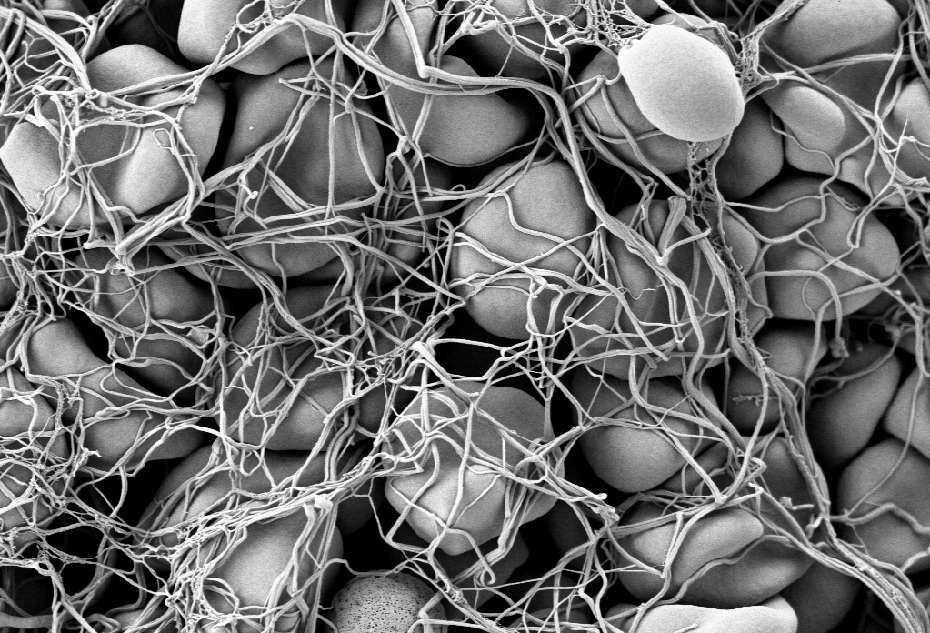
Electronic scanning microscopic image of red blood cells forming a clot
Hemostasis is the process by which blood stops flowing from damaged blood vessels. It is a complex process involving multiple molecules and forces, and our current understanding is limited by our inability to test these factors simultaneously in the laboratory. Some tests, for instance, can tell us much about clotting — a part of hemostasis — but little about the other elements at play. In particular, the role in hemostasis of the endothelium, which is the cell layer that lines the blood vessels, has generally been omitted from previous studies.
However, a new article in Nature Communications details the use of microfluidics technology, which is often used to model organ systems outside the body, to engineer a more complete model of hemostasis. Led by Wilbur A. Lam, M.D., Ph.D., Assistant Professor in the Wallace H. Coulter Department of Biomedical Engineering at Georgia Tech, the study authors fabricated microfluidics devices and then seeded vascular channels in the devices with human aortic and umbilical vein cells to simulate the endothelium.
Using the device, the authors were able to visualize hemostatic plug formation with whole blood and with blood from subjects with hemophilia. Although the authors concede that their model best represents capillary bleeding, rather than bleeding from larger vessels, they are confident that their model reliably represents the interaction of the endothelium with multiple varieties of blood cells.
Shedding Light on Cancer Response and Resistance
Penn’s founder, Benjamin Franklin, has a famous axiom: “an ounce of prevention is worth a pound of cure.” If Franklin were alive today, he would likely agree with two common axioms in cancer treatment: 1) if you can’t see it, you can’t treat it; and 2) if you treat it, treat all of it. Recent publications from investigators at Columbia and the University of Maryland reveal how imaging technologies can help predict to outcomes and how nanotechnology is offering a new therapeutic tools for fighting cancer.
Using diffuse optical tomography (DOT), which employs near-infrared spectroscopy to obtain three-dimensional images, scientists at Columbia have shown in an article in Radiology that treatment response could be predicted as early as two weeks after the start of therapy. The authors, led by Andreas H. Hielscher, PhD, Professor of Biomedical Engineering, Electrical Engineering, and Radiology at Columbia, applied DOT in 40 women with breast cancer undergoing chemotherapy. They found that DOT imaging features were associated, some very strongly, with treatment outcomes at 5 months. Given their positive findings, the authors intend to continue testing DOT in a larger cohort prospective study.
Another major issue in cancer chemotherapy is multidrug resistance (MDR), a highly frustrating complication resulting from lengthy treatment. In MDR, cancer types can find ways to overcome the effects of chemotherapy, resulting in relapse, often with deadly consequences. Therefore, among the challenges that oncologists face is the need to predict MDR, preferably before treatment even begins.
Based on the knowledge that adenosine triphosphate (ATP), a common organic molecule in energy generation, is involved in MDR, scientists at the University of Maryland engineered nanoparticles that could target cancer cells and, when exposed to near-infrared laser irradiation, reduce the amount of ATP in the cells . The scientists, led by Xiaoming Shawn He, Ph.D., Professor of Bioengineering at Maryland, published their findings in Nature Communications.
Dr. He’s team tested their nanoparticles in vitro and subsequently in mice and found decreased tumor sizes in mice treated with the particles, as well as more deaths of cancer cells. In addition, two of seven mice treated with the nanoparticles plus light experienced complete tumor eradication. The findings offer hope that MDR could be overcome with direct delivery of targeted treatment to resistant tumors.
Preserving the Tooth
A frustrating problem often encountered by dentists is the growth of new cavities around existing fillings. Microbes are often critical catalysts for these new cavities. Using antimicrobial agents at cavity-repair sites could make a real difference. However, mesoporous silica has proved suboptimal for this purpose.
However, help might be on the way. A study in a recent issue of Scientific Reports, written by a trio of authors led by Benjamin D. Hatton, Ph.D., Assistant Professor at the Institute of Biomaterials & Biomedical Engineering of the University of Toronto, reports the successful synthesis of 500-nm nanocomposite spheres combining silica with octenidine dihydrochloride, a common antiseptic. The newly synthesized nanospheres outperformed earlier attempts with mesoporous silica. The authors will continue to develop these nanoparticles to deliver other drugs for longer periods of time.
Cell signaling and the proteins involved in it participate in virtually every process in the body, whether normal or pathological. Much of this signaling involves proteins called cytokines, and of particular interest among them are tumor necrosis factors (TNFs), whose job it is to carry out apoptosis — the process by which cells die at predetermined time points as part of their normal life cycle. Among this family of cytokines, TNF-related apoptosis-inducing ligand (TRAIL) has been of particular interest to oncologists.
The process by which TRAIL combines with or binds to other molecules that modulate the life cycle of cancer cells can interfere with the ability of these molecules to facilitate the growth of cancer cells into tumors. However, attempts to deploy the cytokine to interfere in the process that produces cancer have been unsuccessful because of issues regarding inefficient delivery of TRAIL to the relevant sites, poor circulation of the cytokine in the blood, and the development of resistance to TRAIL. Bioengineers have been hard at work attempting to overcome these barriers.
In a new article published in ACS Nano coauthored by Michael J. Mitchell, Ph.D., Skirkanich Assistant Professor of Innovation at Penn Bioengineering, and Robert Langer, Ph.D., David H. Koch Institute Professor at MIT, these engineered solutions are reviewed and assessed. The review covers nanoparticle technologies with potential to solve the problems encountered thus far, including a range of materials (polymers, lipids, inorganic), cell-nanoparticle hybrids, and therapeutic cells genetically engineered using nanoparticles.
“The TRAIL protein is a essential component of our immune system,” Dr. Mitchell says, “and it kills tumor cells without harming normal ones. However, it remains challenging to deliver the protein into tumors, and tumors can also be resistant to the protein. We and others are now exploiting nanotechnology, genetic engineering, and immune cell-biomaterial hybrids to overcome these key biological barriers to cancer therapy.”
The introduction of morphine in the 19th century to alleviate pain revolutionized medicine in a way few innovations do, but it brought with it a grave unintended consequence: addiction. In today’s society, opioid addiction is creating the biggest health crisis of the last half century. Affecting nearly 1 in 100 people, opioid addiction occurs more than type 1 diabetes, multiple sclerosis, or a number of other diseases. The addiction crisis also appears in global affairs and impacts our national security: heroin production in Afghanistan over the last 40 years has been critical to funding military actions by insurgent groups against both the US and, in the past, the Soviet Union.
However, bioengineers at Stanford have begun to tackle the issue of production and might have begun to tackle the issue of addiction. In the lab of Christine Smolke, Ph.D., Professor of Bioengineering at Stanford, they have been genetically engineering yeast to produce opioids. They described the process in a 2015 article from Science. Now, in a recent interview in Fast Company, Dr. Smolke discusses the possibility of using the yeast producing method she pioneered to produce opioids without addiction potential. These alternative drugs are very expensive to produce, and Dr. Smolke’s process could provide safer, less addictive compounds to people in need.
Breaking the Barrier
Among the challenges faced by bioengineers working on therapies for brain disease is the blood-brain barrier (BBB), a tightly regulated boundary between the circulatory system and the brain that prevents all but the tiniest molecules from getting into the brain. The poor permeability of the BBB to many molecules means that one needs to use higher drug dosages to reach the brain, which is one of the reasons why most psychiatric medications have a broad array of side effects.
One way of circumventing this issue is to deliver the drugs directly to the brain, rather than using oral or intravenous delivery methods that need to cross the BB. Here, the challenge is one of size — unless a needle used to administer such a drug is very small, it will invariably damage brain tissue, which can have devastating consequences. Answering this call has been Robert Langer, Ph.D., David H. Koch Institute Professor at MIT, whose lab has successfully microfabricated delivery cannulas as small as 30 microns, about one-third the diameter of human hair. As they report in Science Translational Medicine, the new cannula can target brain areas as small as 1 cubic millimeter.
Dr. Langer and his colleagues used the new cannula to create an implantable device, called the miniaturized neural drug delivery system (MiNDS), that they subsequently tested in rats and rhesus monkeys. They found that the device could modulate neuronal activity in both animals. In addition, MiNDS could also record and transmit information from the treatment site to enable feedback control. Going forward, the study authors envision the use of non-metallic materials to fashion cannulas and hydrogel coatings to facilitate MR imaging and increase biocompatibility.
Unlocking the Mystery of IPF
Idiopathic pulmonary fibrosis (IPF) is a lung disease that causes permanent scarring of the lung tissue. The disease affects around five million people worldwide, mainly people 50 or older, and the five-year mortality rate is very high. Although risk factors, such as cigarette smoking, have been identified, as the word “idiopathic” implies, the cause is unknown, making it difficult to create effective therapies other than ones that merely slow the progression of the disease.
However, thanks to a new discovery, we might be closer to effective treatments. In an article in the Journal of Clinical Investigation Insight, a team of scientists from Yale University report that the tissue lesions that constitute IPF are made up of roughly one-fifth pericytes — a type of contractile cell that plays an important role in the proper function of capillaries, including those in the lungs.
The study authors, led by Anjelica Gonzalez, Ph.D., Donna L. Dubinsky Associate Professor of Biomedical Engineering at Yale, found that IPF caused pericytes to take on the properties of myofibroblasts, a cell type that is important to the wound-healing process. They found further that treatment of these myofibroblast-like pericytes with nintedanib, a drug approved for IPF treatment, reversed this effect. Armed with this knowledge, we come a step closer to designing and producing more effective therapies for IPF, as well as for diseases with similar effects.
People and Places
Washington University in St. Louis has announced it will launch a Ph.D. program in imaging science, to enroll its first cohort this fall. The program will be headed by Mark Anastasio, Ph.D., Professor of Biomedical Engineering and a 1993 recipient of an MSE from Penn. WashU’s program is only the second such program in the country, following the program at the Rochester Institute of Technology.
Closer to home, Johns Hopkins is the recipient of a $50 million donation from the United Arab Emirates. The money will be used to create the Sheikh Khalifa Stroke Institute, which will unite faculty members from biomedical engineering, neurology, and rehabilitation medicine to advance research into stroke.
George H.W. Bush refused to eat it, but maybe he should start. It turns out that broccoli, combined with bioengineered yogurt, could provide effect cancer prevention. We’ve known for some time that compounds in certain fresh vegetables can increase chemoprevention, but the levels are usually too low to be effective, or they can’t be assimilated optimally by the body. However, scientists in Singapore found that engineered bacteria, when ingested by mice with colorectal cancer, had anticancer effects. The bacteria caused the secretion of an enzyme by the cancer cells that transformed glucosinolates — compounds found in vegetables — into molecules with anticancer efficacy. The scientists report their findings in Nature Biomedical Engineering.
The authors programmed an E. coli cell line to bind to heparan sulfate proteoglycan, a cell surface protein that occurs in colorectal cancer cells. Once the engineered bacteria bound to the cancer cells, the bacteria secreted myrosinase, an enzyme that commonly occurs in many plants to defend them against aphids. In the cell model employed by the authors, myrosinase caused the conversion of glucosinolates into sulforaphane, which in turn could inhibit cancer cell growth.
The scientists then applied their system in a mouse model of colorectal cancer, feeding the mice yogurt infused with the engineered bacteria. They found that the mice fed broccoli plus the yogurt developed fewer and smaller tumors than mice fed broccoli alone. Additional testing is necessary, of course, but the study authors believe that their engineered bacteria could be used both as a preventive tool in high-risk patients and as a supplement for cancer patients after surgery to remove their tumors.
The Gates of CRISPR
About two years ago, software giant Microsoft unveiled Azimuth, a gene-editing tool for CRISPR/Casa9 that it had developed in collaboration with scientists at the Broad Institute. Now, in response to concerns that CRIPR may edit more of the genome than a bioengineer wants, the team has introduced a tool called Elevation. A new article in Nature Biomedical Engineering discusses the new tool.
In the article, the team, co-led by John C. Doench, Ph.D., Institute Scientist at the Broad Institute, describes how it developed Azimuth and Elevation, both of which are machine learning models, and deployed the tools to compare their ability to predict off-target editing with the ability of other approaches. The Elevation model outperformed the other methods. In addition, the team has implemented a cloud-based service for end-to-end RNA design, which should alleviate some of the time and resource handicaps that scientists face in using CRISPR.
Reducing Infant Mortality With an App
Among the challenges still faced in the developing world with regard to health care is high infant mortality, with the most common cause being perinatal asphyxia, or lack of oxygen reaching the infant during delivery. In response, Nigerian graduate student Charles C. Onu, a Master’s student in the computer science lab of Doina Precup, Ph.D., at McGill University in Montreal, founded a company called Ubenwa, an Igbo word that means “baby’s cry.”
With Ubenwa and scientists from McGill, Onu developed a smartphone app and a wearable that apply machine learning to instantly diagnose birth asphyxia based on the sound of a baby’s cry. In initial testing, the device performed well, with sensitivity of more than 86% and specificity of more than 89%. You can read more about the development and testing of Ubenwa at Arxiv.
People and Places
Several universities have announced that they are introducing new centers for research in bioengineering. Purdue University secured $27 million in funding from Semiconductor Research Corp. for its Center for Brain-inspired Computing Enabling Autonomous Intelligence, or C-BRIC, which opened last month. The center will develop, among other technologies, robotics that can operate without human intervention.
In Atlanta, Emory University received a $400 million pledge from the Robert W. Woodruff Foundation for two new centers — the Winship Cancer Institute Tower and a new Health Sciences Research Building. The latter will host five research teams, including one specializing in biomedical engineering. Further north in Richmond, Virginia Commonwealth University announced that it will begin construction on a new $92 million Engineering Research Building in the fall. The uppermost floors of the new building will include labs for the college’s Department of Biomedical Engineering.
Finally, North Carolina’s Elon College will introduce a bachelor’s degree program in engineering in the fall. The program will offer concentrations in biomedical engineering and computer engineering. Sirena Hargrove-Leak, Ph.D., is director of the program.

 Last month, the National Toxicology Program (NTP), a division of the U.S. Department of Health and Human Services, announced the findings of a draft study in which it was shown that high exposure to radiofrequency radiation, similar to that caused by persistent use of cell phones, resulted in the formation of tumors in nerves surrounding the hearts of male rats — but not female rats or mice. These are the final results of the study, the preliminary results of which were released in 2016. The study must still undergo peer review later this month.

 The introduction of morphine in the 19th century to alleviate pain revolutionized medicine in a way few innovations do, but it brought with it a grave unintended consequence: addiction. In today’s society, opioid addiction is creating the biggest health crisis of the last half century. Affecting nearly 1 in 100 people, opioid addiction occurs more than type 1 diabetes, multiple sclerosis, or a number of other diseases. The addiction crisis also appears in global affairs and impacts our national security: heroin production in Afghanistan over the last 40 years has been critical to funding military actions by insurgent groups against both the US and, in the past, the Soviet Union.