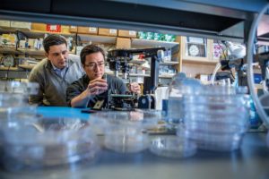
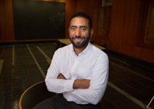
Andrei Georgescu (left) and Dan Huh developed several organ-on-a-chip platforms in Huh’s lab. Their spin-off company, Vivodyne, aims to use the technology as a scalable alternative to animal testing in the pharmaceutical industry.
With Vivodyne, Associate Professor in the Department of Bioengineering Dan Huh is translating the organs-on-chips technology into a promising industry venture. Using microfluidic structures that mimic aspects of human physiology, organs-on-chips allow scientists to test therapies on lab-grown human cells. Vivodyne specifically focuses on designing organs-on-chips to create a scalable alternative for pharmaceutical drug testing on animals.
Fast Company now lists it as one of “the 10 most innovative companies with fewer than 10 employees,” saying “Vivodyne is helping major pharmaceutical companies like GlaxoSmithKline quickly adopt viable alternatives for testing drugs on monkeys.”
Vivodyne, launched in 2021, has created a platform that allows fully automated, complex studies at a far larger scale and lower cost than would be possible with manual experimentation, so pharmaceutical companies can actually test lab-made organs instead of animals in their drug-development processes. When done by hand, only 20 to 40 living tissue samples can be managed in parallel; Vivodyne’s instrument can cultivate, dose, and image more than 2,000 living tissues at once. The company, which raised $4 million in seed funding last year, says its instruments currently play pivotal roles in clinical drug testing for respiratory diseases, cancer treatment, vaccine development, diabetes therapies, and maternal medicine. GlaxoSmithKline, one of Vivodyne’s clients, estimates that for some projects the lab-grown tissues may displace as much as 80% of its animal testing. The company’s ultimate goal? “To supplant the vast majority of animal testing within the next decade,” says CEO Andrei Georgescu.
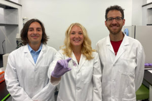
Michael Mitchell, Sarah Shepherd and David Issadore pose with their new device.
The COVID vaccines currently being deployed were developed with unprecedented speed, but the mRNA technology at work in some of them is an equally impressive success story. Because any desired mRNA sequence can be synthesized in massive quantities, one of the biggest hurdles in a variety of mRNA therapies is the ability to package those sequences into the lipid nanoparticles that deliver them into cells.
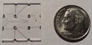
Now, thanks to manufacturing technology developed by bioengineers and medical researchers at the University of Pennsylvania, a hundred-fold increase in current microfluidic production rates may soon be possible.
The researchers’ advance stems from their design of a proof-of-concept microfluidic device containing 128 mixing channels working in parallel. The channels mix a precise amount of lipid and mRNA, essentially crafting individual lipid nanoparticles on a miniaturized assembly line.
This increased speed may not be the only benefit; more precisely controlling the nanoparticles’ size could make treatments more effective. The researchers tested the lipid nanoparticles produced by their device in a mouse study, showing they could deliver therapeutic RNA sequences with four-to-five times greater activity than those made by conventional methods.
The study was led by Michael Mitchell, Skirkanich Assistant Professor of Innovation in Penn Engineering’s Department of Bioengineering, and David Issadore, Associate Professor in Penn Engineering’s Department of Bioengineering, along with Sarah Shepherd, a doctoral student in both of their labs. Rakan El-Mayta, a research engineer in Mitchell’s lab, and Sagar Yadavali, a postdoctoral researcher in Issadore’s lab, also contributed to the study.
They collaborated with several researchers at Penn’s Perelman School of Medicine: postdoctoral researcher Mohamad-Gabriel Alameh, Lili Wang, Research Associate Professor of Medicine, James M. Wilson, Rose H. Weiss Orphan Disease Center Director’s Professor in the Department of Medicine, Claude Warzecha, a senior research investigator in Wilson’s lab, and Drew Weissman, Professor of Medicine and one of the original developers of the technology behind mRNA vaccines.
“We believe that this microfluidic technology has the potential to not only play a key role in the formulation of current COVID vaccines,” says Mitchell, “but also to potentially address the immense need ahead of us as mRNA technology expands into additional classes of therapeutics.”
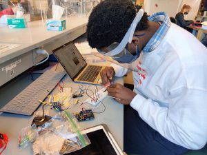
The junior year BE-MAD lab series includes modules on dialysis, drug delivery, insect limb control, microfluidics, cell-cell communication, ECG analysis (pictured here), and spectroscopy. (Image: Bioengineering Educational Lab)
While the majority of courses remained online this spring, a small number of lab-based undergraduate courses were able to resume limited in-person instruction. One course was BE 310, the second semester of the Bioengineering Modeling, Analysis, and Design lab sequence. Better known as BE-MAD, this junior-year bioengineering course was able to bring students back to the teaching lab safely this spring while adapting its curriculum to keep remote learners engaged with hands-on lab modules at home.
An Essential Step Towards Becoming a Bioengineer
After learning the basics of chemistry, physics, biology, and math during freshman year and studying bioengineering fundamentals throughout sophomore year, BE-MAD is designed to provide essential hands-on experience to bioengineering majors during their junior years. In BE-MAD, students integrate what they’ve learned so far in the classroom to addressing complex, real-world problems by breaking down the silos that exist across different STEM fields.
“Usually what we hear from students is that this BE 309/310 sequence is when they really feel like they are engineers,” says Brian Chow, one of the BE 310 instructors. “They can put what they learn in classes to work in some practical setting and applied context.”
BE-MAD is also an important course to prepare students for senior design and is designed to be a “safe space to fail,” allowing students to build confidence through trial and error within a supportive environment, explains Sevile G. Mannickarottu, director of the educational laboratories. “We’re trying to build skills needed for senior year as well as teaching students how to think critically about problems by pulling together the materials they’ve learned all in one place,” he says. “By senior year, we want them to, when presented with a problem, not be afraid.”
Adapting BE-MAD for Both Remote and Hybrid Instruction
Traditionally, the BE-MAD lab is taught in the George H. Stephenson Foundation Educational Laboratory & Bio-MakerSpace, the primary bioengineering teaching lab, and includes modules on dialysis, drug delivery, insect limb control, microfluidics, cell-cell communication, ECG analysis, and spectroscopy. In the fall, the first lab in the series (BE-309) pivoted to remote learning using video tutorials of lab experiments and providing real data to students for analysis.
This spring, with more aspects of on-campus life able to reopen, the Educational Laboratory staff and BE-MAD instructors developed protocols in collaboration with David Meaney, Penn Engineering senior associate dean and an instructor for BE 309, and Penn’s Environmental Health and Radiation Safety office to safely reopen the teaching lab and Bio-MakerSpace for both BE-310 and for bioengineering senior design students.
The BE-MAD lab was also recreated on Gather.Town, an online video chat platform where students can speak with group members or instructors. Student groups also had their own tables where they could meet virtually to work on data analysis and lab report writing.
To continue to meet the needs of remote students, BE 310 instructor Lukasz Bugaj says that the curriculum was adapted to be two parallel courses—one that could be done entirely at home and the other in-person. The challenge was to adjust the content so that it could be completed either in-person or virtually, and could be switched from in-person to virtual at a moment’s notice because of COVID precautions, all while maximizing the hands-on experience, says Bugaj. “That’s a real credit to the lab staff of Sevile and Michael Patterson, who put a lot of work into revamping this entire class.”
Andrei Georgescu (left) and Dan Huh are the co-founders of Vivodyne, a spin-off of Huh’s BIOLines lab.
Dan Huh, Associate Professor in the Department of Bioengineering, has been steadily growing a collection of organs-on-chips. These devices incorporate human cells into precisely engineered microfluidic channels that mimic an organ’s natural environment, providing a way to conduct experiments that would not otherwise be feasible.
Huh’s previous research has involved using a placenta-on-a-chip to study which drugs are able to reach a developing fetus; investigating microgravity’s effect on the immune system by sending one of his chips to the International Space Station; and testing treatments for dry eye disease using an eye-on-a-chip, complete with a mechanical blinking eyelid.
Now, he and his colleagues are taking this technology out of their lab and into industry with their company, Vivodyne.
Andrei Georgescu, Huh’s lab-member and co-founder of Vivodyne, recently spoke with Technical.ly Philly’s Paige Gross about the growth of their company.
Research into potential drugs is usually performed first on mice, and success is only found in a fraction of humans once implemented in clinical trials, Andrei Georgescu, cofounder and CEO of Vivodyne, told Technical.ly. The genetic makeup just isn’t similar enough. But technology that allows scientists to test therapies on lab-grown human organs called “organs on chip” is allowing for testing without human subjects.
The organs on chip allow for a drug to react to tissue in a more similar way to the body than it would in a petri dish, Georgescu said. Cells sense their environment very well, he added.
“We’re making the environment more complicated, making its spacial features complicated enough to match the native complexity of the organs,” he said. “When [cells] sense a softer environment, they start to behave more realistically. Their response to the drug is more realistic.”
Speaker: Weihua Guan, Ph.D.
Assistant Professor
Department of Electrical Engineering & Department of Biomedical Engineering (courtesy)
Pennsylvania State University, University Park
Date: Thursday, April 8, 2021
Time: 3:00-4:00 PM EDT
Zoom – check email for link or contact ksas@seas.upenn.edu
Abstract:
Due to their conceptual simplicity, the nanopore sensors have attracted intense research interest in electronic single molecule detection. While considerable success has been achieved, the solid-state nanopores still face three significant challenges, including repeatable nanopore size control, introduction sensing specificity, and prolonged sensor response time at low concentrations. In this talk, I will discuss a calibration-free solid-state nanopore counting method and two representative applications in nucleic acid testing. One is an isothermal amplification-coupled nanopore counting for malaria analysis. The other is the CRISPR-cas12a-coupled nanopore counting for HIV analysis. Finally, I will also discuss how we can develop a fully integrated ‘sample-to-result’ nucleic acid testing device using the solid-state counting strategy. I believe the reaction-coupled solid-state nanopore digital counting could open a new avenue towards compact, robust, low-cost electronic nucleic acid testing at the point of care.
Weihua Guan Bio:
Weihua Guan received his Ph.D. degree in Electrical Engineering from Yale University in 2013 and did his postdoctoral training at Johns Hopkins University from 2013 to 2014. He joined the Department of Electrical Engineering at Pennsylvania State University in Jan 2015. He also held a courtesy appointment in the Department of Biomedical Engineering at Penn State. Dr. Guan’s research interests are in the multidisciplinary areas of micro- and nanotechnology, micro/nanofluidics, bioMEMS, lab-on-a-chip devices, and point-of-care devices. Dr. Guan’s research group at Penn State focuses on developing micro and nanoscale devices as well as novel sensing principles towards advanced medical diagnostics and testing. Dr. Guan is a member of IEEE, Engineering in Medicine and Biology Society, Biophysical Society, and American Physics Society. Among other honors, Dr. Guan is a recipient of the HHMI International Research Fellowship and NSF CAREER award.
The Penn Bioengineering virtual seminar series continues on October 8th.
Aaron Streets, PhD
Speaker: Aaron Streets, Ph.D.
Associate Professor of Bioengineering
University of California, Berkeley
Date: Thursday, October 8, 2020
Time: 2:00-3:00 pm (note the change from our regular seminar time)
Zoom – check email for link or contact ksas@seas.upenn.edu
Title: “Imaging and Sequencing Single Cells”
Abstract:
Recent advances in microfluidics and high-throughput sequencing technology have enabled rapid profiling of genomic material in single cells. Valve- and droplet-based microfluidic platforms can precisely and efficiently manipulate, sort, and process cells to generate indexed sequencing libraries, allowing for high-throughput single-cell analysis of the genome, transcriptome, proteome, and epigenome. Such technology has been instrumental in the global effort to create a human cell atlas, with the ambitious goal of identifying and cataloging all human cell types and cell states in health and disease. However, not all cell phenotypes are directly encoded in the genome and high-throughput sequencing cannot probe the full space of cellular identity. Therefore, microscopy remains one of the most powerful and versatile tools for characterizing cells. Fluorescent imaging and quantitative non-linear optical imaging can reveal morphological characteristics, protein localization, chromatin organization, and chemical composition in single cells. Both single-cell genomics and microscopy can uncover heterogeneity in cellular populations that would otherwise be obscured in ensemble measurement. In this talk, I will discuss a suite of new microfluidic platforms for coupling genomic measurements and optical measurements of the same single cell, and some novel computational approaches to grapple with these new datasets. With a combination of new hardware and software, our goal is to converge on a quantitative and comprehensive understanding of cellular identity.
Bio:
Aaron received a Bachelor of Science in Physics and a Bachelor of Arts in Art at UCLA. He completed his PhD in Applied Physics at Stanford with Dr. Stephen Quake. Aaron then went to Beijing, China as a Whitaker International Postdoctoral Fellow and a Ford postdoctoral fellow and worked with Dr. Yanyi Huang in the Biodynamic Optical Imaging Center (BIOPIC) at Peking University. Aaron joined the faculty of UC Berkeley as an Assistant Professor in Bioengineering in 2016 and is currently a core member of the Biophysics Program and the Center for Computational Biology and he is a Chan Zuckerberg Biohub investigator. Aaron has received the NSF Early Career award and was recently named a Pew Biomedical Scholar.
See the full list of upcoming Penn Bioengineering fall seminars here.
University of Washington Researchers Engineer a New Way to Study Circulatory Obstruction
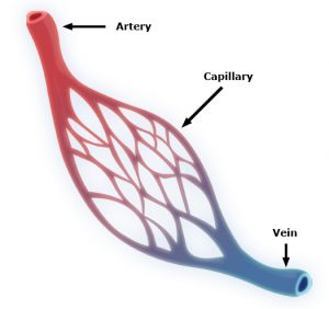
Capillaries are one of the most important forms of vasculature in our body, as they allow our blood to transfer nutrients to other parts of our body. But for how much effect capillary functionality can have on our health, their small size makes them extremely difficult to engineer into models for a variety of diseases. Now, researchers at the University of Washington led by Ying Zheng, Ph. D., engineered a three-dimensional microvessel model with living cells to study the mechanisms of microcirculatory obstruction involved with malaria.
Rather than just achieving a physical model of capillaries, these researchers created a model that allowed them to study typical flow and motion through capillaries, before comparing it to deficiencies in this behavior involved with diseases like malaria. The shape of the engineered model is similar to that of an hourglass, allowing the researchers to study instances where red blood cell transit may encounter bottlenecks between the capillaries and other vessels. Using multiphoton technology, Zheng and her team created 100mm capillary models with etched-in channels and a collagen base, to closely model the typical size and rigidity of the vessels. Tested with malaria-infected blood cells, the model showed similar circulatory obstructive behavior to that which occurs in patients, giving hope that this model can be transferred to other diseases involving such obstruction, like sickle cell anemia, diabetes, and cardiovascular conditions.
Understanding a Cell Membrane Protein Could Be the Key to New Cancer Treatments
Almost every cell in the body has integrins, a form of proteins, on its membrane, allowing cells to sense biological information from beyond their membranes while also using this feedback information to initiate signals within cells themselves. Bioengineers at the Imperial College of London recently looked at the way another membrane protein, called syndecan-4, interacts with integrins as a potential form of future cancer treatment. Referred to as “cellular hands” by lead researcher of the study Armando del Rio Hernandez, Ph.D., syndecan-4 sometimes controls the development of diseases or conditions like cancer and fibrosis. Hernandez and his team specifically studied the ties of syndecan-4 to yes-associated protein (YAP) and enzyme called P13K, both of which are affiliated with qualities of cancer progression like halted apoptosis or cell stiffening. Knowing this, Hernandez and his team hope to continue research into understanding the mechanisms of syndecan-4 throughout the cell, in search of new mechanisms and targets to focus on with future developments of cancer treatments.
A New Medical Device Could Improve Nerve Functionality After Severe Damage
Serious nerve damage remains difficult to repair surgically, often involving the stretching of nerves for localized damage, or the transfer of healthy nerve cells from another part of the body to fill larger gaps in nerve damage. But these imperfect solutions limit the return of full nerve function and movement to the damaged part of the body, and in more serious cases with large areas of nerve damage, can also risk damage in other areas of the body that healthy nerves are borrowed from for treatment. A new study from the University of Pittsburgh published in Science Translational Medicine led by Kacey Marra, Ph. D., has successfully repaired nerve damage in mice and monkeys using a biodegradable tube that releases growth factors called glial-cell-derived neurotrophic factors over time.
Marra and her team showed that this new device restored nerve function up to 80% in nonhuman primates, where current methods of nerve replacement often only achieve 50-60% functionality restoration. The device might have an easier time getting FDA-approval, since it doesn’t involve the use of stem cells in its repair mechanisms. Hoping to start human clinical trials in 2021, Marra and her team hope that the device will help both injured veterans and typical patients with nerve damage, and see potential future applications in facial nerve damage as well.
A New Computational Model Could Improve Treatments for Cancer, HIV, and Autoimmune Diseases
With cancer, HIV, and other autoimmune diseases, the best treatment options for patients are often determined with trial-and-error methods, leading to prolonged instances of ineffective approaches and sometimes unnecessary side effects. A group of researchers led by Wesley Errington, Ph.D., at the University of Minnesota decided to take a computational approach this problem, in an effort to more quickly and efficiently determine the most appropriate treatment for a given patient. Based on parameters controlling interactions between molecules with multiple binding sites, the team’s new model looks primarily at binding strength, linkage rigidity, and size of linkage arrays. Because diseases can often involve issues in molecular binding, the model aimed to model the 78 unique binding configurations for cases of when interacting molecules only have three binding sites, which are often difficult to observe experimentally. This new approach will allow for faster and easier determination of treatments for patients with diseases involving these molecular interactions.
Improved Drug Screening for Glioblastoma Patients
A new microfluidic brain chip from researchers at the University of Houston could help improve treatment evaluations for brain tumors. Glioblastoma patients, who have a five-year survival rate of a little over 5%, are some of the most common patients suffering from malignant brain tumors. This new chip, developed by the lab of Yasemin Akay, Ph.D., can quickly determine cancer drug effectiveness by analyzing a piece of cultured tumor biopsy from a patient by incorporating different chemotherapy treatments through the microfluidic vessels. Overall, Akay and her team found that this new chip holds hope as a future efficient and inexpensive form of drug screening for glioblastoma patients.
People and Places
The brain constructs maps to guide people, not just of physical spaces but also to connect stimuli around them, like conversations and other people. It’s long been known that the brain area responsible for this spatial navigation—the medial temporal lobe—is also involved in recalling memories.
Michael Kahana (left) is principal investigator in the Defense Advanced Research Projects Agency’s RAM program and a professor in the Department of Psychology. Ethan Solomon is an M.D./Ph.D. student in the Department of Bioengineering of the School of Engineering and Applied Science and in the Perelman School of Medicine.
Now, neuroscientists at the University of Pennsylvania have discovered that the signals the brain produces during spatial navigation and episodic memory recall look similar. Low-frequency brain waves called the theta rhythm appear as people jump from one memory to the next, as many prior studies looking only at human navigation have shown. The new findings, which suggest that the brain structures responsible for helping people navigate the world may also “navigate” a mental map of prior experiences, appear in the Proceedings of the National Academy of Sciences.
The Florida Institute of Technology recently announced plans to start construction in spring 2020 on a new Health Sciences Research Center, set to further establish biomedical engineering and pre-medical coursework and research at the institute. With plans to open the new center in 2022, Florida Tech anticipates increased enrollment in the two programs, and hopes that the center will offer more opportunities in a growing professional field.
Anson Ong, Ph.D., the Associate Dean of Administration and Graduate Programs at the University of Texas at San Antonio, was recently elected to the International College of Fellows of Biomaterials Science and Engineering. With a focus on research in biomaterial implants for orthopaedic applications, Ong’s election to the college honors his advancement and contribution to the field of biomaterials research.
Speaker: Sumita Pennathur, Ph.D.
Professor of Mechanical Engineering
University of California, Santa Barbara
Date: Thursday, November 21, 2019
Time: 12:00-1:00 pm
Location: Room 337, Towne Building
Title: “Nanofluidic Technologies for Biomolecule Manipulation”
Abstract:
In the last 20 years, microfabrication techniques have allowed researchers to miniaturize tools for a plethora of bioanalytical applications. In addition to better sensitivity, accuracy and precision, scaling down the size of bioanalytical tools has led to the exploitation of new technologies to further manipulate biomolecules in ways that has never before been achieved. For example, when microfluidic channels are on the same order of magnitude of the electric double layers that form due to localized charge at the surfaces, there exists unique physics that create different flow phenomenon, such as analyte concentration and/or separation, mainly due to the couples physics of electrostatics and fluid dynamics. This talk will outline the basis of such interesting phenomena, such as nanofluidic separation and concentration, and well as probe the applications of such coupled systems, for example, handheld DNA detection. Most importantly, we will focus on the most recent work in the Pennathur lab in this field — biopolar electrode (BPE)-based phenomenon. Bipolar electrodes (BPE) have been studied in microfluidic systems over the past few decades, and through rigorous experimentally-validated modeling of the rich combined physics of fluid dynamics, electrokinetics, and electrochemistry at BPEs, I will show the potential of utilizing microfluidic-based BPEs for the design and development of low power, accurate, low volume fluid pumping mechanisms, with the ultimate goal of integration into wearable drug delivery and µTAS systems.
Bio:
Professor Pennathur has been a Professor of Mechanical Engineering at University of California, Santa Barbara in 2007, specializing in the fields of MEMS, nanofludics, and electrokinetics. Her most significant contributions include: 1) unearthing a novel mechanism for separation and concentration of analytes for bioanalytical applications, 2) developing a label-free detection mechanism for nucleic acids (that has since spun off into a point-of-care diagnostic company), 3) developing commercial medical diagnostic products, 4) building optical and acoustic biosensors and 5) developing revolutionized methods for measuring blood glucose for patients with diabetes. She received her B.S. and M.S. from MIT and PhD. From Stanford University.
David Issadore (center) was announced as the awardee of the JPOD @ Philadelphia QuickFire Challenge by Katherine Merton (right), head of JLABS New York City, Boston, and Philadelphia, at last week’s BIO 2019 International convention. (Photo: Johnson & Johnson Innovation)
Chip Diagnostics is a Philadelphia-based device company founded in 2016 based on research from the lab of David Issadore, Assistant Professor of Bioengineering and Electrical and Systems Engineering in the School of Engineering and Applied Science. The startup combines microelectronics, microfluidics, and nanomaterials with the aim to better diagnose cancer. The company is developing technologies and digital assays for minimally-invasive early cancer detection and screening that can be done using mobile devices.
There has been a long interest in diagnosing cancer using blood tests by looking for proteins, cells, or DNA molecules shed by tumors, but these tests have not worked well for many cancers since the molecules shed tend to be either nonspecific or very rare.
Issadore’s group aims to target different particles called exosomes: Tiny particles shed by cells that contain similar proteins and RNA as the parent cancer cell. The problem, explains Issadore, is that because of the small size of the exosomes, conventional methods such as microscopy and flow cytometry wouldn’t work. “As an engineering lab, we saw an opportunity to build devices on a nanoscale that could specifically sort the cancer exosomes versus the background exosomes of other cells,” he explains.
After Issadore was approached by the IP group at PCI Ventures in the early stages of their research, Chip Diagnostics has since made huge strides as a company. Now, as the awardee of the JPOD @ Philadelphia QuickFire Challenge, Chip Diagnostics will receive $30,000 in grant funding to further develop the first-in-class, ultra-high-definition exosomal-based cancer diagnostic. The award also includes one year of residency at Pennovation Works as well as access to educational programs and mentoring provided by Johnson & Johnson Family of Companies global network of experts.
A New Microscopy Technique Could Reduce the Risk of LASIK Surgery
Though over ten million Americans have undergone LASIK vision corrective surgery since the option became available about 20 years ago, the procedure still poses some risk to patients. In addition to the usual risks of any surgery however, LASIK has even more due to the lack of a precise way to measure the refractive properties of the eye, which forces surgeons to make approximations in their measurements during the procedure. In an effort to eliminate this risk, a University of Maryland team of researchers in the Optics Biotech Laboratory led by Giuliano Scarcelli, Ph. D., designed a microscopy technique that would allow for precise measurements of these properties.
Using a form of light-scattering technology called Brillouin spectroscopy, Scarcelli and his lab found a way to directly determine a patient’s refractive index – the quantity surgeons need to know to be able to measure and adjust the way light travels through the eye. Often used as a way to sense mechanical properties of tissues and cells, this technology holds promise for taking three-dimensional spatial observations of these structures around the eye. Scarcelli hopes to keep improving the resolution of the new technique, to further understanding of the eye, and reduce even more of the risks involved with LASIK surgery.
Taking Tissue Models to the Final Frontier
Space flight is likely to cause deleterious changes to the composition of bacterial flora, leading to an increased risk of infection. The environment may also affect the susceptibility of microorganisms within the spacecraft to antibiotics, key components of flown medical kits, and may modify the virulence of bacteria and other microorganisms that contaminate the fabric of the International Space Station and other flight platforms.
“It has been known since the early days of human space flight that astronauts are more prone to infection,” says Dongeun (Dan) Huh, Wilf Family Term Assistant Professor in Bioengineering at Penn Engineering. “Infections can potentially be a serious threat to astronauts, but we don’t have a good fundamental understanding of how the microgravity environment changes the way our immune system reacts to pathogens.”
In collaboration with G. Scott Worthen, a physician-scientist in neonatology at the Children’s Hospital of Philadelphia, Huh will attempt to answer this question by sending tissues-on-chips to space. Last June, the Center for the Advancement of Science in Space (CASIS) and the National Center for Advancing Translational Sciences (NCATS), part of the National Institutes of Health (NIH), announced that the duo had received funding to study lung host defense in microgravity at the International Space Station.
Huh and Worthen aim to model respiratory infection, which accounts for more than 30 percent of all infections reported in astronauts. The project’s goals are to test engineered systems that model the airway and bone marrow, a critical organ in the immune system responsible for generating white blood cells, and to combine the models to emulate and understand the integrated immune responses of the human respiratory system in microgravity.
Sappi Limited Teams Up with the University of Maine to Develop Paper Microfluidics
At the Westbrook Technology Center of Sappi, a global pulp and paper company, researchers found ways to apply innovations in paper texture for medical use. So far, these include endeavors in medical test devices and patches for patient diagnostics. In collaboration with the Caitlin Howell, Ph.D., Assistant Professor of Chemical and Biomedical Engineering at the University of Maine, Sappi hopes to continue advances in these unconventional uses of their paper, especially as the business in paper for publishing purposes declines.
Sappi’s projects with the university focus on the development of paper microfluidics devices as what’s now becoming a widespread solution for obstacles in point-of-care diagnostics. One project in particular, called Sharklet, uses a paper that mimics shark skin as a way to impede unwanted microbial growth on the device – a key characteristic needed for its transition into commercial use. Beyond this example, Sappi’s work in developing paper microfluidics underscores the benefits of these devices in their mass producibility and adaptability.
New Observations of the WNT Pathway Deepen the Understanding of Protein Signaling in Cellular Development
Scientists at Rice University recently found that a protein signaling pathway called WNT, typically associated with its role in early organism development, can both listen for signals from a large amount of triggers and influence cell types throughout embryonic development. These new findings, published in PNAS, add to the already known functions of WNT, deepening our understanding of it and opening the doors to new potential applications of it in stem cell research.
Led by Aryeh Warmflash, Ph. D., researchers discovered that the WNT pathway is different between stem cells and differentiated cells, contrary to prior belief that it was the same for both. Using CRISPR-Cas9 gene editing technology, the Warmflash lab observed that the WNT signaling pathway is actually context-dependent throughout the process of cellular development. This research brings a whole new understanding to the way the WNT pathway operates, and could open the doors to new forms of gene therapy and treatments for diseases like cancers that involve genetic pathway mutations.
People and Places
In a recent article from Technical.ly Philly, named Group K Diagnostics on a list of ten promising startups in Philadelphia. Group K Diagnostics founder Brianna Wronko graduated with a B.S.E. from Penn’s Department of Bioengineering in 2017, and her point-of-care diagnostics company raised over $2 million in funding last year. Congratulations Brianna!
We would also like to congratulate Pamela K. Woodward, M.D., on her being named as the inaugural Hugh Monroe Wilson Professor of Radiology at the Washington University School of Medicine in St. Louis. Also a Professor of Biomedical Engineering at the university, Dr. Woodward leads a research lab with a focus on cardiovascular imaging, including work on new standards for diagnosis of pulmonary blood clots and on an atherosclerosis imaging agent.
Lastly, we would like to congratulate all of the following researchers on their election to the National Academy of Engineering:
David Bishop, Ph. D., a professor at the College of Engineering at Boston University whose current research involves the development of personalized heart tissue as an all-encompassing treatment for patients with heart disease.
Joanna Aizenberg, Ph. D., a professor of chemistry and chemical biology at Harvard University who leads research in the synthesis of biomimetic inorganic materials
Gilda Barabino, Ph. D., the dean of the City College of New York’s Grove School for Engineering whose lab focuses on cartilage tissue engineering and treatments for sickle cell disease.
Karl Deisseroth, M.D., Ph. D., a professor of bioengineering at Stanford University whose research involves the re-engineering of brain circuits through novel electromagnetic brain stimulation techniques.
Rosalind Picard, Ph.D., the founder and director of the Affective Computing Research Group at the Massachusetts Institute of Technology’s Media Lab whose research focuses on the development of technology that can measure and understand human emotion.
And finally, Molly Stevens, Ph. D., the Research Director for Biomedical Material Sciences at the Imperial College of London with research in understanding biomaterial interfaces for biosensing and regenerative medicine.


 Capillaries are one of the most important forms of vasculature in our body, as they allow our blood to transfer nutrients to other parts of our body. But for how much effect capillary functionality can have on our health, their small size makes them extremely difficult to engineer into models for a variety of diseases. Now, researchers at the University of Washington led by
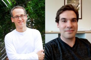

 In the last 20 years, microfabrication techniques have allowed researchers to miniaturize tools for a plethora of bioanalytical applications. In addition to better sensitivity, accuracy and precision, scaling down the size of bioanalytical tools has led to the exploitation of new technologies to further manipulate biomolecules in ways that has never before been achieved. For example, when microfluidic channels are on the same order of magnitude of the electric double layers that form due to localized charge at the surfaces, there exists unique physics that create different flow phenomenon, such as analyte concentration and/or separation, mainly due to the couples physics of electrostatics and fluid dynamics. This talk will outline the basis of such interesting phenomena, such as nanofluidic separation and concentration, and well as probe the applications of such coupled systems, for example, handheld DNA detection. Most importantly, we will focus on the most recent work in the Pennathur lab in this field — biopolar electrode (BPE)-based phenomenon. Bipolar electrodes (BPE) have been studied in microfluidic systems over the past few decades, and through rigorous experimentally-validated modeling of the rich combined physics of fluid dynamics, electrokinetics, and electrochemistry at BPEs, I will show the potential of utilizing microfluidic-based BPEs for the design and development of low power, accurate, low volume fluid pumping mechanisms, with the ultimate goal of integration into wearable drug delivery and µTAS systems.
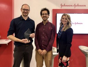
 Though over ten million Americans have undergone LASIK vision corrective surgery since the option became available about 20 years ago, the procedure still poses some risk to patients. In addition to the usual risks of any surgery however, LASIK has even more due to the lack of a precise way to measure the refractive properties of the eye, which forces surgeons to make approximations in their measurements during the procedure. In an effort to eliminate this risk, a University of Maryland team of researchers in the Optics Biotech Laboratory led by