Chlorine gas is a commonly used industrial chemical. It is also highly toxic and potentially deadly; it was used as a chemical weapon in both World War I and the Syrian Civil War and has led to multiple deaths from industrial accidents. Mixing certain household cleaners can also produce the toxic gas, leading to lasting lung injuries for which there are currently no effective treatments.
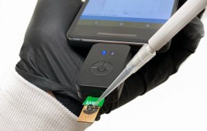
Now, researchers at Penn Engineering and Penn’s Perelman School of Medicine are collaborating with BARDA, the U.S. Office of Health and Human Services’ Biomedical Advanced Research and Development Authority, to address this need using their lung-on-a-chip technology.
The laboratory of Dan Huh, associate professor in the Department of Bioengineering, has developed a series of organ-on-a-chip platforms. These devices incorporate human cells into precisely engineered microfluidic channels that mimic an organ’s natural environment, providing a way to conduct experiments that would not otherwise be feasible.
Huh’s previous research has involved using a placenta-on-a-chip to study which drugs are able to reach a developing fetus; investigating microgravity’s effect on the immune system by sending one of his chips to the International Space Station; and testing treatments for dry eye disease using an eye-on-a-chip, complete with a mechanical blinking eyelid.
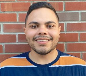
As an undergraduate studying bioengineering, Gabriel DeSantis spent a semester abroad at ETH Zurich in Zurich, Switzerland. Now in the Bioengineering master’s program, DeSantis is also a Penn Abroad Leader, serving on the office’s student advisory board and supporting fellow students interested in global experiences.
Penn Abroad’s Selene Li recently interviewed DeSantis about what drew him to Switzerland and his time there:
How did you discover the Switzerland abroad program?
I think it was probably in my sophomore year when I started seriously thinking about going abroad as something I could do during my time at Penn. I had cousins and other friends who had gone abroad, and just heard great things about it, so it seemed like something worth looking into. So as a bioengineer, I remember I first looked at what programs even offer bioengineering credit, and there’s really only a handful of options. I think I really wanted to be in Europe, and the Zurich program was at a super strong school. There was also someone in one of my classes who was a year older than me that was planning on going and was going through the whole application process. They were telling me about their whole application process and that really showed me how feasible going abroad was as a bioengineer.
What kinds of classes did you take?
I went in as a Health Sciences and Technology major. I had a decent amount of freedom in terms of the courses I could take because of that. While there, I took four credits that I ended up bringing back, and then two courses that were a little smaller that I didn’t do for credit but were just sort of interesting.
What was a really good or favorite memory from going abroad?
I really like hiking. During the first few weeks, we would do day trips to mountains nearby, but we did one weekend trip where we did two nights in Interlaken and had two full days to do some more intense hikes. Interlaken is just one of the most beautiful places in the world. We were staying at a nice little hostel, so I had an amazing day doing an 18-mile hike, and then coming back and hanging out with a bunch of people who I was also abroad with, as well as other people who were at the hostel. I think it was just being around constant beauty and around such great people and feeling accomplished because of the hike that made it such a great memory.
Nader Engheta, H. Nedwill Ramsey Professor in Electrical and Systems Engineering, Bioengineering and Materials Science and Engineering, has been awarded the 2020 Isaac Newton Medal and Prize by the Institute of Physics (IOP). The IOP is the professional body and scholarly society for physics in the UK and Ireland.
Engheta has been recognized for ” groundbreaking innovation and transformative contributions to electromagnetic complex materials and nanoscale optics, and for pioneering development of the fields of near-zero-index metamaterials, and material-inspired analogue computation and optical nanocircuitry.”
The Perelman School of Medicine has announced the winners of the 2020 Penn Medicine Awards of Excellence. The Office of the Dean says:
“These awardees exemplify our profession’s highest values of scholarship, teaching, innovation, commitment to service, leadership, professionalism and dedication to patient care. They epitomize the preeminence and impact we all strive to achieve. The awardees range from those at the beginning of their highly promising careers to those whose distinguished work has spanned decades.
Each recipient was chosen by a committee of distinguished faculty from the Perelman School of Medicine or the University of Pennsylvania. The contributions of these clinicians and scientists exemplify the outstanding quality of patient care, mentoring, research, and teaching of our world-class faculty.”
Two faculty members affiliated with Penn Bioengineering are among this year’s recipients.
Yale Cohen, PhD, Professor of Otorhinolaryngology with secondary appointments in Neuroscience and Bioengineering, is the recipient of the Jane M. Glick Graduate Student Teaching Award. Cohen is an alumnus of the Penn Bioengineering doctoral program and is currently the department’s Graduate Chair.
“Dr. Cohen’s commitment to educating and training the next generation of scientists exemplifies the type of scientist and educator that Jane Glick represented. His students value his highly engaging and supportive approach to teaching, praising his enthusiasm, energy, honesty, and compassion.”
Douglas H. Smith, MD, Robert A. Groff Endowed Professor of Research and Teaching in Neurosurgery and member of the Penn Bioengineering Graduate Group, is the recipient of this year’s William Osler Patient Oriented Research Award:
“Dr. Smith is the foremost authority on diffuse axonal injury (DAI) as the unifying hypothesis behind the short- and long-term consequences of concussion. After realizing early in his career that concussion, or mild traumatic brain injury (TBI), was a much more serious event than broadly appreciated, Dr. Smith and his team have used computer biomechanical modeling, in vitro and in vivo testing in parallel with seminal human studies to elucidate mechanisms of concussion.”
Speaker: Audrey Bowden, Ph.D.
Dorothy J. Wingfield Phillips Chancellor’s Faculty Fellow and Associate Professor of Biomedical Engineering and Electrical Engineering & Computer Science
Vanderbilt University
Date: Thursday, November 19, 2020
Time: 3:00-4:00 PM EST
Zoom – check email for link or contact ksas@seas.upenn.edu
Title: “Emerging Technologies for Detection of Early Stage Bladder Cancer”
Abstract:
Bladder cancer (BC) — the 4th most common cancer in men and the most expensive cancer to treat over a patient’s lifetime — is a lifelong burden to BC patients and a significant economic burden to the U.S. healthcare system. The high cost of BC stems largely from its high recurrence rate (>50%); hence, BC management involves frequent surveillance. Unfortunately, the current in-office standard-of-care tool for BC surveillance, white light cystoscopy (WLC), is limited by low sensitivity and specificity for carcinoma in situ (CIS), a high-grade carcinoma with high potential to metastasize. Early detection and complete eradication of CIS are critical to improve treatment outcomes and to minimize recurrence. The most promising macroscopic technique to improve sensitivity to CIS detection, blue light cystoscopy (BLC), is costly, time-intensive, has low availability and a high false-positive rate. Given the limitations of WLC, we aim to change the paradigm around how BC surveillance is performed by validating new tools with high sensitivity and specificity for CIS that are appropriate for in-office use. In this seminar, I discuss our innovative solutions to improve mapping the bladder for longitudinal tracking of suspicious lesions and to create miniature tools for optical detection based on optical coherence tomography (OCT). OCT and its functional variant, cross-polarized OCT, can detect early-stage BC with better sensitivity and specificity than WLC. We discuss the critical technical innovations necessary to make OCT and CP-OCT a practical tool for in-office use, and new results from recent explorations of human bladder samples that speak to the promise of this approach to change the management of patient care.
Bio:
Audrey K. Bowden is the Dorothy J. Wingfield Phillips Chancellor Faculty Fellow and Associate Professor of Biomedical Engineering (BME) and of Electrical Engineering and Computer Science (EECS) at Vanderbilt University. Prior to this, she served as Assistant and later Associate Professor of Electrical Engineering and Bioengineering at Stanford University. Dr. Bowden received her BSE in Electrical Engineering from Princeton University, her PhD in BME from Duke University and completed her postdoctoral training in Chemistry and Chemical Biology at Harvard University. During her career, Dr. Bowden served as an International Fellow at Ngee Ann Polytechnic in Singapore. From 2007-2008, she was the Arthur H. Guenther Congressional Fellow sponsored by the OSA and SPIE and served as a Legislative Assistant in the United States Senate through the AAAS Science and Technology Policy Fellows Program. Dr. Bowden is a Fellow of SPIE, a Fellow of AIMBE and is the recipient of numerous awards, including the Air Force Young Investigator Award, the NSF Career Award, the Hellman Faculty Scholars Award, the Phi Beta Kappa Teaching Award, Ford Foundation Postdoctoral Fellowship, and the NSBE Golden Torch Award. She is a former Associate Editor of IEEE Photonics Journal, former Lead Guest Editor of a Biomedical Optics Express Special Issue and is a member of numerous professional committees. Her research interests include biomedical optics – particularly optical coherence tomography and near infrared spectroscopy – microfluidics, and point of care diagnostics.
Using a magnetic field and hydrogels, a team of researchers in the Perelman School of Medicine have demonstrated a new possible way to rebuild complex body tissues, which could result in more lasting fixes to common injuries, such as cartilage degeneration. This research was published in Advanced Materials.
“We found that we were able to arrange objects, such as cells, in ways that could generate new, complex tissues without having to alter the cells themselves,” says the study’s first author, Hannah Zlotnick, a graduate student in bioengineering who works in the McKay Orthopaedic Research Laboratory at Penn Medicine. “Others have had to add magnetic particles to the cells so that they respond to a magnetic field, but that approach can have unwanted long-term effects on cell health. Instead, we manipulated the magnetic character of the environment surrounding the cells, allowing us to arrange the objects with magnets.”
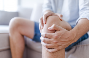
In humans, tissues like cartilage can often break down, causing joint instability or pain. Often, the breakdown isn’t in total, but covers an area, forming a hole. Current fixes are to fill those holes in with synthetic or biologic materials, which can work but often wear away because they are not the same exact material as what was there before. It’s similar to fixing a pothole in a road by filling it with gravel and making a tar patch: The hole will be smoothed out but eventually wear away with use because it’s not the same material and can’t bond the same way.
What complicates fixing cartilage or other similar tissues is that their makeup is complex.
“There is a natural gradient from the top of cartilage to the bottom, where it contacts the bone,” Zlotnick explains. “Superficially, or at the surface, cartilage has a high cellularity, meaning there is a higher number of cells. But where cartilage attaches to the bone, deeper inside, its cellularity is low.”
So the researchers, which included senior author Robert Mauck, PhD, director of the McKay Lab and a professor of Orthopaedic Surgery and Bioengineering, sought to find a way to fix the potholes by repaving them instead of filling them in. With that in mind, the research team found that if they added a magnetic liquid to a three-dimensional hydrogel solution, cells, and other non-magnetic objects including drug delivery microcapsules, could be arranged into specific patterns that mimicked natural tissue through the use of an external magnetic field.
Speaker: Daniel Palanker, Ph.D.
Director of the Hansen Experimental Physics Laboratory and Professor of Ophthalmology
Stanford University
Date: Wednesday, November 18, 2020
Time: 1:00-2:00 PM EST
Zoom – check email for link or contact eprince@seas.upenn.edu
Title: “Photovoltaic Restoration of Sight in Age-related Macular Degeneration”
Abstract:
Retinal degenerative diseases lead to blindness due to loss of the “image capturing” photoreceptors, while neurons in the “image-processing” inner retinal layers are relatively well preserved. Information can be reintroduced into the visual system using electrical stimulation of the surviving inner retinal neurons. We developed a photovoltaic substitute of photoreceptors which convert light into pulsed electric current, stimulating the secondary retinal neurons. Visual information captured by a camera is projected onto the retina from augmented-reality glasses using pulsed near-infrared (~880nm) light. This design avoids the use of bulky electronics and wiring, thereby greatly reducing the surgical complexity. Optical activation of the photovoltaic pixels allows scaling the number of electrodes to thousands. In preclinical studies, we found that prosthetic vision with subretinal implants preserves many features of natural vision, including flicker fusion at high frequencies (>30 Hz), adaptation to static images, antagonistic center-surround organization and non-linear summation of subunits in receptive fields, providing high spatial resolution. Results of the clinical trial with our implants (PRIMA, Pixium Vision) having 100μm pixels, as well as preclinical measurements with 75 and 55μm pixels, confirm that spatial resolution of prosthetic vision can reach the pixel pitch. Remarkably, central prosthetic vision in AMD patients can be perceived simultaneously with peripheral natural vision. For broader acceptance of this technology by patients who lost central vision due to agerelated macular degeneration, visual acuity should exceed 20/100, which requires pixels smaller than 25μm. I will describe the fundamental limitations in electro-neural interfaces and 3-dimensional configurations which should enable such a high spatial resolution. Ease of implantation of these wireless arrays, combined with high resolution opens the door to highly functional restoration of sight.
Bio:
Daniel Palanker is a Professor of Ophthalmology and Director of the Hansen Experimental Physics Laboratory at Stanford University. He received MSc in Physics in 1984 from the State University of Armenia in Yerevan, and PhD in Applied Physics in 1994 from the Hebrew University of Jerusalem, Israel. Dr. Palanker studies interactions of electrical field with biological cells and tissues, and develops optical and electronic technologies for diagnostic, therapeutic, surgical and prosthetic applications, primarily in ophthalmology. In the range of optical frequencies, his studies include laser-tissue interactions with applications to ocular therapy and surgery, and interferometric imaging of neural signals. In the field of electro-neural interfaces, he is developing highresolution photovoltaic retinal prosthesis for restoration of sight and implants for electronic control of organs. Several of his developments are in clinical practice world-wide: Pulsed Electron Avalanche Knife (PEAK PlasmaBlade, Medtronic), Patterened Scanning Laser Photocoagulator (PASCAL, Topcon), Femtosecond Laser-assisted Cataract Surgery (Catalys, J&J), and Neural Stimulator for enhancement of tear secretion (TrueTear, Allergan). Photovoltaic retinal prosthesis for restoration of sight (PRIMA, Pixium Vision) is in clinical trials.
See the full list of upcoming Penn Bioengineering events here.
Title: “Engineering Next-Generation CAR-T Cells for Cancer Immunotherapy”
Abstract:
The adoptive transfer of T cells expressing chimeric antigen receptors (CARs) has demonstrated clinical efficacy in the treatment of advanced cancers, with anti-CD19 CAR-T cells achieving up to 90% complete remission among patients with relapsed B-cell malignancies. However, challenges such as antigen escape and immunosuppression limit the long-term efficacy of adoptive T-cell therapy. Here, I will discuss the development of next-generation T cells that can target multiple cancer antigens and resist immunosuppression, thereby increasing the robustness of therapeutic T cells against tumor defense mechanisms. Specifically, I will discuss the development of multi-input receptors and T cells that can interrogate intracellular antigens. I will also discuss the engineering of T cells that can effectively convert TGF-beta from a potent immunosuppressive cytokine into a T-cell stimulant. This presentation will highlight the potential of synthetic biology in generating novel mammalian cell systems with multifunctional outputs for therapeutic applications.
Bio:
Dr. Yvonne Chen is an Associate Professor of Microbiology, Immunology, and Molecular Genetics at the University of California, Los Angeles. She is also a faculty, by courtesy, in the Department of Chemical and Biomolecular Engineering. The Chen Laboratory focuses on applying synthetic biology and biomolecular engineering techniques to the development of novel mammalian-cell systems. The Chen Lab’s work on engineering next-generation T-cell therapies for cancer has been recognized by the NIH Director’s Early Independence Award, the NSF CAREER Award, the Hellman Fellowship, the ACGT Young Investigator Award in Cell and Gene Therapy for Cancer, the Mark Foundation Emerging Leader Award, and the Cancer Research Institute Lloyd J. Old STAR Award. Prior to joining UCLA in 2013, Yvonne was a Junior Fellow in the Harvard Society of Fellows. She received postdoctoral training at the Center for Childhood Cancer Research within the Seattle Children’s Research Institute, and in the Department of Systems Biology at Harvard Medical School. Yvonne received her B.S. in Chemical Engineering from Stanford University and her Ph.D. in Chemical Engineering from the California Institute of Technology.
And while innovation in health care usually brings to mind new treatments and medicines, the efforts of clinicians, engineers, and IT specialists demonstrate the importance technological infrastructure for rapidly deployable, tech-based solutions so clinicians can provide the best care to patients amid social distancing and coronavirus restrictions.
The telemedicine revolution
In late March, telemedicine was key for allowing Penn Medicine clinicians to deliver care while avoiding potentially risky in-person interactions. Chief Medical Information Officer C. William Hanson III and his team helped set up the IT infrastructure for scaling up telemedicine capabilities and provided guidance to clinicians. Thanks to the quick pivot, Penn Medicine went from 300 telemedicine visits in February to more than 7,500 visits per day in a matter of weeks.
But far from seeing telemedicine as a temporary solution during the pandemic, Hanson has been a long-time advocate for this approach to health care. In his role as liaison between clinicians and the IT community in the past 10 years Hanson, helped establish remote ICU monitoring protocols and broadened opportunities for televisits with specialists. Now, with the pandemic removing many of the previous barriers to entry, be they technical, insurance-based, or simply a lack of familiarity, Hanson believes that telemedicine is here to stay.
“As the pandemic evolved, people were aware that telemedicine could help the health care system, as well as doctors and patients, during this crisis,” he says. “Now, there are definitely places where telemedicine makes good sense, and we will continue to use that as part of our way of handling a problem.” Other benefits include removing geographic barriers to entry for new patients, reduced appointment times, increased patient satisfaction, and reduced health care provider burnout.
Simple solutions for COVID-19 challenges
As the director of Penn’s Telestroke Program, neurologist Michael Mullen has experience diagnosing from a distance. This spring, telemedicine carts his group uses were repurposed in COVID ICUs. At the same time, Mullen and group wanted to expand their ability to assess stroke patients remotely, so he reached out to Brian Litt, faculty director of Penn Health-Tech, to see how he could collaborate to create an analogous telemedicine station using readily available, cost-effective components.
Rapid and simple solutions are at the heart of Penn’s ModLab, a subgroup of the GRASP lab focused on robots made of configurable individual components. As part of a COVID-19 rapid response initiative, engineers worked with Mullen to figure out a viable solution in record time. “The idea was to make it as simple and as fast as possible,” says graduate student Caio Mucchiani. “With robotics, usually you want to make things more sophisticated, however, given the situation, we needed to know how we could use off-the-shelf components to make something.”
Fellow graduate student Ken Chaney, postdoc Bernd Pfrommer, and Mucchiani came up with a plan that replicated the required specs of the existing telemedicine carts, including state-of-the-art cameras for detailed imaging as well as a reliable, easily rechargeable battery. The team then put together 10 telemedicine carts, assembling the prototypes with social distancing and masks at the GRASP lab in early April.
While changes to treatment approaches mean that these carts still require additional field testing, Mullen is still eager to expand the program, be it for diagnosing patients safely or educating medical students in an era of social distancing. “In the setting of COVID, when everything was getting crazy, it was remarkable to see the energy that GRASP brought to help,” adds Mullen. “Everyone was really busy, and it was amazing to see this group of people who wanted to use their expertise to help.”
Pain may be a universal experience, but what actually causes that experience within our brains is still poorly understood. Pain often continues long after the relevant receptors in the body have stopped being stimulated and can persist even after those receptors cease to exist, as is the case with “phantom limb” pain.
The exact experience an individual will have after a painful incident comes down to the complex, variable connections formed between several different parts of the brain. The inability to predict how those connections will form and evolve can make pain management a tricky, frustrating endeavor for both healthcare providers and patients.
Now, a team of Penn researchers has shown a way to make such predictions from the pattern of neural connections that begin to take shape soon after the first onset of pain. Though their study was conducted in rats, it suggests that similar brain imaging techniques could be used to guide treatment decisions in humans, such as which individuals are most likely to benefit from different drugs or therapies.
The study, published in the journal Pain, was led by Beth Winkelstein, Eduardo D. Glandt President’s Distinguished Professor in Penn Engineering’s Department of Bioengineering and Deputy Provost of the University of Pennsylvania, along with Megan Sperry, then a graduate student in her lab. Eric Granquist, Director of the Center for Temporomandibular Joint Disease at the Hospital of the University of Pennsylvania in the Department of Oral & Maxillofacial Surgery, and assistant professor of Oral & Maxillofacial Surgery in Penn’s School of Dental Medicine, also contributed to the research.
“Our findings provide the first evidence that brain networks differ between acute and persistent pain states, even before those different groups of rats actually show different pain symptoms,” says Winkelstein.