Week in BioE (May 29, 2018)

Virtual Biopsy for Melanoma

virtual biopsy
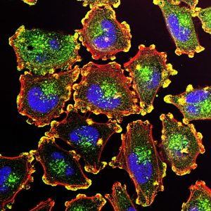
Melanoma cells stained to show cell nuclei (blue), podosomes (yellow), actin (red), and an actin regulator (green).

Melanoma is a common form of skin cancer that is most often successfully treated by removal of the cancerous cells. However,  malignant forms of melanoma can metastasize and become deadly. The significance of malignant melanoma is evident in its incidence – melanoma is the fifth most common cause of deaths from cancer in the US. Treating melanoma relies on using biopsy samples to determine the virulence of the cancer. However, the biopsy process is invasive and painful, and it can even be disfiguring.

Addressing this issue, Jesse Wilson, PhD, Assistant Professor in the Department of Electrical and Computer Engineering and in the School of Biomedical Engineering at Colorado State University (CSU), is developing a virtual biopsy for the disease. Funded by a Young Investigator Award from the Melanoma Research Alliance and a grant from the Colorado Clinical and Translational Sciences Institute, Dr. Wilson’s virtual biopsy uses multiphoton microscopy, which normally requires the use of a costly short-pulse laser for optimal visualization; his research seeks to obviate the need for laser, thus rendering the process more broadly available. 

Dr. Wilson intends to begin testing of his biopsy device on dogs from CSU’s veterinary school. Dogs also develop malignant melanoma, so the device will be used to gather data about each lesion that a dog develops. Once the imaging data are collected, the dogs will undergo normal biopsy and, if needed, treatment. In parallel, Dr. Wilson’s imaging algorithm will process the microscopy data collected prior to the biopsy, score it as malignant or not, and compare the predictions with the actual biopsy results to determine the new technique’s accuracy.

A Clue to Consciousness

Among the great mysteries in neuroscience is the nature of consciousness — that aspect of our psyche that allows us to observe that we are aware. We know that we have consciousness, but we aren’t sure why we do, nor do we fully understand the biological mechanisms that underlie consciousness.

A new study from scientists at Washington University in St. Louis might offer some clues, however. In the study, published in Neuron, the authors used a combination of calcium and hemoglobin imaging in mice to detect infra-slow spatiotemporal trajectories — essentially brain waves that are qualitatively different from other traditional electrical activity waves measured in the brain. These new waveforms were much slower than the activity of other traditional activity waves, and they traveled through different areas of the animals’ brains. The direction of the waves, moreover, changed on the basis of the level of consciousness of the mice.

Closer to home (and to humans), in a new article in Frontiers in Human Neuroscience, Hasan Ayaz, PhD, Associate Research Professor in the
School of Biomedical Engineering, Science and Health Systems at Drexel University, in collaboration with scientists from France, reports that the cognitive load of airline pilots differs significantly between pilots in the actual cockpit, compared to those using flight simulators. Dr. Ayaz and his colleagues used functional near infrared spectroscopy (fNIRS) for their comparisons. A future step for this research will be to integrate flight data recordings with the fNIRS data.

3D Printing Now Sweeter

Three-dimensional printing has become a vital resource in tissue engineering. However, the ability of commercial 3D printing technology to produce water-soluble glass — a key compound used in many tissue engineering processes — has been elusive because of the specific properties of the carbohydrates used to create this glass, which do not work with the technology used in available 3D printers.

However, this issue could be closer to a solution. In a new article in Additive Manufacturing, a team of scientists led by Rohit Bhargava, PhD, Founder Professor of Engineering in the Department of Bioengineering at the University of Illinois in Urbana-Champaign, reports that they have solved some of these problems. Using isomalt, a type of sugar alcohol, for their experiments, the authors were able to determine the characteristics inherent in the material necessary for 3D printing, as well as modeling the type of machinery necessary to use isomalt in a 3D printing process. Work on creating the 3D printing model recently published is still under way, but video of a bridge model has been published online here.

Seeing Like a Bat

Earlier this month, CLEO (the Conference on Lasers and Electro-Optics) held its annual meeting in San Jose, with a bioengineering contingent out in full force. Nader Engheta, PhD, the H. Nedwill Ramsey Professor with appointments in the Departments of Bioengineering, Electrical and Systems Engineering, and Materials Science and Engineering, was there and gave an interview with Optics & Photonics News. In the interview, Dr. Engheta discusses, among other things, bioinspired polarization — a developing field that seeks to enable people to see polarized light, which is visible to some animals, such as bats, but not to the human eye.

People and Places

Elon University in North Carolina will expand its current offerings in engineering in the coming year. In addition to a dual-degree program, Elon will offer for the first time an undergraduate degree program in engineering with an available concentration in biomedical engineering.  Sirena Hargrove-Leak, PhD, has been named director of the new program.

APOC Ghana 2018: Day 5

By Yasmina Al Ghadban, Bioengineering ’20; and Rebecca Zappala, Bioengineering ‘21

Ghana 2018
From left to right: KNUST second-year medical student Muti Agyekum, Penn students Sheldon Amoo-Mitchual, Amber Figueroa, Rebecca Zappala, and Ethan Zhao.

This morning, after a breakfast of eggs, sausage, and toast, we headed to the Maternal and Child Welfare Center (at the Komfo Anokye teaching hospital) to visit the malnutrition ward. We learned about two types of severe acute malnutrition: marasmus, which is characterized by severe wasting due to starvation and a lack of both protein and energy nutrients; and kwashiorkor, which is characterized by swelling of the belly, cheeks, and limbs due to a lack of protein in the diet. We also learned about the process of treatment starting from diagnosis by measuring the mid-upper arm circumference (MUAC), as well as the weight. After the specific case of malnutrition has been diagnosed, healthcare providers look for underlying conditions, such as infection, anemia, HIV, or malaria, by running lab tests. For treatment, depending on the severity of the case and the age of the child, they administer F75 or F100, which are different kinds of ready-to-use therapeutic agents. After learning about the third case that we will be working on, we went to the nutrition center where families are sent for outpatient care and follow-up. This is also the first point of contact between the family and the healthcare system; if the case is severe, the patient is referred to the malnutrition ward that we first visited. At first, we had questions about how the follow-up process works. We discovered that mothers are supposed to come back weekly to collect more food and check the weight and MUAC of their children to track their progress. In cases in which mothers do not come back for their follow-up, they are called and sometimes even visited at their homes by nurses.

Although we had visited this same ward yesterday, it is still so hard to look at the children there. The clinic seems to be doing everything they can; however, it is difficult to ignore that there is a clear lack of resources, funds, and accessibility. For example, the malnutrition center had to move from being a 2-room clinic to a 1-room clinic due to a lack of funds. This resulted in having the waiting area, the food distribution, and the assessment of the child in the same confined space, which then limits the number of patients they can care for at once.

Ghana 2018
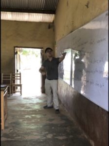
Penn student Ethan Zhao teaches a grade 8 class at Kokoben Municipal Assembly School about the importance of a balanced diet and its components.

Later in the afternoon, after a drive through the market and a stop to get coconuts, we headed to Kokoben Municipal Assembly school (kindergarten through grade 9) for service. We were divided into pairs, which each taught a class about something the grade was currently working on or wanted to learn. The experiences ranged from singing with 5-year-olds and struggling to communicate in English, as most children only know Twi, to teaching about heart diseases and the circulatory system. There was a lot of shock at first since it was not easy to stand in front of 40 students and teach an unprepared lesson. Overall, the kids seemed very excited and fascinated by our presence. Although we were glad we were able to spend some time with them and share an (infinitely) small part of knowledge, we were shocked by their fascination and overwhelming joy to see us.

As on our fourth day in Ghana, we feel like we have learned so much — both about the healthcare system and the culture, and we look forward to continuing to learn and grow tomorrow.

APOC Ghana 2018: Day 4

By Xuanjie (Lucas) Gong, Biotechnology MS ’19; Shihan Dong, Biotechnology MS ’19; and Princess Aghayere, Health & Societies ‘19

Ghana 2018
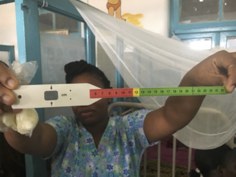
MUAC tape

Today, we all went to the Maternal and Child Health Hospital in Rural Kumasi. The main purpose of the visit was to observe malnutrition cases in children and to interact with doctors and nurses who treat children suffering from malnutrition.

Ghana 2018
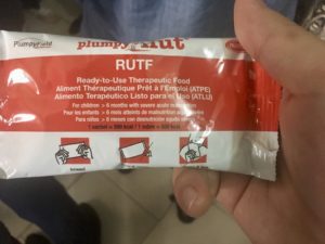
RUTF

While visiting the clinic, the nutrition group asked the questions that we prepared yesterday. It turns out that the situations here match up with our research. Doctors and nurses use WHO standards to determine the nutrition status of the kids. Also, they use MUAC tape to determine the severe acute malnutrition. Children who have MUAC less than 11.5 cm will be sorted into a severe acute malnutrition group.

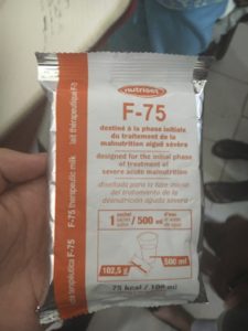
As for solutions or treatments, they do know about and have RUTF (Ready-to-Use Therapeutic Food). We also learned that they have therapeutic milk, F-75 and F-100, to treat malnourished children in different phases. They have F-75 to use at the starting phase of treatment. If F-75 helps to stabilize the children, they move onto F-100, and they use diluted F-100 for children under 6 months.

We had 2 cases that we mainly focused on. The first case was of a 3-year-old girl who suffered from Kwashiorkor, Marasmus, and Marasmic Kwashiorkor. All 3 diseases are signs of severe acute malnutrition. She had been there for 3 weeks for treatment, and her condition was not improving. The doctors attributed her declining condition to poverty and the mother’s psychiatric problems. The patient’s mother has already lost two children to the same condition. The doctors describe the girl’s status as unstable because she often vomits and isn’t gaining weight. The second patient is a child exposed to HIV as a result of her mother being positive for the disease. Because she is only 3 months old, blood cannot be drawn, and testing cannot be done to ensure her HIV status.

Ghana 2018
F-75 therapeutic milk

In the evening, we went to a church to give a presentation to local women with little to no education. We started by asking them what they eat everyday, and luckily we received a lot of responses. From their responses, we could clearly see that their diet lacked components of vegetables and fruits. Then, we delivered a brief speech containing basic nutrition knowledge, mainly based on the six essential nutrients. We explained the function of these nutrients and some local sources to obtain them. Surprisingly, one of the female audience members said that this was her first time hearing about vitamins and minerals.

During the question and answer segment of our presentation, some women asked about the different types of sources for minerals and vitamins. In particular, one woman asked about the foods that she could eat to help with her hypertension. To our surprise, another women asked whether her intake of fruits was excessive. This question made us think about people’s awareness of obesity and other diseases related to overeating. At the end of the presentation, the audience was happy about what we presented today and looked forward to learning more on our next visit.

APOC Ghana 2018: Day 3

By Xuanjie (Lucas) Gong, Biotechnology MS ’19; Shihan Dong, Biotechnology MS ’19; and Princess Aghayere, Health & Societies ‘19

This morning, we had our official kickoff in the morning. It was a great meeting with Dr. Ellis, the Dean of School of Public Health. In the afternoon, professors from different schools delivered three lectures about the state of mental health, maternal and child nutrition, and health systems in Ghana.

Ghana 2018The mental health lecture, given by Dr. Emma Adjaottor, was impressive and surprising. We are so lucky that we met one of the psychiatric physicians among a hundred across the country. The doctor frankly introduced mental health development in Ghana. Though they have lagged behind, they have made a lot of progress, with the number of psychiatrists in Ghana  recently increasing from single digits to the double digits. Dr. Adjattor emphasized that the epidemiology of mental health in Ghana is nearly identical to the rest of the world, particularly in terms of incidence and prevalence, even though it is severely under-reported. The doctor explained that, although the epidemiology is the same, the terms used to describe these issues are often different, relying on more local ideas, such as spirits and witchcraft.

The maternal and child nutrition lecture, given by Dr. Samuel Newton, was even more astonishing. Kangaroo mother care (KMC), in which a prematurely born infant is given constant skin-to-skin contact with the mother, is undergoing study in Ghana, and it has shown promising outcomes. Even with limited studies, KMC has been found to greatly increase the likelihood of survival even when using “surrogates,” such as grandmothers and even fathers. The professor also introduced another study that found that the application of oxytocin during the third stage of labor in mothers at risk of postpartum hemorrhage (PPH) greatly decreased the likelihood of PPH.

Finally, Professor Ellis Owusu-Dabo gave an extensive lecture covering the health systems of Ghana, which are logically and hierarchically managed, with administrations at the national, local, district, sub-district, and community levels. We also learned about the insurance program in Ghana. For formally employed workers, national insurance participation is mandatory, as it is funded by a mandatory tax on their income, like in many socialist countries. However, insurance coverage is a more complicated matter in Ghana, in which a very large proportion of the population is not formally employed and instead earn its living through trading and cash-paying jobs. For these people, other than the free public health services provided by the government such as malaria, tuberculosis, and HIV care, they must pay out of pocket for many other services.

Tomorrow will be the very first clinic visit, where we can observe children suffering from severe acute malnutrition. So the three of us in the nutrition group, Princess, Shihan, and Xuanjie, gathered together after dinner to discuss what we would ask the doctors and nurses during the visit. We agreed that, at first, we should ask whether we can take pictures to record. As for nutrition, we will ask how healthcare workers there define malnutrition.

To our knowledge, malnutrition standards are normally based on BMI, which is a composition of weight, height, and age-related data. We want to know whether Ghanaian healthcare professions use the same standards here, as well as how the local standards differ from the established WHO standards. Furthermore, we will ask for access to their local data sets. In addition to the results of malnutrition, we also want to know what the major causes of malnutrition are. We assumed that the principal cause is poverty, and based on this assumption, we want to ask them whether there are any welfare systems or NGOs helping to resolve the problem. We hope to find out what the clinics’ initial steps are to resolve malnutrition. Our last question involves how macronutrients are measured.

Tomorrow evening, we will give an open presentation in a local church to female market workers. We first planned to deliver the presentation in a discussion form, but considering the language barrier and other factors, we decided to give a speech, but we will ask simple questions. We thought that the scope of our research was not appropriate for local life in Ghana because much of the research was done in the U.S.; thus, it was necessary to get the input of local people. By asking them questions about what they eat on a daily basis, we want to make our research as appropriate as possible. In relation to our presentation, we will briefly introduce what nutrition is and explain the 6 essential nutrients through definitions and explanation. For example, when introducing carbohydrates, we will refer to fufu or banku, which are local main dishes basically consisting of starch. Also, we came up with an educational idea for the future. If we have a projector, we could possibly show some dishes to let the audience choose which is more nutritious, to provide general idea of a nutritious diet.
APOC Ghana 2018: Day 2

By Sandy Tang, Bioengineering ’19; Eliza Culp, Fine Arts ’20; and Jessica Fan, Biotechnology MS ‘18

Ghana 2018
The mouthwatering jollof and fried tilapia we ordered at the swimming pool

This morning, we had a chance to sleep in a bit and recover from our busy travel day. We had a delicious breakfast that consisted of porridge with sugar cubes, sausage, hard boiled eggs, a locally specialized donut made of beans, watermelon, butter bread, and a variety of hot drinks: yum. After our breakfast, a group of us went on a walk around campus, and another group went to a church service at First Love Church.

The church service started with praise worship, followed by numerous student performances. There was a dance group, a drama group, a praise group, and a traditional Twi praise group, and the congregation members were bursting with energy. The pastor then gave her sermon and afterward greeted the congregation. We made a few friends who happened to be students at the university we are staying at: KNUST.

For the other group, we explored KNUST’s expansive campus. We started to wander, and only when we noticed excessive nature did we realize we were no longer on campus. Once back on track, we saw some lizards, baby goats, and plenty of flora and returned to the residential area of campus. We accidentally entered a boy’s dorm but caught a glimpse of what it would be like to be a student at KNUST. Inside the dorm, a pastor was giving a fiery sermon, and many students were hand washing laundry in the courtyard. After leaving the dorms a few of us bought some amazing meat pies, for only 1 Cedi (about a quarter)!

Around 1 p.m., we all got into our bathing suits to head to the pool! The ride was short, and we were excited to have some relief from the heat. We ordered some food before entering the water; there was a choice of jollof or fried rice with either fish or chicken. After ordering our food, we applied sunscreen and dove into the water. There were many other people in the pool already, so we made some friends with the swimmers our age, many of whom were instructors. Shortly after being in the water, Genevieve and Estabelle, two young girls, joined us in the pool, and we played with them and carried them around in the pool. It was such a great time.

We stayed until the pool closed at 6 p.m. and then dried off and took the bus back home. A few of us bought sweet treats from the little shop in our graduate student hostel, Tek Credit. Then, dinner was brought to us by Nana Yaa: a meal of yams, spinach fish stew, rice, fried fish, beef and water melon. We had a brief Twi lesson from Nana Yaa on basic greetings. Our night ended with a quick debrief from Dr. Wattenbarger about our upcoming week.

Excited for the days that lie ahead!

APOC Ghana 2018: Day 1

By Ethan Zhao, Bioengineering ’19 and Sheldon Amoo-Mitchual, Biological Basis of Behavior ‘20

Ghana 2018
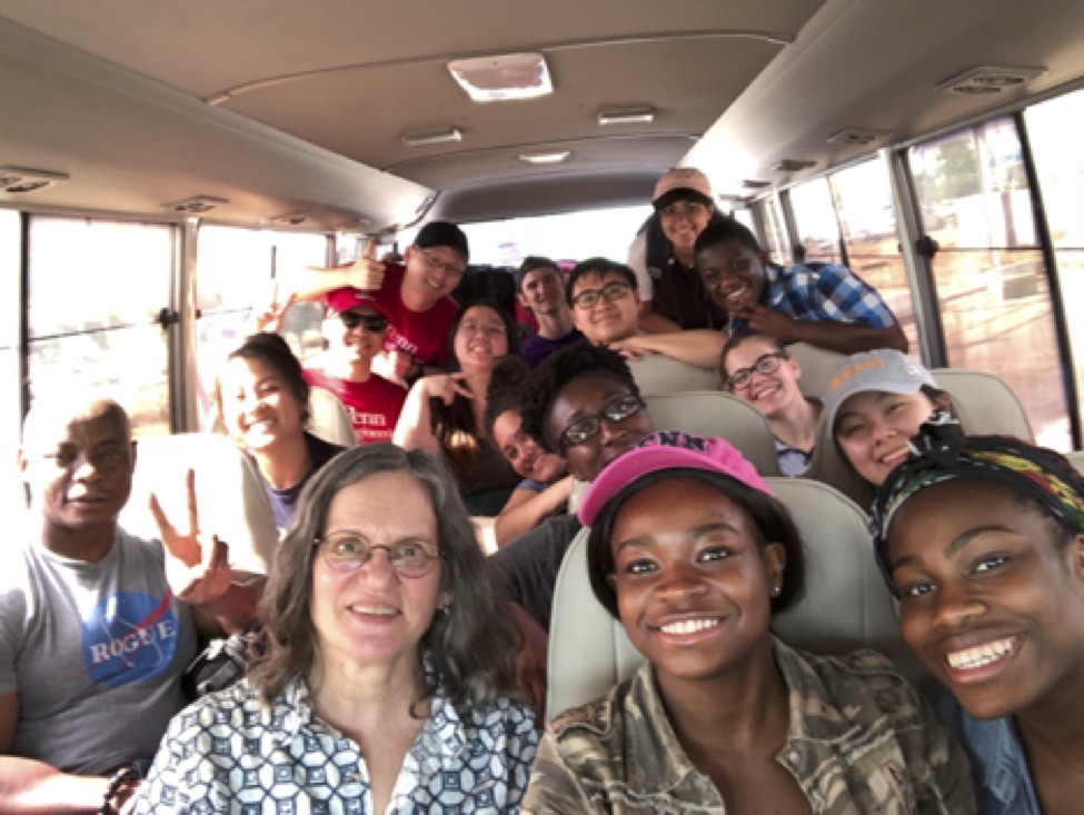
The APOC team! From left to right, then bottom to top: Dr. Ocek Eke, Dr. Miriam Wattenbarger, Summer Kollie, Princess Aghayere, Eliza Culp, Bosede Ajiboye, Jessica Fan, Amber Figueroa, Xuanjie (Lucas) Gong, Sandy Tang, Ethan Zhao, Sheldon Amoo-Mitchual, Shihan Dong, Adam Yablonski, Yasmina Al Ghadban

Like last year, a group of Penn Engineering undergrads enrolled in ENGR566 – Appropriate Point of Care Diagnostics, a large number of them Bioengineering majors, have gone to Ghana to get some hand-on experience. They’re blogging their experiences daily.

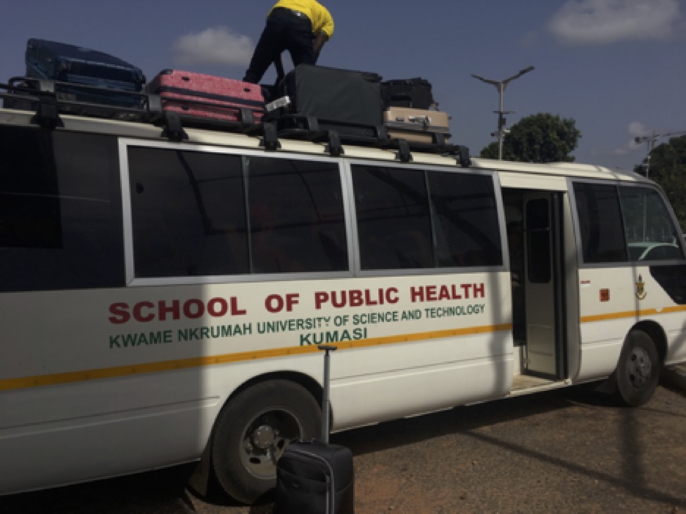
Today we touched down in Accra bright and early at 8 a.m. We went through immigration and baggage claim without a hitch, and we met up with Nana Ya Awua-Boateng, a PhD student at the Kwame Nkrumah University of Science and Technology (KNUST), who helped us with arrangements as a translator. We loaded up the bus with luggage (both inside and out) and set off for the 5-hour bus ride to Kumasi. Along the way, we slept (a lot), given that we touched down at the equivalent of 4 a.m. EDT. We stopped in the middle for lunch and our first taste of Ghanaian jollof rice and grilled tilapia, which were incredible.

Afterward, we finished our journey to Kumasi and arrived at KNUST, where we unpacked our belongings in the hostel. We also met Salim, a medical student, and Nana Ya’s children, Nana and Genevieve. Soon after, we all took a quick trip down to Big Chinese Restaurant (its literal name) to taste Ghana’s version of Chinese food. We then went back to campus, where we finally went to bed after a long day’s journey.

Ghana 2018
Loading up the bus for our drive to Kumasi at the Accra airport! The bus was so full we had to strap luggage to the to the roof of the bus.

Winkelstein Named to World Council of Biomechanics

Winkelstein
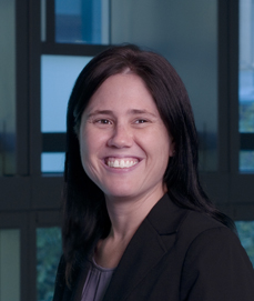
Beth Winkelstein, PhD

The University of Pennsylvania Department of Bioengineering is proud to announce that our senior faculty member Beth Winkelstein, PhD, who is also Vice Provost for Education and the newly named Eduardo D. Glandt President’s Distinguished Professor, was elected as a councilor to the World Council of Biomechanics (WCB).  In the words of Dominique Barthes-Biesel, PhD, Chair of the WCB, and Roger Kamm, PhD, Chair of the Nominating Committee, Dr. Winkelstein’s election comes in recognition of her “distinguished contributions to and leadership in the field of biomechanics at an international level.” The appointment will be recognized at the WCB General Assembly, to be held at the 8th World Congress of Biomechanics in Dublin, Ireland on July 8.

Instituted in 1990, the WCB is an international academic and professional forum of engineers and scientists from five continents.  With her appointment, Dr. Winkelstein joins colleagues from MIT, Columbia, and Georgia Tech, among others. “I’m honored to be included as a representative among the impressive world leaders in biomechanics,” Dr. Winkelstein says, “and I look forward to helping shape the upcoming World Congresses and meetings.

Week in BioE (May 22, 2018)

Ultrasound Helmet Provides Perioperative Images

ultrasound helmet
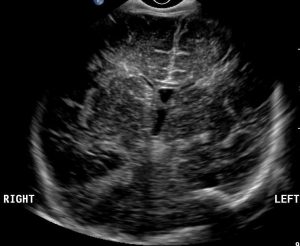
Normal ultrasound image of an infant’s brain

As we’ve mentioned here before, surgery on the brain is particularly difficult because of the limited visibility afforded to the surgical field and the complexity of the organ. Because the brain’s gray matter can be easily damaged, a false move by a surgeon can have a lifetime of consequences. Better visualization during surgery could go a long way toward preventing accidental damage by the surgeon and minimize the removal of healthy brain tissue during tumor removal. However, ultrasound imaging of the brain has remained difficult because of the tendency of ultrasound waves to bounce off the skull.

To help solve this problem, a biomedical engineer at Vanderbilt University  developed an ultrasound helmet to create perioperative ultrasound images of the brain. It could also provide a new variety of platform for brain-machine interfaces. According to Brett Byram, PhD, Assistant Professor of Biomedical Engineering at Vanderbilt, the helmet will eventually combine ultrasound with electroencephalography (EEG) to simultaneously visualize the brain and record its activity.  Dr. Byram used a machine learning-based technique called aperture domain model image reconstruction (ADMIRE) to overcome the technical obstacle of ultrasound waves transmitting through the skull. 

Although the initial thought of how to apply this technology was surgical, Dr. Bryram believes that the ability to detect blood flow to different parts of the brain in real time using ultrasound could facilitate the creation of technologies that would use this blood flow information, smoothed using ADMIRE, and EEG data to communicate with implants or robotic extensions to perform tasks.

A Roach Motel for Cancer

One key to curing cancer is preventing its spread, called metastasis. The mechanisms underlying metastasis are becoming clearer after years of research. Typically, the spread of cancer is the result of cancerous cells shed by a tumor affecting another organ after traveling via the bloodstream or lymphatic system. Unfortunately, sometimes this shedding is caused by the surgical procedure to remove the tumor. Therefore, preventing metastasis requires preventing these cells from circulating during and after the surgical procedure.

At the University of Texas at Arlington (UTA), Liping Tang, Ph.D., Professor of Biomedical Engineering at the University of Texas at Arlington, has patented what he calls a “roach motel” for cancer cells. Dr. Tang’s device, which is implanted under the skin, circulates cells of its own that attract circulating metastatic cells. The result of the device is the trapping of the cancer cells within the device and preventing them from traveling further. In vitro testing has been quite successful in a variety of cancers. Preclinical testing in animals will be the next step.

Injectable Alcohol Sensor Could Augment Treatment Programs

A few weeks ago, we detailed here how a scientist is developing DNA-based drug and alcohol screening tests. Recently a group of bioengineers at the University of California–San Diego (UCSD), led by Drew A. Hall, PhD, Assistant Professor of Electrical and Computer Engineering and an affiliate professor in the Department of Bioengineering at UCSD, has developed an injectable biosensor that can communicate blood alcohol levels to a wearable device. The sensor is a complementary metal–oxide semiconductor approximately 1 square millimeter in size and is designed for implantation under the skin surface. If in vivo testing proves successful, the system could be used as part of holistic approaches to preventing alcohol abuse among recovering alcoholics.

A Temperature-measuring Microscope

If you’ve used a microscope, then you’ve probably noticed that the samples viewed using microscopes are almost always on glass slides placed beneath the lens of the device. Now, in an article recently published in Nature Communications, an engineering team reports on their invention of a slide that can also measure temperature fluctuations in samples while maintaining microscopic imaging capability. Ruogang Zhao, PhD, assistant professor in the University at Buffalo Department of Biomedical Engineering, along with colleagues from our sister Departments of Electrical and Systems Engineering and Materials Science and Engineering here at Penn, coated a normal slide with 20-nanometer layers of gold activated by an external laser. Applications of the technology are numerous, and will be accelerated through mass production of slides, which the authors estimate would cost less than 10 cents each.

People and Places
Two large donations make our news this week. First, the University of Southern California received a $10 million gift from a retired ophthalmologist and his wife. The Dr. Allen and Charlotte Ginsburg Institute for Biomedical Therapeutics is being led by Mark S. Humayun, MD, PhD., Professor of Ophthalmology, Biomedical Engineering, and Cell and Neurobiology at USC. Across the country, the University of Maryland School of Medicine will establish the Robert E. Fischell Center for Biomedical Innovation with a $20 million gift from Robert Fischell, an inventor and holder of 200 patents. Distinguished University Professor and founding chair of the Fischell Department of Bioengineering William E. Bentley, PhD, will head the Fischell Center.

Also, it’s May, which means graduate news. Two special congratulations are in order. First, we congratulate Rowan University in New Jersey for graduating its first cohort of three newly minted PhDs in Biomedical Engineering. Also at the University of California at Davis, Tanishq Abraham will graduate next month with a Bachelor’s degree in Biomedical Engineering. In case that doesn’t sound like big news, bear in mind that Tanishq is only 14 years old. Tanisq will continue at Davis in studying in an MD/PhD program, which he hopes to finish before finishing his second decade of life.

Finally, we congratulate Brian Holland, MD, who has been named the new chief of pediatric cardiology at the University of Louisville. Dr. Holland is an alumnus of Penn Bioengineering, graduating summa cum laude in 1996.

You Do Belong in Science-stravaganza!

You Do Belong in Science

Sally and Kayla wrap up the You Do Belong in Science series with listener stories and lessons learned from this series. Listeners write in with stories about the importance of professors’ LGBTQ allyship and dealing with chronic illness in graduate school. Sally and Kayla reveal who does not belong in science (spoiler alert/content advisory: it’s sexual harassers). They also welcome allyship correspondent Jon Muncie to discuss actions everyone can take to prevent and respond to sexual harassment in the workplace, fairly judge peers’ research, and increase representation and promote the inclusion of people from underrepresented groups in STEM. He reminds Double Shelix that we need to get comfortable being uncomfortable when it comes to discussing and addressing these important issues facing our science workplaces.

Resources
* Resources for LGBTQ+ students, staff, faculty, and allies at Berkeley, at UCSF, and at U of Pennsylvania
* Proud and Prepared: A Guide for LGBT Students Navigating Graduate Training – resource from the American Psychological Association. Preview it here
* Dr. Kate Clancy’s congressional testimony video (starts ~41:30) and transcript
* Dr. Kate Clancy’s amazing podcast, Period Podcast
* Sexual harassment videos and NYT analysis: https://nyti.ms/2Gg4NHT
* Resources for dealing with sexual harassment: rainn.org/thatsharassment

Sally and Kayla thank the Berkeley Student Tech Fund, as well as Gustavo Villarreal @wikirascals for their graphics. Get your Double Shelix and You Do Belong in Science stickers at doubleshelix.com/stickers.

Share your thoughts on this episode — or your belonging story — on voicemail 415-895-0850 or email Double Shelix doubleshelixpodcast@gmail.com. Sally and Kayla are on Twitter @doubleshelixpod and coming soon to Instagram @doubleshelixpodcast — give them a follow!

Week in BioE (May 10, 2018)

Advances in Cancer Detection

glioblastoma
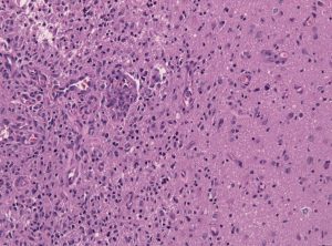
Tumor-brain-interface in a glioblastoma biopsy specimen.

Among the deadliest and most difficult to treat types of cancer is glioblastoma, an especially aggressive form of brain cancer. Widely available imaging techniques can diagnose the tumor, but often the diagnosis is too late to treat the cancer effectively. Although blood-based cancer biomarkers can provide for earlier detection of cancer, these markers face the difficult task of crossing the blood-brain barrier (BBB), which prevents all but the tiniest molecules from moving from the brain to the bloodstream.

A study recently published in Scientific Reports, coauthored by Hong Chen, PhD, Assistant Professor of Biomedical Engineering at Washington University in St. Louis (WUSTL), reports of successful deployment of a strategy consisting of focused ultrasound (FUS), enhanced green fluorescent protein (eGFP), and systemically injected microbubbles to see if the BBB could be opened temporarily to allow biomarkers to pass from the brain into the bloodstream. The authors used eGFP-activated mouse models of glioblastoma, injecting the microbubbles into the mice and then exposing the mice to varying acoustic pressures of FUS. They found that circulating blood levels of eGFP were several thousand times higher in the FUS-treated mice compared to non-treated mice, which would significantly facilitate the detection of the marker in blood tests.

The method has some way to go before it can be used in humans. For one thing, the pressures used in the Scientific Reports study would damage blood vessels, so it must be determined whether lower pressures would still provide detectable transmission of proteins across the BBB. In addition, the authors must exclude the possibility of FUS unexpectedly enhancing tumor growth.

In other body areas, with easier access from tissue to the bloodstream, engineers have developed a disease-screening pill that, when ingested and activated by infrared light, can indicate tumor locations on optical tomography. The scientists, led by Greg M. Thurber, PhD, Assistant Professor of Biomedical and Chemical Engineering at the University of Michigan, reported their findings in Molecular Pharmaceutics.

The authors of the study used negatively charged sulfate groups to facilitate absorption by the digestive system of molecular imaging agents. They tested a pill consisting of a combination of these agents and found that their model tumors were visible. The next steps will include optimizing the imaging agent dosage loaded into the pill to optimize visibility. The authors believe their approach could eventually replace uncomfortable procedures like mammograms and invasive diagnostic procedures.

Liquid Assembly Line to Produce Drug Microparticles

Pharmaceuticals owe their effects mostly to their chemical composition, but the packaging of these drugs into must be done precisely. Many drugs are encapsulated in solid microparticles, and engineering consistent size and drug loading in these particles is key. However, common drug manufacturing techniques, such as spray drying and ball milling, produce uneven results. 

University of Pennsylvania engineers developed a microfluidic system in which more than ten thousand of these devices run in parallel, all on a silicon-and-glass chip that can fit into a shirt pocket, to produce a paradigm shift in microparticle manufacturing. The team, led by David Issadore, Assistant Professor in the Department of Bioengineering, outlined the design of their system in the journal Nature Communications.

The Penn team first tested their system by making simple oil-in-water droplets, at a rate of more than 1 trillion droplets per hour. Using materials common to current drug manufacturing processes, they manufactured polycapralactone  microparticles at a rate of ‘only’ 328 billion particles per hour. Further testing backed by pharma company GlaxoSmithKline will follow.

Preventing Fungal Infections of Dental Prostheses

Dental prostheses are medical devices that many people require, particularly as they age. One of the chief complications with prostheses is fungal infections, with an alarming rate of two-thirds among people wearing dentures. These infections can cause a variety of problems, spreading to other parts of the digestive system and affecting nutrition and overall well-being. Fungal infections can be controlled in part by mouthwashes, microwave treatments, and light therapies, but none of them have high efficacy.

To address this issue, Praveen Arany, DDS, PhD, Assistant Professor, Department of Oral Biology and Biomedical Engineering at SUNY Buffalo, combined 3D printing technology and polycaprolactone microspheres containing amphotericin-B, an antifungal agent. Initial fabrication of the prostheses is described in an article in Materials Today Communications, along with successful in vitro testing with fungal biofilm. If further testing proves effective, these prostheses could be used in dental patients in whom the current treatments are either ineffective or contraindicated.

People and Places

West Virginia University has announced that it will launch Master’s and doctoral programs in Biomedical Engineering. The programs will begin enrolling students in the fall. The graduate tracks augment a Bachelor’s degree program begun in 2014.