
Week in BioE (July 27, 2017)

The Brain in Focus

BrainAt Caltech, scientists are exploiting the information generated by body movements, determining how the brain codes these movements in the anterior intraparietal cortex — a part of the brain beneath the top of the skull. In a paper published in Neuron, Richard A. Andersen, James G. Boswell Professor of Neuroscience at Caltech, and his team tested how this region coded body side, body part, and cognitive strategy, i.e., intention to move vs. actual movement. They were able determine specific neuron groups activated by different movements. With this knowledge, more effective prosthetics for people experiencing limb paralysis or other kinds of neurodegenerative conditions could benefit enormously.

Elsewhere in brain science, findings of chronic traumatic encephalopathy in football players have raised significant controversy. Seeking to better understand head impact exposure in young football players, scientists from Wake Forest University led by biomedical engineer Joel D. Stitzel, fitted athletes with telemetric devices and collected four years of data and more than 40,000 impacts. They report in the Journal of Neurotrauma that, while all players experienced more high magnitude impacts during games compared to practices, younger football players experienced a greater number of such impacts during practices than the other groups, and older players experienced a greater number during actual games. The authors believe their data could contribute to better decision-making in the prevention of football-related head injuries.

Up in Canada, a pair of McGill University researchers in the Department of Neurology and Neurosurgery — Professor Christopher Pack and Dave Liu, a grad student in Dr. Pack’s lab — found that neuroplasticity might apply to more parts of the brain than previously thought. They report in Neuron that the middle temporal area of the brain, which contributes to motion discrimination and can be inactivated by certain drugs, could become relatively impervious to such inactivation if pretrained. Their findings could have impacts on both prevention of and cures for certain types of brain injury. 

The Virtues of Shellfish

If you’ve ever had a diagnostic test performed at the doctor’s office, you’ve had your specimen submitted to bioassay, a test in which living cells or tissue is used to test the sampled material. University of Washington bioengineer Xiaohu Gao and his colleagues used polydopamine, an enzyme occurring in shellfish, to increase the sensitivity of bioassays by orders of magnitude. As reported in Nature Biomedical Engineering, they tested the technology, called enzyme-accelerated signal enhancement (EASE), in HIV detection, finding that it was able to help bioassays identify the virus in tiny amounts. This advance could lead to earlier diagnosis of HIV, as well as other conditions.

Mussels are also contributing to the development of new bioadhesives. Julie Liu, associate professor of chemical engineering at Purdue, modeled an elastin-like polypeptide after a substance produced naturally by mussels, reporting her findings in Biomaterials. With slight materials, Dr. Liu and her colleagues produced a biomaterial with moderate adhesive strength that demonstrated the greatest strength yet among these materials when tested under water. The authors hope to develop a “smart” underwater adhesive for medical and other applications.

Science in Motion

Discussions of alternative forms of energy have focused on the big picture, such as alleviating our dependence on fossil fuels with renewable forms of energy, like the sun and wind. On a much smaller level, however, engineers are finding smaller energy sources — specifically people.

Reporting in ACS Energy Letters, a research team led by Vanderbilt’s Cary Pint, assistant professor in the Department of Mechanical Engineering and head of Vanderbilt’s Nanomaterials and Energy Devices Laboratory Nanomaterials and Energy Devices Laboratory, designed a battery in the form of an ultrathin black phosphorous device that can generate electricity as it is bent. Dr. Pint describes the device in a video here. Although it can’t yet power an iPhone, the possibility isn’t far away.

Moving Up

Two BE/BME departments have named new chairs. At the University of Utah, David Grainger, who previously chaired the Department of Pharmaceutics and Pharmaceutical Chemistry, will become chair of the Department of Bioengineering. Closer to home, Michael I. Miller became the new chair of the Department of Biomedical Engineering on July 1. Congratulations to them both!

This Week in BioE (July 6, 2017)

Bioengineering of Genes and DNA

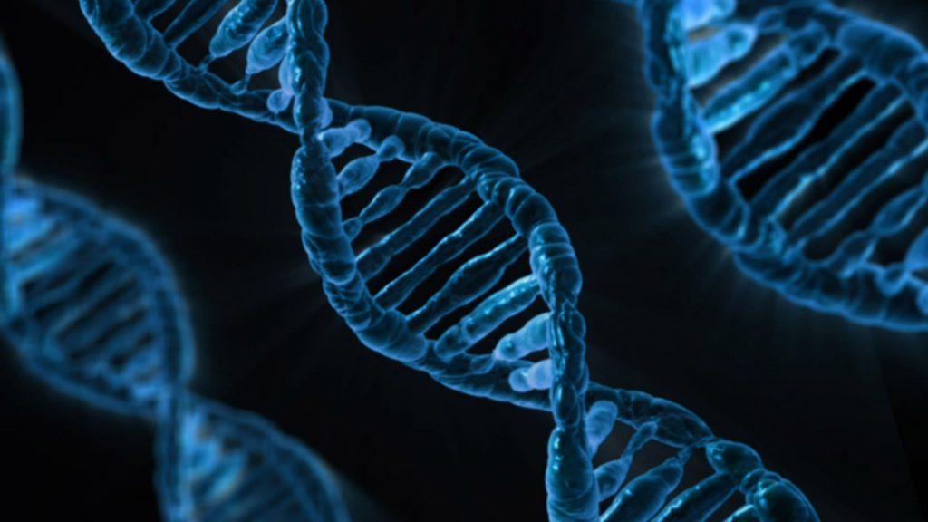
Since Watson and Crick published their initial studies detailing the double helix structure of DNA in the early 1960s, what we know about genetics and the nucleic acids underlying them has grown enormously. Consequently, what bioengineering can do with DNA and genes continually expands.

One fascinating bioengineering field that emerged in the past decade was DNA origami, which uses the well-established binding across DNA elements to create three-dimensional structures out of linear DNA sequences. Recent work has utilized this feature of DNA construction to make machines, rather than just parts, out of DNA.

Yonggang Ke, Ph.D., of Georgia Tech/Emory’s Department of Biomedical Engineering, constructed machines made of DNA that consist of arrays of units that can “switch” between “settings” by changing shape. A change in shape of one unit of an array can cause the other units in the array to shift; these changes are stimulated by inserting a previously deleted strand of DNA into the array. Although it has been known for some time that DNA could be used to store and transmit information, Dr. Ke’s research team proved for the first time that these arrays could be shaped physically into machines in the shapes of rectangles and tubes.

genes dna
DNA under a microscope

While we learn more about how to make DNA-based devices, we are also creating new technologies to manipulate DNA more rapidly.  Scientists at Rutgers and Harvard developed a process whereby thousands of genes could be cloned at one time to create enormous libraries of proteins. To achieve this goal, the authors used a technology called LASSO (long-adapter single-strand oligonucleotide) probes, which they have already used to clone a library using a human microbiome sample.

Instead of the traditional process of cloning one gene at a time, the team led by Professor Biju Parekkadan, Ph.D. at Rutgers, invented a technology to clone hundred of genes simultaneously. These cloned DNA segments are much longer than the length of DNA cloned with standard techniques, allowing us to test the functional significance of these much longer DNA segments.  The technology could impact a number of scientific fields because we will finally learn how long stretches of protein function — some parts may degrade other proteins, while other parts will interact and modify other proteins (e.g., phosphorylation, a key process in epigenetics). These new discoveries can be key for discovering new ways to engineer proteins and to manufacture new drugs that mimic the function of nature’s DNA products.

Using Sweat as a Biosensor

While the field learns more about the molecular-level control of DNA, we are also taking advantage of new micro- and nanoscale manufacturing processes to capture diagnostic information from easily accessible body fluids. Many clinical diagnostics use chemical measurements from blood to diagnose a disease or to take corrective action. This is not an ideal procedure because it requires either the collection of blood at a laboratory or the repeated collection of small blood volumes through a pinprick.  Either one hurts.

Bioengineers at the University of Texas at Dallas developed a wearable diagnostic device to detect cortisol, glucose, and IL-6 in body sweat, eliminating any painful needle sticks.  Its transmissions vary, but if optimized, the device could replace the painful and inconvenient practice of sticking one’s finger to obtain a drop of blood for glucose testing, which many patients with diabetes must do several times per day. Although insulin pumps have been available for some time, these are invasive devices that must be worn at all times.

This Week in BioE (June 22, 2017)

Diversifying the Field

One of the ongoing issues in STEM (science, technology, engineering, and medicine) fields is a lack of diversity among students and faculty. Bioengineering stands out among other engineering fields because it enjoys terrific gender diversity. For example, about half of Penn Bioengineers are women, a feature of our class that goes back decades.

diversifyingHowever, diversity extends well beyond gender. For example, the National Research Mentoring Network (NRMN) has been working to increase diversity, including among students with disabilities. A consortium of people and groups providing mentors for science students, the MRMN recently highlighted the American Association for the Advancement of Science’s (AAAS) Entry Point! program, which focuses on helping students with physical disabilities. Mentoring, it turns out is a big part of helping these students succeed.
Another recent development that should help to increase diversity in the field is the awarding of a $1 million grant from the National Science Foundation’s Directorate of Engineering to the University of Wisconsin, Madison, and the College of Menominee Nation (CMN), a native American college in Wisconsin, to collaborate in engineering research and education. The new grant builds on a program begun in 2010 between the colleges to build labs and facilitate the transfer of pre-engineering students from CMN to UWM.

Brain Science Developments

Speaking of education, three recent news stories discuss how we might be able to expedite the learning process, increase intelligence, and reward ourselves when we create art. In one of the stories, a company called Kernel is investing $100 million in research at the University of Southern California to determine whether using brain implants, which have been helpful in some patients with epilepsy, can be used to increase or recover memory. If successful, this may bridge one critical treatment gap in neurology. About one out of every three people with epilepsy don’t respond to drug treatment.

In the second story, scientists at the University of Texas at Dallas were awarded a $5.8 million contract from DARPA to investigate the role of vagus nerve stimulation in accelerated learning of foreign languages. Stimulating the peripheral nervous system to activate and train areas of the brain is one more example that our nervous system is connected in ways that we do not yet understand completely. The Department of Defense hopes to use the technology to more quickly train intelligence operatives and code breakers.

Finally, in a third story involving the brain, a professor at Drexel University used functional near-infrared spectroscopy to determine which parts of the brain were activated while participants were making art. Dr. Girija Kaimal’s team found that creative endeavors activate the brain’s rewards pathway, as well as elevating the participants’ self-opinion. So making art always made people feel good about themselves; now we know more of the reasons why.

Phytoplankton Research Earns Award

phytoplankton
Phytoplankton

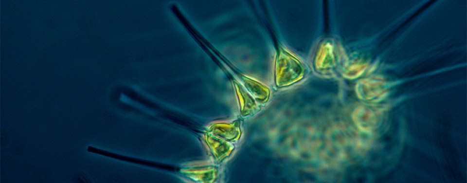
The Scripps Institution of Oceanography at the University of California, San Diego, announced last week that one of its faculty members, Andrew Barton, PhD, received a Simons Foundation Early Career Award to study phytoplankton — a type of algae that requires sunlight to survive and that serves as the basis for much of the marine food chain.

Dr. Barton’s research will use the Scripps Plankton Camera System, which provides real-time photographic images to monitor these phytoplankton. While not exactly offering the excitement or cuteness factor of the Golden Retriever Puppy Cam, this sort of technology is incredibly important to better understanding certain aspects of marine biology.

“This is an interesting project that brings cutting edge image-processing technology to the natural habitat to study complex organismal dynamics in the real-world setting,” says Brian Chow, PhD, assistant professor of bioengineering at the University of Pennsylvania. “Establishing the critical interplay between an organism’s form and function and the forces of its local and global environments are important problems in physical biology in general. Diatoms have long been studied by bioengineers interested in self-assembly, programmed assembly, biomineralization, and biomimicry, so the work may lead to some novel insights for our field.”

Congratulations to Dr. Barton on receiving this prestigious award.

Tissue Engineering Makes Spinach Leaf Beat Like a Heart

tissue
Bestill my beating spinach leaf!

One of the more interesting tissue engineering stories to emerge this past month was the successful finding of a team at Worcester Polytechnic Institute (WPI), which used the veins in spinach leaves as a scaffold that was then recellularized with stem cells that produce heart muscle cells. After three weeks, the transplanted cells showed the ability to contract like the heart does when it beats.

“Proper vascularization of artificial living tissues has been one of the most critical challenges of tissue engineering for decades. This is particularly problematic when the size of the engineered tissue increases.,” said Dongeun (Dan) Huh, PhD, Wilf Family Term Assistant Professor in the Department of Bioengineering at the University of Pennsylvania “This work takes an unusual yet ingenious approach to solving this long-standing problem.”

Below you can watch a short video of some of the investigators on the study talking about it.