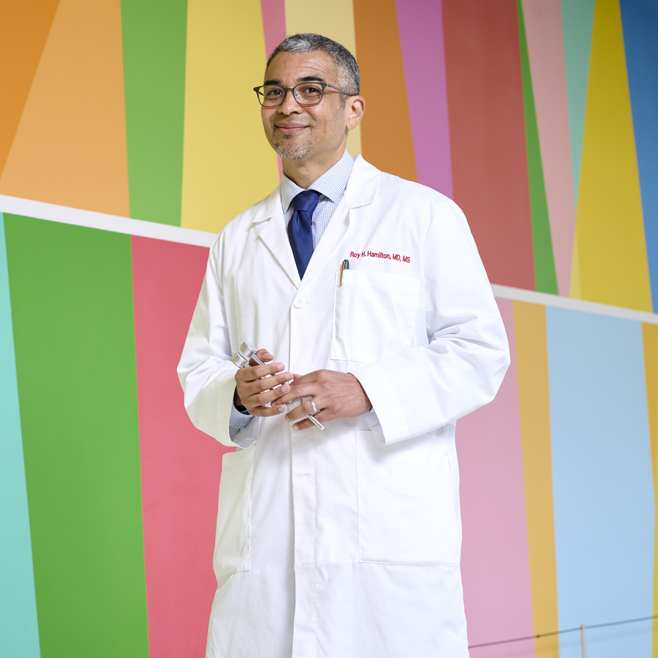
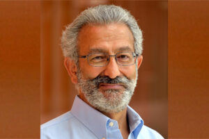
A multiracial Black and Asian self-described secular humanist, who was raised as one of Jehovah’s Witnesses and is now in an interracial, interfaith marriage, walked into a Passover seder.
It’s not the setup for a groaner of a joke, or an epic fail of an evening. Rather, as Roy H. Hamilton, MD, tells it, this was his experience this spring as his Jewish in-laws—the family he has loved as his own for over two decades—came together to commemorate the universal human themes of freedom and deliverance from oppression reflected in the Passover narrative. Though he does this every year, this year he had some trepidation. In a time marked by tragic conflict and with tensions both abroad and at home, it seemed like having a frank discussion of these themes might invite acrimony. But what emerged instead was a profound opportunity to listen, to appreciate each other’s perspective, and to “exercise empathy for trauma that’s happening to everyone.”
It was a bit of a revelation for Hamilton, Penn Medicine’s new vice dean for Inclusion, Diversity, and Equity. “In the moment that you would have thought would be the worst to open up certain topics, we all ended up having a great dialogue across differences,” he said. Why? “Because we all felt connected enough to give each other respect, compassion, and grace, even when our thoughts and opinions differed. It made me think about how we can further cultivate a culture of empathy at Penn too.”
Today, as Hamilton begins his third decade on the faculty at Penn’s Perelman School of Medicine, he is devoted to making academia a safe, supportive space for students and colleagues alike. He serves as a professor of Neurology, with secondary appointments in Psychiatry and Physical Medicine and Rehabilitation. Hamilton is also director of both the Laboratory for Cognition and Neural Stimulation; and the Penn Brain Science, Translation and Modulation (BrainSTIM) Center. Previously, he was the Perelman School of Medicine’s assistant dean for Cultural Affairs and Diversity for almost a decade, and launched similar efforts in his field, serving as Penn Neurology’s vice chair for Diversity and Inclusion from 2017 until his recent elevation to the role for Penn Medicine as a whole.
Given his own diverse background and personal life, Hamilton wants everyone—trainees, faculty, patients—to feel valued and included. “I touch enough spaces in my personal life that when groups are being clearly systematically disadvantaged, it often feels like it’s touching on some piece of my own identity,” he said, in discussing his background and hope for his new leadership position. “I bring a lot of myself to this role.”
In a recent discussion, Hamilton shared perspectives on why supporting inclusion, diversity, and equity matters—particularly for an institution training future doctors—and what Penn Medicine is doing in this sphere.
Let’s say you typically eat eggs for breakfast but were running late and ate cereal. As you crunched on a spoonful of Raisin Bran, other contextual similarities remained: You ate at the same table, at the same time, preparing to go to the same job. When someone asks later what you had for breakfast, you incorrectly remember eating eggs.
This would be a real-world example of a false memory. But what happens in your brain before recalling eggs, compared to what would happen if you correctly recalled cereal?
In a paper published in Proceedings of the National Academy of Sciences, University of Pennsylvania neuroscientists show for the first time that electrical signals in the human hippocampus differ immediately before recollection of true and false memories. They also found that low-frequency activity in the hippocampus decreases as a function of contextual similarity between a falsely recalled word and the target word.
“Whereas prior studies established the role of the hippocampus in event memory, we did not know that electrical signals generated in this region would distinguish the imminent recall of true from false memories,” says psychology professor Michael Jacob Kahana, director of the Computational Memory Lab and the study’s senior author. He says this shows that the hippocampus stores information about an item with the context in which it was presented.
Researchers also found that, relative to correct recalls, the brain exhibited lower theta and high-frequency oscillations and higher alpha/beta oscillations ahead of false memories. The findings came from recording neural activity in epilepsy patients who were already undergoing invasive monitoring to pinpoint the source of their seizures.
Noa Herz, lead author and a postdoctoral fellow in Kahana’s lab at the time of the research, explains that the monitoring was done through intracranial electrodes, the methodology researchers wanted to use for this study. She says that, compared to scalp electrodes, this method “allowed us to more precisely, and directly, measure the neural signals that were generated in deep brain structures, so the activity we are getting is much more localized.”
Michael Kahana is the Edmund J. and Louise W. Kahn Term Professor of Psychology in the School of Arts & Sciences and director of the Computational Memory Lab at the University of Pennsylvania. He is a member of the Penn Bioengineering Graduate Group.
Traumatic brain injury (TBI) has disabled 1 to 2% of the population, and one of their most common disabilities is problems with short-term memory. Electrical stimulation has emerged as a viable tool to improve brain function in people with other neurological disorders.
Now, a new study in the journal Brain Stimulation shows that targeted electrical stimulation in patients with traumatic brain injury led to an average 19% boost in recalling words.
Led by University of Pennsylvania psychology professor Michael Jacob Kahana, a team of neuroscientists studied TBI patients with implanted electrodes, analyzed neural data as patients studied words, and used a machine learning algorithm to predict momentary memory lapses. Other lead authors included Wesleyan University psychology professor Youssef Ezzyat and Penn research scientist Paul Wanda.
“The last decade has seen tremendous advances in the use of brain stimulation as a therapy for several neurological and psychiatric disorders including epilepsy, Parkinson’s disease, and depression,” Kahana says. “Memory loss, however, represents a huge burden on society. We lack effective therapies for the 27 million Americans suffering.”
Michael Kahana is the Edmund J. and Louise W. Kahn Term Professor of Psychology at the University of Pennsylvania. He is a member of the Penn Bioengineering Graduate Group.
Brain technology offers all kinds of exciting possibilities — from treating conditions like epilepsy or depression, to simply maximizing brain health. But medical ethicists are concerned about potential dangers and privacy concerns. Roy Hamilton, Professor of Neurology in the Perelman School of Medicine, Director of the Penn Brain Science, Translation, Innovation, and Modulation (BrainSTIM) Center, and member of the Penn Bioengineering Graduate Group, spoke with WHYY about how brain stimulation is being used.
Two faculty affiliated with the Department of Bioengineering at the University of Pennsylvania have been elected to the American Academy of Arts & Sciences. They join nearly 270 new members honored in 2023, recognized for their excellence, innovation, leadership, and broad array of accomplishments.
Nader Engheta, the H. Nedwill Ramsey Professor.
Nader Engheta is the H. Nedwill Ramsey Professor, with affiliations in the departments of Electrical and Systems Engineering (primary appointment), Bioengineering (secondary appointment) and Materials Science and Engineering (secondary appointment) in the School of Engineering and Applied Science; and Physics and Astronomy (secondary appointment) in the School of Arts & Sciences. His current research activities span a broad range of areas including optics, photonics, metamaterials, electrodynamics, microwaves, nano-optics, graphene photonics, imaging and sensing inspired by eyes of animal species, microwave and optical antennas, and physics and engineering of fields and waves. He has received numerous awards for his research, including the 2023 Benjamin Franklin Medal in Electrical Engineering, the 2020 Isaac Newton Medal and Prize from the Institute of Physics (U.K.), the 2020 Max Born Award from OPTICA (formerly OSA), induction to the Canadian Academy of Engineering as an International Fellow (2019), U.S. National Academy of Inventors (2015), and the Ellis Island Medal of Honor from the Ellis Island Honors Society (2019). He joins four other Penn faculty elected to the Academy this year.
Read the announcement and the full list of Penn electees in Penn Today.
Susan Margulies, Ph.D. (Photo: Jack Kearse)
Susan Margulies, Professor in the Wallace H. Coulter Department of Biomedical Engineering in the College of Engineering at Georgia Tech, was also elected. Margulies is both Professor Emeritus in Penn Bioengineering and an alumna of the program, having earned her Ph.D. with the department in 1987. Margulies is an expert in pediatric traumatic brain injury and lung injury. She previously served as Chair of Biomedical Engineering at Georgia Tech/Emory University and in 2021 became the first biomedical engineer selected to lead the National Science Foundation’s (NSF) Directorate of Engineering.
Read the announcement of Margulies’ elected to the Academy at Georgia Tech.
Title: “Mapping emotions: discovering structure in mesoscale electrical brain recordings”
Kafui Dzirasa, MD, PhD
Speaker: Kafui Dzirasa,MD, PhD
K. Ranga Rama Krishnan Endowed Associate Professor
Department of Psychiatry and Behavioral Sciences
Duke University Medical Center
Date: Wednesday, September 16, 2020
Time: 4:00-5:30 PM Eastern Time
This event will be held virtually via Bluejeans (link here)
In a ‘Wired’ feature, Bassett helps explain the growing field of network neuroscience and how the form and function of the brain are connected.
Danielle Bassett, Ph.D.
Early attempts to understand how the brain works included the pseudoscience of phrenology, which theorized that various mental functions could be determined through the shape of the skull. While those theories have long been debunked, modern neuroscience has shown a kernel of truth to them: those functions are highly localized to different regions of the brain.
Now, Danielle Bassett, Professor of J. Peter Skirkanich Professor of Bioengineering and Electrical and Systems Engineering, is pioneering a new subfield that goes even deeper into the connection between the brain’s form and function: network neuroscience.
In a recent feature article in Wired, Bassett explains the concepts behind this new subfield. While prior understanding has long relied on the idea that certain areas of the brain control certain functions, Bassett and other network neuroscientists are using advances in imaging and machine learning to reveal the role the connections between those areas play.
For Bassett, one of the first indicators that these connections mattered more than previously realized was the shape of the neurons themselves.
Speaking with Wired’s Grace Huckins, Bassett says:
“Neurons are not spherical — neurons have a cell body, and then they have this long tail that allows them to connect to many other cells. You can even look at the morphology of the neuron and say, ‘Oh, well, connectivity has to matter. Otherwise, it wouldn’t look like this.’”
Read more about Bassett and the field of network neuroscience in Wired.
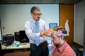
Penn’s brainSTIM center will study neuromodulation to repair and enhance human brain function
Penn Medicine has launched a new center to study the brain, one of the most complex systems in the body:
The Penn Brain Science, Translation, Innovation, and Modulation (brainSTIM) Center brings together a team of leading neuroscientists, neurologists, psychiatrists, psychologists, and engineers at Penn using neuromodulation techniques to research, repair, and enhance human brain function—the first translational center of its kind in the region.
Among the key faculty involved in this new center is J. Peter Skirkanich Professor of Bioengineering Danielle Bassett. Bassett’s Complex Systems Lab studies biological, physical, and social systems by using and developing tools from network science and complex systems theory. Bassett, along with Assistant Professor of Psychiatry Desmond Oathes, will work to:
understand how TMS [i.e. transcranial magnetic stimulation] might improve working memory in healthy adults and those with ADHD by combining network control theory (a set of concepts and principles employed in engineering), magnetic stimulation of the brain, and functional brain imaging.
A new approach uses “mechanoceuticals” to treat pain.
Drugs are commonly injected directly into an injury site to speed healing. For chronic pain, clinicians can inject drugs to reduce inflammation in painful joints, or can inject nerve blockers to block the nerve signals that cause pain. In a recent study, a group from UCLA developed a technique to deform a material surrounding nerve fibers to trigger a response in the fibers that would relieve pain. The combination of mechanics and treatment – i.e., ‘mechanoceuticals’ – is a clever way to trick fibers and reverse painful symptoms. Done without any injections and simply controlling magnetic fields outside the body, this approach can be reused as necessary.
The design of this mechanoceutical was completed by Dino Di Carlo, PhD, Professor of Bioengineering, and his team at UCLA’s Sameuli School of Engineering. By encasing tiny, magnetic nanoparticles within a biocompatible hydrogel, the group used magnetic force to stimulate nerve fibers and cause a corresponding decrease in pain signals. This promising development opens up a new approach to pain management, one which can be created with different biomaterials to suit different conditions, and delivered “on demand” without worrying about injections or, for that matter, any prescription drugs.
Understanding the Adolescent Brain
It’s no surprise that adults and adolescents often struggle to understand one another, but the work of neurologists and other researchers provides a possible physical reason for why that might be. Magnetic resonance elastrography (MRE) is a tool used in biomedical imaging to estimate the mechanical properties, or stiffness, of tissue throughout the body. Unexpectedly, a recent study suggests that brain stiffness correlates with cognitive ability, suggesting MRE may provide insight into patients’ behavior, psychology, and psychiatric state.
A new paper in Developmental Cognitive Neuroscience published the results of a study using MRE to track the relative “stiffness” vs. “softness” of adult and adolescent brains. The University of Delaware team, led by Biomedical Engineering Assistant Professor Curtis Johnson, PhD, and his doctoral student Grace McIlvain, sampled 40 living subjects (aged 12-14) and compared the properties to healthy adult brains.
The study found that children and adolescent brains are softer than those of adults, correlating to the overall malleability of childhood development. The team hopes to continue their studies with younger and older children, looking to demonstrate exactly when and how the change from softness to stiffness takes place, and how these properties correspond to individual qualities such as risk-taking or the onset of puberty. Eventually, establishing a larger database of measurements in the pediatric brain will help further studies into neurological and cognitive disorders in children, helping to understand conditions such as multiple sclerosis, autism, and cerebral palsy.
Can Nanoparticles Replace Stents?
Researchers and clinicians have made amazing advances in heart surgery. Stents, in particular, have become quite sophisticated: they are used to both prop open clogged arteries as well as deliver blood-thinning medication slowly over days to weeks in the area of the stent. However, the risk of blood clotting increases with stents and the blood vessels can constrict over time after the stent is placed in the vessel.
A recent NIH grant will support the design of a stent-free solution to unclog blood vessels. Led by Shaoqin Gong, PhD, Vilas Distinguished Professor of Biomedical Engineering at UW-Madison, the team used nanoparticles (or nanoclusters) to directly target the affected blood vessels and prevent regrowth of the cells post-surgery, eliminating the need for a stent to keep the pathways open. These nanoclusters are injected through an intravenous line, further reducing the risks introduced by the presence of the stent. As heart disease affects millions of people worldwide, this new material has far-reaching consequences. Their study is published in the September edition of Biomaterials.
NIST Grant Supports
The National Institute of Standards and Technology (NIST) awarded a $30 million grant to Johns Hopkins University, Binghamton University, and Morgan State University as part of their Professional Research Experience Program (PREP). Over five years, this award will support the collaboration of academics from all levels (faculty, postdoc, graduate, and undergraduate) across the three universities, enabling them to conduct research and attend NIST conferences.
The principal investigator for Binghamton U. is Professor and Chair of the Biomedical Engineering Department, Kaiming Ye, PhD. Dr. Ye is also the Director of the Center of Biomanufacturing for Regenerative Medicine (CBRM), which will participate in this collaborative new enterprise. Dr. Ye hopes that this grant will create opportunities for academics and researchers to network with each other as well as to more precisely define the standards for the fields of regenerative medicine and biomaterial manufacturing.
The gift honors the late A. James Clark, former CEO of Clark Enterprises and Clark Construction Group LLC, one of the country’s largest privately-held general building contractors. It is designed to prepare future engineering and business leaders, with an emphasis on low income families and first-generation college students. Clark never forgot that his business successes began with an engineering scholarship. This has guided the Clark family’s longstanding investments in engineering education and reflects its commitment to ensure college remains accessible and affordable to high-potential students with financial need.
We are proud to say that three incoming Clark Scholars from the Freshman Class of 2022 will be part of the Bioengineering Department here at Penn.
And finally, our congratulations to the new Dean of the School of Engineering at the University of Mississippi: David A. Puleo, PhD. Dr. Puleo earned his bachelor’s degree and doctorate in Biomedical Engineering from Rensselaer Polytechnic Institute. Most recently he served as Professor of Biomedical Engineering and Associate Dean for Research and Graduate Studies at the University of Kentucky’s College of Engineering. Building on his research in regenerative biomaterials, he also founded Regenera Materials, LLC in 2014. Over the course of his career so far, Dr. Puleo received multiple teaching awards and oversaw much departmental growth within his previous institution, and looks poised to do the same for “Ole Miss.”
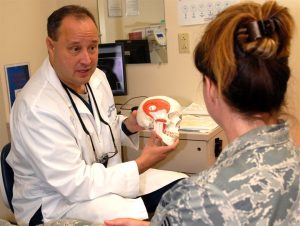
TMD is a common condition affecting movement of the jaw
Medical researchers have long been baffled by the need to find safe and effective treatment for a common condition called temporomandibular joint dysfunction (TMD). Affecting around twenty-five percent of the adult population worldwide, TMD appears overwhelmingly in adolescent, premenopausal women. Many different factors such as injury, arthritis, or grinding of the teeth can lead to the disintegration of or damage to the temporomandibular joint (TMJ), which leads to TMD, although the root cause is not always clear. A type of temporomandibular disorder, TMD can result in chronic pain in the jaw and ears, create difficulty eating and talking, and even cause occasional locking of the joint, making it difficult to open or close one’s mouth. Surgery is often considered a last resort because the results are often short-lasting or even dangerous.
The state of TMD treatment may change with the publication of a study in Science Translational Medicine. With contributions from researchers at the University of California, Irvine (UCI), UC Davis, and the University of Texas School of Dentistry at Houston, this new study has successfully implanted engineered discs made from rib cartilage cells into a TMJ model. The biological properties of the discs are similar enough to native TMJ cells to more fully reduce further degeneration of the joint as well as potentially pave the way for regeneration of joints with TMD.
Senior author Kyriacos Athanasiou, PhD, Distinguished Professor of Biomedical Engineering at UCI, states the next steps for the team of researchers include a long-term study to ensure ongoing effectiveness and safety of the implants followed by eventual clinical trials. In the long run, this technique may also prove useful and relevant to the treatment of other types of arthritis and joint dysfunction.
Advances in Autism Research
Currently, diagnosis of autism spectrum disorders (ASD) has been limited entirely to clinical observation and examination by medical professionals. This makes the early identification and treatment of ASD difficult as most children cannot be accurately diagnosed until around the age of four, delaying the treatment they might receive. A recent study published in the journal of Bioengineering & Translational Medicine, however, suggests that new blood tests may be able to identify ASD with a high level of accuracy, increasing the early identification that is key to helping autistic children and their families. The researchers, led by Juergen Hahn, PhD, Professor and Department Head of Biomedical Engineering at the Rensselaer Polytechnic Institute, hope that after clinical trials this blood test will become commercially available.
In addition to work that shows methods to detect autism earlier, the most recent issue of Nature Biomedical Engineering includes a study to understand the possible causes of autism and, in turn, develop treatments for the disease. The breakthrough technology of Cas9 enzymes allowed researchers to edit the genome, correcting for symptoms that appeared in mice which resembled autism, including exaggerated and repetitive behaviors. This advance comes from a team at the University of California, Berkeley, which developed the gene-editing technique known as CRISPR-Gold to treat symptoms of ASD by injecting the Cas9 enzyme into the brain without the need for viral delivery. The UC Berkeley researchers suggest in the article’s abstract that these safe gene-editing technologies “may revolutionize the treatment of neurological diseases and the understanding of brain function.” These treatments may have practical benefits for the understanding and treatment of such diverse conditions as addiction and epilepsy as well as ASD.
Penn Professor’s Groundbreaking Bioengineering Technology
Our own D. Kacy Cullen, PhD, was recently featured in Penn Today for his groundbreaking research which has led to the first implantable tissue-engineered brain pathways. This technology could lead to the reversal of certain neurodegenerative disorders, such as Parkinson’s disease.
With three patents, at least eight published papers, $3.3 million in funding, and a productive go with the Penn Center for Innovation’s I-Corps program this past fall, Dr. Cullen is ready to take this project’s findings to the next level with the creation of a brand new startup company: Innervace. “It’s really surreal to think that I’ve been working on this project, this approach, for 10 years now,” he says. “It really was doggedness to just keep pushing in the lab, despite the challenges in getting extramural funding, despite the skepticism of peer reviewers. But we’ve shown that we’re able to do it, and that this is a viable technology.” Several Penn bioengineering students are involved in the research conducted in Dr. Cullen’s lab, including doctoral candidate Laura Struzyna and recent graduate Kate Panzer, who worked in the lab all four years of her undergraduate career.
In addition to his appointment as a Research Associate Professor of Neurosurgery at the Perelman School of Medicine at the University of Pennsylvania, Dr. Cullen also serves as a member of Penn’s Department of Bioengineering Graduate Group Faculty, and will teach the graduate course BE 502 (From Lab to Market Place) for the BE Department this fall 2018 semester. He also serves as the director for the Center of Neurotrauma, Neurodegeneration, and Restoration at the VA Medical Center.
New Prosthetics Will Have the Ability to Feel Pain
New research from the Department of Biomedical Engineering at Johns Hopkins University (JHU) has found a way to address one of the difficult aspects of amputation: the inability for prosthetic limbs to feel. This innovative electronic dermis is worn over the prosthetic, and can detect sensations (such as pain or even a light touch), which are conveyed to the user’s nervous system, closing mimicking skin. The findings of this study were recently published in the journal ScienceRobotics.
While one might wonder at the value of feeling pain, both researchers and amputees verify that physical sensory reception is important both for the desired realism of the prosthetic or bionic limb, and also to alert the wearer of any potential harm or damage, the same way that heat can remind a person to remove her hand from a hot surface, preventing a potential burn. Professor Nitish Thakor, PhD, and his team hope to make this exciting new technology readily available to amputees.
People and Places
Women are still vastly outnumbered in STEM, making up only twenty percent of the field, and given the need for diversification, researchers, educators, and companies are brainstorming ways to proactively solve this problem by promoting STEM subjects to young women. One current initiative has been spearheaded by GE Healthcare and Milwaukee School of Engineering University (MSOE) who are partnering to give middle school girls access to programs in engineering during their summer break at the MSOE Summer STEM Camp, hoping to reduce the stigma of these subjects for young women. GE Girls also hosts STEM programs with a number of institutions across the U.S.
The National Science Policy Network (NSPN) “works to provide a collaborative resource portal for early-career scientists and engineers involved in science policy, diplomacy, and advocacy.” The NSPN offers platforms and support including grant funding, internships, and competitions. Chaired and led by emerging researchers and professors from around the country, including biomedical engineering PhD student Michaela Rikard of the University of Virginia, the NSPN seeks to provide a network for young scientists in the current political climate in which scientific issues and the very importance of the sciences as a whole are hotly contested and debated by politicians and the public. The NSPN looks to provide a way for scientists to have a voice in policy-making. This new initiative was recently featured in the Scientific American.
Upon its original founding in 2000, the Bill and Melinda Gates Foundation has included the eradication of malaria as part of its mission, pledging around $2 billion to the cause in the years since. One of its most recent initiatives is the funding of a bioengineering project which targets the type of mosquitoes which carry the deadly disease. Engineered mosquitoes (so-called “Friendly Mosquitoes”) would mate in the wild, passing on a mosquito-killing gene to their female offspring (only females bite humans) before they reach maturity. While previous versions of “Friendly Mosquitoes” have been met with success, concerns have been raised about the potential long-term ecological effects to the mosquito population. UK-based partner Oxitec expects to have the new group ready for trials in two years.


 Penn’s brainSTIM center will study neuromodulation to repair and enhance human brain functio