Neuroscientists frequently say that neural activity ‘represents’ certain phenomena, PIK Professor Konrad Kording and postdoc Ben Baker led a study that took a philosophical approach to tease out what the term means.
Neuroscientists use the word “represent” to encompass multifaceted relationships between brain activity, behavior, and the environment.
One of neuroscience’s greatest challenges is to bridge the gaps between the external environment, the brain’s internal electrical activity, and the abstract workings of behavior and cognition. Many neuroscientists rely on the word “representation” to connect these phenomena: A burst of neural activity in the visual cortex may represent the face of a friend or neurons in the brain’s memory centers may represent a childhood memory.
But with the many complex relationships between mind, brain, and environment, it’s not always clear what neuroscientists mean when they say neural activity “represents” something. Lack of clarity around this concept can lead to miscommunication, flawed conclusions, and unnecessary disagreements.
To tackle this issue, an interdisciplinary paper takes a philosophical approach to delineating the many aspects of the word “representation” in neuroscience. The work, published in Trends in Cognitive Sciences, comes from the lab of Konrad Kording, a Penn Integrates Knowledge University Professor and senior author on the study whose research lies at the intersection of neuroscience and machine learning.
“The term ‘representation’ is probably one of the most common words in all of neuroscience,” says Kording, who has appointments in the Perelman School of Medicine and School of Engineering and Applied Science. “But it might mean something very different from one professor to another.”
Also coauthor on the paper is Benjamin Lansdell, a data scientist in the Department of Developmental Neurobiology at St. Jude Children’s Hospital and former postdoctoral researcher in the Kording lab.
Funding for this study came from the National Institutes of Health (awards 1-R01-EB028162-01 and R01EY021579) and the University of Pennsylvania Office of the Vice Provost for Research.
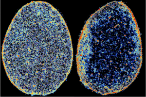
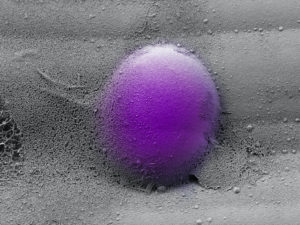
In these super-resolution images of tendon cell nuclei, the color coding represents chromatin density map, from low density in blue to high density in red. Comparing a healthy human tendon cell nucleus (left) to one diagnosed with tendinosis (right) shows that disease alters the spatial localization and compaction of chromatin.
In a recent study published in Nature Biomedical Engineering, the team detailed what they found when they closely observed the nucleus of cells inside connective tissues deteriorating as a result of tendinosis, which is the chronic condition that results from a tendon repeatedly suffering small injuries that don’t heal correctly. Using the latest super-resolution imaging techniques, they found that the tendon cells involved in maintaining the tissue’s structure in a diseased microenvironment improperly reorder their chromatin — the DNA-containing material that chromosomes are composed of — when attempting to repair.
This and other findings highlighted in the report point to the possibility of new treatments, such as small-molecule therapies, that could restore order to the affected cells.
“Interestingly, we were able to explain the role of mechanical forces on the 3-D organization of chromatin by developing a theory that integrates fundamental thermodynamic principles (physics) with the kinetics of epigenetic regulation (biology),” said study co-author and CEMB Director Vivek Shenoy in a news release from Penn Medicine News.
The CEMB, one of 18 active interdisciplinary research centers funded by the National Science Foundation’s Science and Technology Center (STC) program, brings together dozens of researchers from Penn Engineering and the Perelman School of Medicine, as well as others spread across campus and at partner institutions around the world.
With its funding recently renewed for another five years, the CEMB has entered into a new phase of its mission, centered on the nascent concept of “mechanointelligence,” which is exemplified by studies like this one. While mechanobiology is the study of the physical forces that govern the behavior of cells and their communication with their neighbors, mechanointelligence adds another layer of complexity: attempting to understand the forces that allow cells to sense, remember and adapt to their environments.
Ultimately, harnessing these forces would allow researchers to help multicellular organisms — plants, animals and humans — better adapt to their environments as well.
This story originally appeared in Penn Engineering Today.
Vivek Shenoy is Eduardo D. Glandt President’s Distinguished Professor in Materials Science and Engineering, Bioengineering, and in Mechanical Engineering and Applied Mechanics.
Michael Mitchell, J. Peter and Geri Skirkanich Assistant Professor of Innovation in the Department of Bioengineering, is one of this year’s recipients of the National Science Foundation’s CAREER Award. The award is given to early-career faculty researchers who demonstrate the potential to be role models in their field and invest in the outreach and education of their work.
Mitchell’s award will fund research on techniques for “immunoengineering” macrophages. By providing new instructions to these cells via nanoparticles laden with mRNA and DNA sequences, the immune system could be trained to target and eliminate solid tumors. The award will also support graduate students and postdoctoral fellows in his lab over the next five years.
The project aligns with Mitchell’s larger research goals and the current explosion of interest in therapies that use mRNA, thanks to the technological breakthroughs that enabled the development of COVID-19 vaccines.
“The development of the COVID vaccine using mRNA has opened doors for other cell therapies,” says Mitchell. “The high-priority area of research that we are focusing on is oncological therapies, and there are multiple applications for mRNA engineering in the fight against cancer.”
A new wave of remarkably effective cancer treatments incorporates chimeric antigen receptor T-cell (CAR-T) therapy. There, a patient’s T-cells, a type of white blood cell that fights infections, are genetically engineered to identify, target and kill individual cancer cells that accumulate in the circulatory system.
However, despite CART-T therapy’s success in treating certain blood cancers, the approach is not effective against cancers that form solid tumors. Because T-cells are not able to penetrate tumors’ fibrous barriers, Mitchell and his colleagues have turned to another part of the immune system for help.
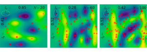
Developing new soft materials requires new data-driven research techniques, such as autonomous experimentation. Data regarding nanometer-scale material structure, taken by X-ray measurements at a synchrotron, can be fed into an algorithm that identifies the most relevant features, represented here as red dots. The algorithm then determines the optimum conditions for the next set of measurements and directs their execution without human intervention. Brookhaven National Laboratory’s Kevin Yager, who helped develop this technique, will co-teach a course on it as part of a new Penn project on Data Driven Soft Materials Research.
The National Science Foundation’s Research Traineeship Program aims to support graduate students, educate the STEM leaders of tomorrow and strengthen the national research infrastructure. The program’s latest series of grants are going toward university programs focused on artificial intelligence and quantum information science and engineering – two areas of high priority in academia, industry and government.
Chinedum Osuji, Eduardo D. Glandt Presidential Professor and Chair of the Department of Chemical and Biomolecular Engineering (CBE), has received one of these grants to apply data science and machine learning to the field of soft materials. The grant will provide five years of support and a total of $3 million for a new Penn project on Data Driven Soft Materials Research.
Osuji will work with co-PIs Russell Composto, Professor and Howell Family Faculty Fellow in Materials Science and Engineering, Bioengineering, and in CBE, Zahra Fakhraai, Associate Professor of Chemistry in Penn’s School of Arts & Sciences (SAS) with a secondary appointment in CBE, Paris Perdikaris, Assistant Professor in Mechanical Engineering and Applied Mechanics, and Andrea Liu, Hepburn Professor of Physics and Astronomy in SAS, all of whom will help run the program and provide the connections between the multiple fields of study where its students will train.
These and other affiliated faculty members will work closely with co-PI Kristin Field, who will serve as Program Coordinator and Director of Education.
CPE4H is one of the focal points of Penn Engineering signature initiative on Engineering Health.
The Penn Center for Precision Engineering for Health (CPE4H) was established late last year to accelerate engineering solutions to significant problems in healthcare. The center is one of the signature initiatives for Penn’s School of Engineering and Applied Science and is supported by a $100 million commitment to hire faculty and support new research on innovative approaches to those problems.
Acting on that commitment, CPE4H solicited proposals during the spring of 2022 for seed grants of $80K per year for two years for research projects that address healthcare challenges in several key areas of strategic importance to Penn: synthetic biology and tissue engineering, diagnosis and drug delivery, and the development of innovative devices. While the primary investigators (PIs) for the proposed projects were required to have a primary faculty appointment within Penn Engineering, teams involving co-PIs and collaborators from other schools were eligible for support. The seed program is expected to continue for the next four years.
“It was a delight to read so many novel and creative proposals,” says Daniel A. Hammer, Alfred G. and Meta A. Ennis Professor in Bioengineering and the Inaugural Director of CPE4H. “It was very hard to make the final selection from a pool of such promising projects.”
Judged on technical innovation, potential to attract future resources, and ability to address a significant medical problem, the following research projects were selected to receive funding.
Evolving and Engineering Thermal Control of Mammalian Cells
Led by Lukasz Bugaj, Assistant Professor in Bioengineering, this project will engineer molecular switches that can be toggled on and off inside mammalian cells at near-physiological temperatures. Successful development of these switches will provide new ways to communicate with cells, an advance that could be used to make safer and more effective cellular therapies. The project will use directed evolution to generate and find candidate molecular tools with the desired properties. Separately, the research will also develop new technology for manipulating cellular temperature in a rapid and programmable way. Such devices will enhance the speed and sophistication of studies of biological temperature regulation.
A Quantum Sensing Platform for Rapid and Accurate Point-of-Care Detection of Respiratory Viral Infections
Combining microfluidics and quantum photonics, PI Liang Feng, Professor in Materials Science and Engineering and Electrical and Systems Engineering, Ritesh Agarwal, Professor in Materials Science Engineering, and Shu Yang, Joseph Bordogna Professor in Materials Science and Engineering and Chemical and Biomolecular Engineering, are teaming up with Ping Wang, Professor of Pathology and Laboratory Medicine in Penn’s Perelman School of Medicine, to design, build and test an ultrasensitive point-of-care detector for respiratory pathogens. In light of the COVID-19 pandemic, a generalizable platform for rapid and accurate detection of viral pathogenesis would be extremely important and timely.
Versatile Coacervating Peptides as Carriers and Synthetic Organelles for Cell Engineering
PI Amish Patel, Associate Professor in Chemical and Biomolecular Engineering, and Matthew C. Good, Associate Professor of Cell and Developmental Biology in the Perelman School of Medicine and in Bioengineering, will design and create small proteins that self-assemble into droplet-like structures known as coacervates, which can then pass through the membranes of biological cells. Upon cellular entry, these protein coacervates can disassemble to deliver cargo that modulates cell behavior or be maintained as synthetic membraneless organelles. The team will design new chemistries that will facilitate passage across cell membranes, and molecular switches to sequester and release protein therapeutics. If successful, this approach could be used to deliver a wide range of macromolecule drugs to cells.
Towards an Artificial Muscle Replacement for Facial Reanimation
Cynthia Sung, Gabel Family Term Assistant Professor in Mechanical Engineering and Applied Mechanics and Computer Information Science, will lead a research team including Flavia Vitale, Assistant Professor of Neurology and Bioengineering, and Niv Milbar, Assistant Instructor in Surgery in the Perelman School of Medicine. The team will develop and validate an electrically driven actuator to restore basic muscle responses in patients with partial facial paralysis, which can occur after a stroke or injury. The research will combine elements of robotics and biology, and aims to produce a device that can be clinically tested.
“These novel ideas are a great way to kick off the activities of the center,” says Hammer. “We look forward to soliciting other exciting seed proposals over the next several years.”
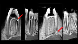
Controlled and actuated by magnetic fields, these mircrorobots are capable of precisely targeting the apical region — the opening where blood vessels and nerve enter the tooth — in a root canal.
With its irregularities and anatomical complexities, the root canal system is one of the most clinically challenging spaces in the oral cavity. As a result, biofilm not fully cleared from the nooks and crannies of the canals remains a leading cause of treatment failure and persistent endodontic infections, and there are limited means to diagnose or assess the efficacy of disinfection. One day, clinicians may have a new tool to overcome these challenges in the form of microrobots.
In a proof-of-concept study, researchers from Penn Dental Medicine and its Center for Innovation & Precision Dentistry (CiPD), have shown that microrobots can access the difficult to reach surfaces of the root canal with controlled precision, treating and disrupting biofilms and even retrieving samples for diagnostics, enabling a more personalized treatment plan. The Penn team shared their findings on the use of two different microrobotic platforms for endodontic therapy in the August issue of the Journal of Dental Research;the work was selected for the issue’s cover.
“The technology could enable multimodal functionalities to achieve controlled, precision targeting of biofilms in hard-to-reach spaces, obtain microbiological samples, and perform targeted drug delivery, ” says Dr. Alaa Babeer, lead author of the study and a Penn Dental Medicine Doctor of Science in Dentistry (DScD) and endodontics graduate, who is now within the lab of Dr. Michel Koo, co-director of the CiPD .
In both platforms, the building blocks for the microrobots are iron oxide nanoparticles (NPs) that have both catalytic and magnetic activity and have been FDA approved for other uses. In the first platform, a magnetic field is used to concentrate the NPs in aggregated microswarms and magnetically control them to the apical area of the tooth to disrupt and retrieve biofilms through a catalytic reaction. The second platform uses 3D printing to create miniaturized helix-shaped robots embedded with iron oxide NPs. These helicoids are guided by magnetic fields to move within the root canal, transporting bioactives or drugs that can be released on site.
“This technology offers the potential to advance clinical care on a variety of levels,” says Dr. Koo, co-corresponding author of the study with Dr. Edward Steager, a senior research investigator in Penn’s School of Engineering and Applied Science. “One important aspect is the ability to have diagnostic as well as therapeutic applications. In the microswarm platform, we can not only remove the biofilm, but also retrieve it, enabling us identify what microorganisms caused the infection. In addition, the ability to conform to the narrow and difficult-to-reach spaces within the root canal allows for a more effective disinfection in comparison to the files and instrumentation techniques presently used.”
Michel Koo is a professor in the Department of Orthodontics and divisions of Community Oral Health and Pediatric Dentistry in Penn Dental Medicine and co-director of the Center for Innovation & Precision Dentistry. He is a member of the Penn Bioengineering Graduate Group.
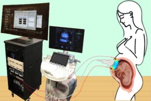
Combining optical measurements with ultrasound, an interdisciplinary team from the School of Arts & Sciences, Perelman School of Medicine, and CHOP developed a device to better measure blood flow and oxygenation in the placenta. (Image: Lin Wang)
A study published in Nature Biomedical Engineering details a novel method for imaging the placenta in pregnant patients as well as the results of a pilot clinical study. By combining optical measurements with ultrasound, the findings show how oxygen levels can be monitored noninvasively and provides a new way to generate a better understanding of this complex, crucial organ. This research was the result of a collaboration of the groups of the University of Pennsylvania’s Arjun Yodh and Nadav Schwartz with colleagues from the Children’s Hospital of Philadelphia (CHOP) and was led by postdoc Lin Wang.
Schwartz describes the placenta as the “engine” of pregnancy, an organ that plays a crucial role in delivering nutrients and oxygen to the fetus. Placental dysfunction can lead to complications such as fetal growth restriction, preeclampsia, and stillbirth. To increase knowledge about this crucial organ, the National Institute of Child Health and Human Development launched the Human Placenta Project in 2014. One focus of the program is to develop tools to assess human placental structure and function in real time, including optical devices.
For three years, the researchers optimized the design of their instrument and tested it in preclinical settings. The process involved integrating optical fibers with ultrasound probes, exploring various ultrasound transducers, and improving the multimodal technology so that measurements were stable, accurate, and reproducible while collecting data at the bedside. The resulting instrumentation now enables researchers to study the anatomy of the placenta while also collecting detailed functional information about placenta blood flow and oxygenation, capabilities that existing commercially devices do not have, the researchers say.
Because the placenta is located far below the body’s surface, one of the key technical challenges addressed by Wang, a postdoc in Yodh’s lab, was reducing background noise in the opto-electronic system. Light is scattered and absorbed when it travels through thick tissues, Yodh says, and the key for success was to reduce background interference so that the small amount of light that penetrates deep into the placenta and then returns is still large enough for a high-quality measurement.
“We’re sending a light signal that goes through the same deep tissues as the ultrasound. The extremely small amount of light that returns to the surface probe is then used to accurately assess tissue properties, which is only possible with very stable lasers, optics, and detectors,” says Yodh. “Lin had to overcome many barriers to improve the signal-to-noise ratio to the point where we trusted our data.”
The authors are Lin Wang, Jeffrey M. Cochran, Kenneth Abramson, Lian He, Venki Kavuri, Samuel Parry, Arjun G. Yodh, and Nadav Schwartz from Penn; Tiffany Ko, Wesley B. Baker, and Rebecca L. Linn from the Children’s Hospital of Philadelphia, and David R. Busch, previously a research associate at Penn and now at the University of Texas Southwestern Medical School.
This research was supported by National Institutes of Health grants F31HD085731, R01NS113945, R01NS060653, P41EB015893, P41EB015893, T32HL007915, and U01HD087180.
Acollaborative team developed an alginate-based hydrogel system that mimics the viscoelasticity of the natural extracellular matrix in bone marrow. By tweaking the balance between elastic and viscous properties in these artificial ECMs, they could recapitulate the viscoelasticity of healthy and scarred fibrotic bone marrow, and study the effects on human monocytes placed into these artificial ECMs. (Image: Adam Graham/Harvard CNS/Wyss Institute at Harvard University)
Fibrosis is the thickening of various tissues caused by the deposition of fibrillar extracellular matrix (ECM) in tissues and organs as part of the body’s wound healing response to various forms of damage. When accompanied by chronic inflammation, fibrosis can go into overdrive and produce excess scar tissue that can no longer be degraded. This process causes many diseases in multiple organs, including lung fibrosis induced by smoking or asbestos, liver fibrosis induced by alcohol abuse, and heart fibrosis often following heart attacks. Fibrosis can also occur in the bone marrow, the spongy tissue inside some bones that houses blood-producing hematopoietic stem cells (HSCs) and can lead to scarring and the disruption of normal functions.
Chronic blood cancers known as “myeloproliferative neoplasms” (MPNs) are one example, in which patients can develop fibrotic bone marrow, or myelofibrosis, that disrupts the normal production of blood cells. Monocytes, a type of white blood cell belonging to the group of myeloid cells, are overproduced from HSCs in neoplasms and contribute to the inflammation in the bone marrow environment, or niche. However, how the fibrotic bone marrow niche itself impacts the function of monocytes and inflammation in the bone marrow was unknown.
Now, a collaborative team from Penn, Harvard, the Dana-Farber Cancer Institute (DFCI), and Brigham and Women’s Hospital has created a programmable hydrogel-based in vitro model mimicking healthy and fibrotic human bone marrow. Combining this system with mouse in vivo models of myelofibrosis, the researchers demonstrated that monocytes decide whether to enter a pro-inflammatory state and go on to differentiate into inflammatory dendritic cells based on specific mechanical properties of the bone marrow niche with its densely packed ECM molecules. Importantly, the team found a drug that could tone down these pathological mechanical effects on monocytes, reducing their numbers as well as the numbers of inflammatory myeloid cells in mice with myelofibrosis. The findings are published in Nature Materials.
“We found that stiff and more elastic slow-relaxing artificial ECMs induced immature monocytes to differentiate into monocytes with a pro-inflammatory program strongly resembling that of monocytes in myelofibrosis patients, and the monocytes to differentiate further into inflammatory dendritic cells,” says co-first author Kyle Vining, who recently joined Penn’s School of Dental Medicine and School of Engineering and Applied Science as an assistant professor of preventive and restorative sciences. “More viscous fast-relaxing artificial ECMs suppressed this myelofibrosis-like effect on monocytes. This opened up the possibility of a mechanical checkpoint that could be disrupted in myelofibrotic bone marrow and also may be at play in other fibrotic diseases.”
Vining worked on the study as a postdoctoral fellow at Harvard in the lab of David Mooney. “Our study shows that the differentiation state of monocytes, which are key players in the immune system, is highly regulated by mechanical changes in the ECM they encounter,” says Mooney, who co-led the study with DFCI researcher Kai Wucherpfennig. “Specifically, the ECM’s viscoelasticity has been a historically under-appreciated aspect of its mechanical properties that we find correlates strongly between our in vitro and the in vivo models and human disease. It turns out that myelofibrosis is a mechano-related disease that could be treated by interfering with the mechanical signaling in bone marrow cells.”
Kevin Johnson is used to forging his own path in the fields of healthcare and computer science.
A picture of Johnson as a child, from his children’s book “I’m a Biomedical Expert Now!”
If you ask him to locate his niche within these fields, Johnson, David L. Cohen and Penn Integrates Knowledge (PIK) Professor with appointments in Penn Engineering and the Perelman School of Medicine, would say “informatics.” But that doesn’t tell the whole story of the board-certified pediatrician, who has dedicated his career to innovations in how patients’ information is created, documented and shared, all with the goal of improving the quality of healthcare they receive.
Informatics, the study of the structure and behavior of interactions between natural and computational systems, is an umbrella term. Within it, there’s bioinformatics, which applies informatics to biology, and biomedical informatics, which looks at those interactions in the context of healthcare systems. Finally, there is clinical informatics, which further focuses on the settings where healthcare is delivered, and where Johnson squarely places himself.
“But you can just call it ‘informatics,’” says Johnson. “It will be easier.”
He mainly studies how computational systems can improve ambulatory care — sometimes known as outpatient care, or the kind of care hospitals give to patients without admitting them — in real time. If you’ve ever heard your doctor complain about the amount of time it takes them to input the information they get from you during your visit, or wondered why they need to capture this information during the visit in the first place, these are some of the questions Johnson is investigating.
“We’re taking care of patients but we’re getting frustrated by things that we thought these new computers should be able to fix,” says Johnson.” I think there’s a very compelling case for using engineering principles to reimagine electronic health records.”
Kevin Johnson is the David L. Cohen University of Pennsylvania Professor in the Departments of Biostatistics, Epidemiology and Informatics and Computer and Information Science. As a Penn Integrates Knowlegde (PIK) University Professor, Johnson also holds appointments in the Departments of Bioengineering and Pediatrics, as well as in the Annenberg School of Communication. Johnson is the Vice President for Applied Informatics for the University of Pennsylvania Health System and has been elected to the American College of Medical Informatics (2004), the Academic Pediatric Society (2010), the National Academy of Medicine (Institute of Medicine) (2010), and the International Association of Health Science Informatics (2021).
Kevin B. Johnson, M.D., M.S., was featured in Cincinnati Children’s Hospital’s “Envisioning Our Future for Children” speaker series, discussing “the evolution of the EHR and its future directions.” An electronic health record, or EHR, is a digital record of a patient’s chart, recording health information and data, coordinating orders, tracking results, and providing patient support. Johnson “predicts a new wave of transformation in digital health technologies that could make rapid progress” in several areas of medicine, including reducing cost and improving patience outcomes. Johnson is Vice President for Applied Informatics at the University of Pennsylvania Health System and the David L. Cohen University Professor with appointments in Biostatistics, Epidemiology and Informatics and Computer and Information Science and secondary appointments in the Annenberg School for Communication, Pediatrics, and Bioengineering.