A Novel Method for Monitoring the ‘Engine’ of Pregnancy

Combining optical measurements with ultrasound, an interdisciplinary team from the School of Arts & Sciences, Perelman School of Medicine, and CHOP developed a device to better measure blood flow and oxygenation in the placenta. (Image: Lin Wang)

A study published in Nature Biomedical Engineering details a novel method for imaging the placenta in pregnant patients as well as the results of a pilot clinical study. By combining optical measurements with ultrasound, the findings show how oxygen levels can be monitored noninvasively and provides a new way to generate a better understanding of this complex, crucial organ. This research was the result of a collaboration of the groups of the University of Pennsylvania’s Arjun Yodh and Nadav Schwartz with colleagues from the Children’s Hospital of Philadelphia (CHOP) and was led by postdoc Lin Wang.

Schwartz describes the placenta as the “engine” of pregnancy, an organ that plays a crucial role in delivering nutrients and oxygen to the fetus. Placental dysfunction can lead to complications such as fetal growth restriction, preeclampsia, and stillbirth. To increase knowledge about this crucial organ, the National Institute of Child Health and Human Development launched the Human Placenta Project in 2014. One focus of the program is to develop tools to assess human placental structure and function in real time, including optical devices.

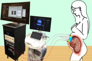
For three years, the researchers optimized the design of their instrument and tested it in preclinical settings. The process involved integrating optical fibers with ultrasound probes, exploring various ultrasound transducers, and improving the multimodal technology so that measurements were stable, accurate, and reproducible while collecting data at the bedside. The resulting instrumentation now enables researchers to study the anatomy of the placenta while also collecting detailed functional information about placenta blood flow and oxygenation, capabilities that existing commercially devices do not have, the researchers say.

Because the placenta is located far below the body’s surface, one of the key technical challenges addressed by Wang, a postdoc in Yodh’s lab, was reducing background noise in the opto-electronic system. Light is scattered and absorbed when it travels through thick tissues, Yodh says, and the key for success was to reduce background interference so that the small amount of light that penetrates deep into the placenta and then returns is still large enough for a high-quality measurement.

“We’re sending a light signal that goes through the same deep tissues as the ultrasound. The extremely small amount of light that returns to the surface probe is then used to accurately assess tissue properties, which is only possible with very stable lasers, optics, and detectors,” says Yodh. “Lin had to overcome many barriers to improve the signal-to-noise ratio to the point where we trusted our data.”

Read the full story in Penn Today.

The authors are Lin Wang, Jeffrey M. Cochran, Kenneth Abramson, Lian He, Venki Kavuri, Samuel Parry, Arjun G. Yodh, and Nadav Schwartz from Penn; Tiffany Ko, Wesley B. Baker, and Rebecca L. Linn from the Children’s Hospital of Philadelphia, and David R. Busch, previously a research associate at Penn and now at the University of Texas Southwestern Medical School.

Arjun Yodh is the James M. Skinner Professor of Science in the Department of Physics and Astronomy in the School of Arts & Sciences at the University of Pennsylvania. He is a member of the Penn Bioengineering Graduate Group.

Nadav Schwartz is an Associate Professor in the Department of Obstetrics and Gynecology in Penn’s Perelman School of Medicine.

Lin Wang is a postdoc in the Department of Physics and Astronomy in Penn’s School of Arts & Sciences.

This research was supported by National Institutes of Health grants F31HD085731, R01NS113945, R01NS060653, P41EB015893, P41EB015893, T32HL007915, and U01HD087180.

Deconstructing the Mechanics of Bone Marrow Disease

by Katherine Unger Baillie

Acollaborative team developed an alginate-based hydrogel system that mimics the viscoelasticity of the natural extracellular matrix in bone marrow. By tweaking the balance between elastic and viscous properties in these artificial ECMs, they could recapitulate the viscoelasticity of healthy and scarred fibrotic bone marrow, and study the effects on human monocytes placed into these artificial ECMs. (Image: Adam Graham/Harvard CNS/Wyss Institute at Harvard University)

Fibrosis is the thickening of various tissues caused by the deposition of fibrillar extracellular matrix (ECM) in tissues and organs as part of the body’s wound healing response to various forms of damage. When accompanied by chronic inflammation, fibrosis can go into overdrive and produce excess scar tissue that can no longer be degraded. This process causes many diseases in multiple organs, including lung fibrosis induced by smoking or asbestos, liver fibrosis induced by alcohol abuse, and heart fibrosis often following heart attacks. Fibrosis can also occur in the bone marrow, the spongy tissue inside some bones that houses blood-producing hematopoietic stem cells (HSCs) and can lead to scarring and the disruption of normal functions.

Chronic blood cancers known as “myeloproliferative neoplasms” (MPNs) are one example, in which patients can develop fibrotic bone marrow, or myelofibrosis, that disrupts the normal production of blood cells. Monocytes, a type of white blood cell belonging to the group of myeloid cells, are overproduced from HSCs in neoplasms and contribute to the inflammation in the bone marrow environment, or niche. However, how the fibrotic bone marrow niche itself impacts the function of monocytes and inflammation in the bone marrow was unknown.

Now, a collaborative team from PennHarvard, the Dana-Farber Cancer Institute (DFCI), and Brigham and Women’s Hospital has created a programmable hydrogel-based in vitro model mimicking healthy and fibrotic human bone marrow. Combining this system with mouse in vivo models of myelofibrosis, the researchers demonstrated that monocytes decide whether to enter a pro-inflammatory state and go on to differentiate into inflammatory dendritic cells based on specific mechanical properties of the bone marrow niche with its densely packed ECM molecules. Importantly, the team found a drug that could tone down these pathological mechanical effects on monocytes, reducing their numbers as well as the numbers of inflammatory myeloid cells in mice with myelofibrosis. The findings are published in Nature Materials.

“We found that stiff and more elastic slow-relaxing artificial ECMs induced immature monocytes to differentiate into monocytes with a pro-inflammatory program strongly resembling that of monocytes in myelofibrosis patients, and the monocytes to differentiate further into inflammatory dendritic cells,” says co-first author Kyle Vining, who recently joined Penn’s School of Dental Medicine and School of Engineering and Applied Science as an assistant professor of preventive and restorative sciences. “More viscous fast-relaxing artificial ECMs suppressed this myelofibrosis-like effect on monocytes. This opened up the possibility of a mechanical checkpoint that could be disrupted in myelofibrotic bone marrow and also may be at play in other fibrotic diseases.”

Vining worked on the study as a postdoctoral fellow at Harvard in the lab of David Mooney. “Our study shows that the differentiation state of monocytes, which are key players in the immune system, is highly regulated by mechanical changes in the ECM they encounter,” says Mooney, who co-led the study with DFCI researcher Kai Wucherpfennig. “Specifically, the ECM’s viscoelasticity has been a historically under-appreciated aspect of its mechanical properties that we find correlates strongly between our in vitro and the in vivo models and human disease. It turns out that myelofibrosis is a mechano-related disease that could be treated by interfering with the mechanical signaling in bone marrow cells.”

Continue reading at Penn Today

Kevin Johnson: Informatics Evangelist

by Ebonee Johnson

Kevin Johnson is used to forging his own path in the fields of healthcare and computer science.

A picture of Johnson as a child, from his children’s book “I’m a Biomedical Expert Now!”

If you ask him to locate his niche within these fields, Johnson, David L. Cohen and Penn Integrates Knowledge (PIK) Professor with appointments in Penn Engineering and the Perelman School of Medicine, would say “informatics.” But that doesn’t tell the whole story of the board-certified pediatrician, who has dedicated his career to innovations in how patients’ information is created, documented and shared, all with the goal of improving the quality of healthcare they receive.

Informatics, the study of the structure and behavior of interactions between natural and computational systems, is an umbrella term. Within it, there’s bioinformatics, which applies informatics to biology, and biomedical informatics, which looks at those interactions in the context of healthcare systems. Finally, there is clinical informatics, which further focuses on the settings where healthcare is delivered, and where Johnson squarely places himself.

“But you can just call it ‘informatics,’” says Johnson. “It will be easier.”

He mainly studies how computational systems can improve ambulatory care — sometimes known as outpatient care, or the kind of care hospitals give to patients without admitting them — in real time. If you’ve ever heard your doctor complain about the amount of time it takes them to input the information they get from you during your visit, or wondered why they need to capture this information during the visit in the first place, these are some of the questions Johnson is investigating.

“We’re taking care of patients but we’re getting frustrated by things that we thought these new computers should be able to fix,” says Johnson.” I think there’s a very compelling case for using engineering principles to reimagine electronic health records.”

Read the full story in Penn Engineering Today.

Kevin Johnson is the David L. Cohen University of Pennsylvania Professor in the Departments of Biostatistics, Epidemiology and Informatics and Computer and Information Science. As a Penn Integrates Knowlegde (PIK) University Professor, Johnson also holds appointments in the Departments of Bioengineering and Pediatrics, as well as in the Annenberg School of Communication. Johnson is the Vice President for Applied Informatics for the University of Pennsylvania Health System and has been elected to the American College of Medical Informatics (2004), the Academic Pediatric Society (2010), the National Academy of Medicine (Institute of Medicine) (2010), and the International Association of Health Science Informatics (2021).

Kevin Johnson Discusses the Future of the Electronic Health Record

Kevin B. Johnson, M.D., Ph.D.

Kevin B. Johnson, M.D., M.S., was featured in Cincinnati Children’s Hospital’s “Envisioning Our Future for Children” speaker series, discussing “the evolution of the EHR and its future directions.” An electronic health record, or EHR, is a digital record of a patient’s chart, recording health information and data, coordinating orders, tracking results, and providing patient support. Johnson “predicts a new wave of transformation in digital health technologies that could make rapid progress” in several areas of medicine, including reducing cost and improving patience outcomes. Johnson is Vice President for Applied Informatics at the University of Pennsylvania Health System and the David L. Cohen University Professor with appointments in Biostatistics, Epidemiology and Informatics and Computer and Information Science and secondary appointments in the Annenberg School for Communication, Pediatrics, and Bioengineering.

Read “What Will It Take to Make EHR a Partner Instead of a Burden?” in the Cincinnati Children’s Hospital Research Horizons blog. View Johnson’s seminar talk on the Envisioning Our Future website.

The Changing Face of Portraiture at Penn

by Katherine Unger Baillie

One of the new portraits in Leidy depicts Jane Hinton, one of the first two Black women to earn a doctorate in veterinary medicine from Penn. Captions on the photos chronicle the achievements of those displayed, but also, in some cases, the challenges they faced due to their race or gender.

A grand split staircase inside the entrance to Leidy Labs invites visitors into the home of the School of Arts & SciencesBiology Department. As students ascend or descend on their way to lab meetings and classes, a set of faces looks down on them—not the old, gilt-framed portraits that long hung in the stairwell, but 14 new photos in chestnut-colored wooden frames, depicting scientists who have close connections to Penn and the department. The gallery now highlights a more diverse suite of individuals, such as Emily Gregory, the first female teaching fellow at Penn, and Roger Arliner Young, the first African American woman to earn a doctorate in zoology.

The new art is part of a collective effort by the department, working with guidance from the University Curator’s office, to rethink how portraiture and representation operate in the halls of their buildings. Many other University departments, schools, and leaders are in the process of undertaking similar initiatives, driven in part by the question: How can the walls of campus buildings better reflect the communities they serve?

“We have about 1,500 to 1,600 portraits in our collection,” says University curator Lynn Marsden-Atlass. “Most of them are paintings by white men of white men. Since I have been the University curator, my goal has really been to bring in more visible diversity to our art collection. And now we’ve been getting increasing numbers of requests, like from the Biology Department, to take on some of this themselves.”

The changes are meant to enhance a sense of inclusion for all at Penn, notably students, says history of art professor Gwendolyn DuBois Shaw. “There are certain contexts that students, in particular, want to assert that they belong,” she says, “that they are not just at Penn, but they’re of Penn.”

Pushing against homogeny

At Penn and many institutions like it, portraits find their way onto walls through a variety of means. Portraits honor department chairs, deans, or others who have ascended to the top ranks of the academy. Sometimes they depict thought leaders in a field, who may or may not have a direct connection to the University. And occasionally donors write into their gift agreement that a portrait will be hung in recognition of their philanthropy.

The result, however, can mean building walls that function like memorials or museums, highlighting the past but not the current community, or a hoped-for future one.

Located at one of the unofficial “entrances” to Penn’s campus at 34th and Walnut streets, the 16-foot-tall bronze form of Brick House, by artist Simone Leigh, makes a statement. Installed in November 2020, it is the first campus sculpture of and by a Black woman.

“I’ve had such an interesting set of conversations about what the walls of Penn are for,” says Dani Bassett, a professor in the School of Engineering and Applied Science. “We as an institution have used the walls to display our history. But there’s a sense in which the students who walk the halls feel that, especially when those faces are not diverse, this kind of art can be really oppressive, saying that, ‘This space is not for me, it’s only for white men.’ So, the question is, how do we venerate our history without hurting our students? Are our walls the place for history or the place for the future?”

In June 2020, amid widespread Black Lives Matter protests, Bassett, together with Junhyong Kim, chair of the Biology Department, as well as other faculty and staff, addressed an open letter requesting institutional and financial support for diversifying portraiture at Penn.

“Many spaces at Penn reflect its history but do not reflect our core values of diversity and inclusion, nor do they accurately reflect the student, staff, and faculty bodies that comprise the Penn of today, or those we envision to comprise the Penn of tomorrow,” they wrote. More than 430 members of the Penn community signed the letter.

Bassett has felt the need to act—and felt it most viscerally—when they interact with students, who have identified the issue of portraiture as an area that makes them feel uncomfortable, even unwelcome. For example, Bassett notes, one room in which students present their thesis proposals (and later defend their Ph.D. theses) is lined with portraits of white men. “The students walk into this room and think, ‘Here is this space where I will be evaluated and I will be evaluated, most likely, by people who are not like me,’” Bassett says. “It was those conversations with students that made me realize this is so important to address.”

Read the full story in Penn Today.

Dani Bassett is the J. Peter Skirkanich Professor, with appointments in the Departments of Bioengineering and Electrical & Systems Engineering in the School of Engineering and Applied Science, the Department of Physics & Astronomy in the School of Arts & Sciences, and the Departments of Neurology and Psychiatry in the Perelman School of Medicine.


Decade-long Remission After CAR T Cell Therapy

Bill Ludwig, left, was the first patient to receive CAR T cells as part of clinical trials at Abramson Cancer Center. Carl June, right, has played a pioneering roll in the therapeutic use of CAR T cells. (Image: Penn Medicine)

Carl H. June, the Richard W. Vague Professor in Immunotherapy in Pathology and Laboratory Medicine at Penn Medicine, director of the Center for Cellular Immunotherapies and the Parker Institute for Cancer Immunotherapy, and member of the Penn Bioengineering Graduate Group at the University of Pennsylvania, has led a new analytical study published in Nature that explains the longest persistence of CAR T cell therapy recorded to date against chronic lymphocytic leukemia (CLL), and shows that the CAR T cells remained detectable at least a decade after infusion, with sustained remission in both patients. June’s pioneering work in gene therapy led to the FDA approval for the CAR T therapy (sold by Novartis as Kymriah) for treating leukemia and transforming the fight against cancer. His lab develops new forms of T cell based therapies.

Read the story in Penn Today

Understanding Optimal Resource Allocation in the Brain

by Erica K. Brockmeier

A processed image representative of the types of images used in this study. Natural landscapes were transformed into binary images, ones made of black and white pixels, that were decomposed into different textures defined by specific statistics. (Image: Eugenio Piasini)

The human brain uses more energy than any other organ in the body, requiring as much as 20% of the body’s total energy. While this may sound like a lot, the amount of energy would be even higher if the brain were not equipped with an efficient way to represent only the most essential information within the vast, constant stream of stimuli taken in by the five senses. The hypothesis for how this works, known as efficient coding, was first proposed in the 1960s by vision scientist Horace Barlow.

Now, new research from the Scuola Internazionale Superiore di Studi Avanzati (SISSA) and the University of Pennsylvania provides evidence of efficient visual information coding in the rodent brain, adding support to this theory and its role in sensory perception. Published in eLife, these results also pave the way for experiments that can help understand how the brain works and can aid in developing novel artificial intelligence (AI) systems based on similar principles.

According to information theory—the study of how information is quantified, stored, and communicated—an efficient sensory system should only allocate resources to how it represents, or encodes, the features of the environment that are the most informative. For visual information, this means encoding only the most useful features that our eyes detect while surveying the world around us.

Vijay Balasubramanian, a computational neuroscientist at Penn, has been working on this topic for the past decade. “We analyzed thousands of images of natural landscapes by transforming them into binary images, made up of black and white pixels, and decomposing them into different textures defined by specific statistics,” he says. “We noticed that different kinds of textures have different variability in nature, and human subjects are better at recognizing those which vary the most. It is as if our brains assign resources where they are most necessary.”

Read the full story in Penn Today.

Vijay Balasubramanian is the Cathy and Marc Lasry Professor in the Department of Physics and Astronomy in the School of Arts & Sciences at the University of Pennsylvania. He is a member of the Penn Bioengineering Graduate Group.

New Lipid Nanoparticles Improve mRNA Delivery for Engineering CAR T Cells

by Melissa Pappas

The Penn researchers’ latest paper on the design of lipid nanoparticles was featured on the cover of the most recent edition of the journal Nano Letters.

From COVID vaccines to cancer immunotherapies to the potential for correcting developmental disorders in utero, mRNA-based approaches are a promising tool in the fight against a wide range of diseases. These treatments all depend on providing a patient’s cells with genetic instructions for custom proteins and other small molecules, meaning that getting those instructions inside the target cells is of critical importance.

The current delivery method of choice uses lipid nanoparticles (LNPs). Thanks to surfaces customized with binding and signaling molecules, they encapsulate mRNA sequences and smuggle them through the cell membrane. But with a practically unlimited number of variables in the makeup of those surfaces and molecules, figuring out how to design the most effective LNP is a fundamental challenge.

Now, in a study featured on the cover of the journal Nano Letters, researchers from the University of Pennsylvania’s School of Engineering and Applied Science and Perelman School of Medicine have now shown how to computationally optimize the design of these delivery vehicles.

Using an established methodology for comparing a wide range of variables known as “orthogonal design of experiments,” the researchers simultaneously tested 256 candidate LNPs. They found the frontrunner was three times better at delivering mRNA sequences into T cells than the current standard LNP formulation for mRNA delivery.

The study was led by Michael Mitchell, Skirkanich Assistant Professor of Innovation in the Department of Bioengineering in Penn’s School of Engineering and Applied Science, and Margaret Billingsley, a graduate student in his lab.

Read the full story in Penn Engineering Today.

Newly Discovered ‘Encrypted Peptides’ Found in Human Plasma Exhibit Antibiotic Properties

by Melissa Pappas

The antimicrobial peptides the researchers studied are “encrypted” in that they are contained within Apolipoprotein B, a blood plasma protein that is not directly involved in the immune response, but are not normally expressed on their own.

The rise of drug-resistant bacteria infections is one of the world’s most severe global health issues, estimated to cause 10 million deaths annually by the year 2050. Some of the most virulent and antibiotic-resistant bacterial pathogens are the leading cause of life-threatening, hospital-acquired infections, particularly dangerous for immunocompromised and critically ill patients. Traditional and continual synthesis of antibiotics will simply not be able to keep up with bacteria evolution.

To avoid the continual process of synthesizing new antibiotics to target bacteria as they evolve, Penn Engineers have looked at a new, natural resource for antibiotic molecules.

César de la Fuente, Ph.D.

A recent study on the search for encrypted peptides with antimicrobial properties in the human proteome has located naturally occurring antibiotics within our own bodies. By using an algorithm to pinpoint specific sequences in our protein code, a team of Penn researchers along with collaborators, led by César de la Fuente, Presidential Assistant Professor in Psychiatry, Bioengineering, Microbiology, and Chemical and Biomolecular Engineering, and Marcelo Torres, a post doc in de la Fuente’s lab, were able to locate novel peptides, or amino acid chains, that when cleaved, indicated their potential to fend off harmful bacteria.

Now, in a new study published in ACS Nano, the team along with Angela Cesaro, the lead author and post doc in de la Fuente’s lab, have identified three distinct antimicrobial peptides derived from a protein in human plasma and demonstrate their abilities in mouse models. Angela Cesaro performed a great part of the activities during her PhD under the supervision of corresponding author, Professor Angela Arciello, from the University of Naples Federico II. The collaborative study also includes Utrecht University in the Netherlands.

“We identified the cardiovascular system as a hot spot for potential antimicrobials using an algorithmic approach,” says de la Fuente. “Then we looked closer at a specific protein in the plasma.”

Read the full story in Penn Engineering Today.

A New Way to Profile T Cells Can Aid in Personalized Immunotherapy

by Melissa Pappas

A scanning electron micrograph of a healthy human T cell. A better understanding the wide variety of antigen receptors that appear on the surfaces of these critical components of the immune system is necessary for improving a new class of therapies. (Credit: NIAID)

Our bodies are equipped with specialized white blood cells that protect us from foreign invaders, such as viruses and bacteria. These T cells identify threats using antigen receptors, proteins expressed on the surface of individual T cells that recognize specific amino acid sequences found in or on those invaders. Once a T cell’s antigen receptors bind to the corresponding antigen, it can directly kill infected cells or call for backup from the rest of the immune system.

We have hundreds of billions of T cells, each with unique receptors that recognize unique antigens, so profiling this T cell antigen specificity is essential in our understanding of the immune response. It is especially critical in developing targeted immunotherapies, which equip T cells with custom antigen receptors that recognize threats they would otherwise miss, such as the body’s own mutated cancer cells.

Jenny Jiang, Ph.D.

Jenny Jiang, Peter and Geri Skirkanich Associate Professor of Innovation in Bioengineering, along with lab members and colleagues at the University of Texas, Austin, recently published a study in Nature Immunology that describes their technology, which simultaneously provides information in four dimensions of T cell profiling. Ke-Yue Ma and Yu-Wan Guo, a former post doc and current graduate student in Jiang’s Penn Engineering lab, respectively, also contributed to this study.

This technology, called TetTCR-SeqHD, is the first to provide such detailed information about single T cells in a high-throughput manner, opening doors for personalized immune diagnostics and immunotherapy development.

There are many pieces of information needed to comprehensively understand the immune response of T cells, and gathering all of these measurements simultaneously has been a challenge in the field. Comprehensive profiling of T cells includes sequencing the antigen receptors, understanding how specific those receptors are in their recognition of invading antigens, and understanding T cell gene and protein expression. Current technologies only screen for one or two of these dimensions due to various constraints.

“Current technologies that measure T cell immune response all have limitations,” says Jiang. “Those that use cultured or engineered T cells cannot tell us about their original phenotype, because once you take a cell out of the body to culture, its gene and protein expression will change. The technologies that address T cell and antigen sequencing with mass spectrometry damage genetic information of the sample. And current technologies that do provide information on antigen specificity use a very expensive binding ligand that can cost more than a thousand dollars per antigen, so it is not feasible if we want to look at hundreds of antigens. There is clearly room for advancement here.”

The TetTCR-SeqHD technology combines Jiang’s previously developed T cell receptor sequencing tool, TetTCR-Seq, described in a Nature Biotechnology paper published in 2018, with the new ability of characterizing both gene and protein expression.

Read the full story in Penn Engineering Today.