Why is Machine Learning Trending in Medical Research but not in Our Doctor’s Offices?

by Melissa Pappas

Illustration of a robot in a white room with medical equipment.Machine learning (ML) programs computers to learn the way we do – through the continual assessment of data and identification of patterns based on past outcomes. ML can quickly pick out trends in big datasets, operate with little to no human interaction and improve its predictions over time. Due to these abilities, it is rapidly finding its way into medical research.

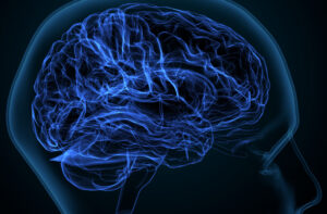
People with breast cancer may soon be diagnosed through ML faster than through a biopsy. Those suffering from depression might be able to predict mood changes through smart phone recordings of daily activities such as the time they wake up and amount of time they spend exercising. ML may also help paralyzed people regain autonomy using prosthetics controlled by patterns identified in brain scan data. ML research promises these and many other possibilities to help people lead healthier lives.

But while the number of ML studies grow, the actual use of it in doctors’ offices has not expanded much past simple functions such as converting voice to text for notetaking.

The limitations lie in medical research’s small sample sizes and unique datasets. This small data makes it hard for machines to identify meaningful patterns. The more data, the more accuracy in ML diagnoses and predictions. For many diagnostic uses, massive numbers of subjects in the thousands would be needed, but most studies use smaller numbers in the dozens of subjects.

But there are ways to find significant results from small datasets if you know how to manipulate the numbers. Running statistical tests over and over again with different subsets of your data can indicate significance in a dataset that in reality may be just random outliers.

This tactic, known as P-hacking or feature hacking in ML, leads to the creation of predictive models that are too limited to be useful in the real world. What looks good on paper doesn’t translate to a doctor’s ability to diagnose or treat us.

These statistical mistakes, oftentimes done unknowingly, can lead to dangerous conclusions.

To help scientists avoid these mistakes and push ML applications forward, Konrad Kording, Nathan Francis Mossell University Professor with appointments in the Departments of Bioengineering and Computer and Information Science in Penn Engineering and the Department of Neuroscience at Penn’s Perelman School of Medicine, is leading an aspect of a large, NIH-funded program known as CENTER – Creating an Educational Nexus for Training in Experimental Rigor. Kording will lead Penn’s cohort by creating the Community for Rigor which will provide open-access resources on conducting sound science. Members of this inclusive scientific community will be able to engage with ML simulations and discussion-based courses.

“The reason for the lack of ML in real-world scenarios is due to statistical misuse rather than the limitations of the tool itself,” says Kording. “If a study publishes a claim that seems too good to be true, it usually is, and many times we can track that back to their use of statistics.”

Such studies that make their way into peer-reviewed journals contribute to misinformation and mistrust in science and are more common than one might expect.

Read the full story in Penn Engineering Today.

Penn Medicine and Independence Blue Cross Eliminate Preapprovals for Imaging Tests

Brian Litt, MD

Brian Litt, Professor in Bioengineering in Penn Engineering and in Neurology in the Perelman School of Medicine, spoke to Neurology Today about the advances in technology for detecting and forecasting seizures.

The Litt Lab for Translational Neuroengineering translates neuroengineering research directly into patient care, focusing on epilepsy and a variety of research initiatives and clinical applications.

“Dr. Litt’s group is working with one of a number of startups developing ‘dry’ electrode headsets for home EEG monitoring. ‘They are still experimental, but they’re getting better, and I’m really optimistic about the possibilities there.'”

Read “How Detecting, Identifying and Forecasting Seizures Has Evolved” in Neurology Today.

Read more stories featuring Litt in the BE Blog.

Study Reveals New Insights on Brain Development Sequence Through Adolescence

by Eric Horvath

3D illustration of a human brain
Image: Courtesy of Penn Medicine News

Brain development does not occur uniformly across the brain, but follows a newly identified developmental sequence, according to a new Penn Medicine study. Brain regions that support cognitive, social, and emotional functions appear to remain malleable—or capable of changing, adapting, and remodeling—longer than other brain regions, rendering youth sensitive to socioeconomic environments through adolescence. The findings are published in Nature Neuroscience.

Researchers charted how developmental processes unfold across the human brain from the ages of 8 to 23 years old through magnetic resonance imaging (MRI). The findings indicate a new approach to understanding the order in which individual brain regions show reductions in plasticity during development.

Brain plasticity refers to the capacity for neural circuits—connections and pathways in the brain for thought, emotion, and movement—to change or reorganize in response to internal biological signals or the external environment. While it is generally understood that children have higher brain plasticity than adults, this study provides new insights into where and when reductions in plasticity occur in the brain throughout childhood and adolescence.

The findings reveal that reductions in brain plasticity occur earliest in “sensory-motor” regions, such as visual and auditory regions, and occur later in “associative” regions, such as those involved in higher-order thinking (problem solving and social learning). As a result, brain regions that support executive, social, and emotional functions appear to be particularly malleable and responsive to the environment during early adolescence, as plasticity occurs later in development.

“Studying brain development in the living human brain is challenging. A lot of neuroscientists’ understanding about brain plasticity during development actually comes from studies conducted with rodents. But rodent brains do not have many of what we refer to as the association regions of the human brain, so we know less about how these important areas develop,” says corresponding author Theodore D. Satterthwaite, the McLure Associate Professor of Psychiatry in the Perelman School of Medicine, and director of the Penn Lifespan Informatics and Neuroimaging Center (PennLINC).

Read the full story in Penn Medicine News.

N.B.: Theodore Satterthwaite in a member of the Penn Bioengineering Graduate Group.

Novel Tools for the Treatment and Diagnosis of Epilepsy

by Nathi Magubane

A neurologist examines an encephalogram of a patient’s brain.
Throughout his career, Brian Litt has fabricated tools that support international collaboration, produced findings that have led to significant breakthroughs, and mentored the next generation of researchers tackling neurological disorders. (Image: iStock Photo/Alona Siniehina)

When Brian Litt of the Perelman School of Medicine and School of Engineering and Applied Science began treating patients as a neurologist, he found that the therapies and treatments for epilepsy were mostly reliant on traditional pharmacological interventions, which had limited success in changing the course of the disease.

People with epilepsy are often prescribed anti-seizure medications, and, while they are effective for many, about 30% of patients still continue to experience seizures. Litt sought new ways to offer patients better treatment options by investigating a class of devices that electronically stimulate cells in the brain to modulate activity known as neurostimulation devices.

Litt’s research on implantable neurostimulation devices has led to significant breakthroughs in the technology and has broadened scientists’ understanding of the brain. This work started not long after he came to Penn in 2002 with licensing algorithms to help drive a groundbreaking device by NeuroPace, the first closed-loop, responsive neurostimulator to treat epilepsy.

Building on this work, Litt noted in 2011 how the implantable neurostimulation devices being used at the time had rigid wires that didn’t conform to the brain’s surface, and he received support from CURE Epilepsy to accelerate the development of newer, flexible wires to monitor and stimulate the brain.

“CURE is one of the epilepsy community’s most influential funding organizations,” Litt says. “Their support for my lab has been incredibly helpful in enabling the cutting-edge research that we hope will change epilepsy care for our patients.”

Read the full story in Penn Today.

Brian Litt is a Professor in Bioengineering and Neurology.

Flavia Vitale is an Assistant Professor in Neurology with a secondary appointment in Bioengineering.

Jonathan Viventi is an Assistant Professor in Biomedical Engineering at Duke University.

A Potential Strategy to Improve T Cell Therapy in Solid Tumors

A new Penn Medicine preclinical study demonstrates a simultaneous ‘knockout’ of two inflammatory regulators boosts T cell expansion to attack solid tumors.

by Meagan Raeke

Image: Courtesy of Penn Medicine News

A new approach that delivers a “one-two punch” to help T cells attack solid tumors is the focus of a preclinical study by researchers from the Perelman School of Medicine. The findings, published in the Proceedings of the National Academy of Sciences, show that targeting two regulators that control gene functions related to inflammation led to at least 10 times greater T cell expansion in models, resulting in increased anti-tumor immune activity and durability.

CAR T cell therapy was pioneered at Penn Medicine by Carl H. June, the Richard W. Vague Professor in Immunotherapy at Penn and director of the Center for Cellular Immunotherapies (CCI) at Abramson Cancer Center, whose work led to the first approved CAR T cell therapy for B-cell acute lymphoblastic leukemia in 2017. Since then, personalized cellular therapies have revolutionized blood cancer treatment, but remained stubbornly ineffective against solid tumors, such as lung cancer and breast cancer.

“We want to unlock CAR T cell therapy for patients with solid tumors, which include the most commonly diagnosed cancer types,” says June, the new study’s senior author. “Our study shows that immune inflammatory regulator targeting is worth additional investigation to enhance T cell potency.”

One of the challenges for CAR T cell therapy in solid tumors is a phenomenon known as T cell exhaustion, where the persistent antigen exposure from the solid mass of tumor cells wears out the T cells to the point that they aren’t able to mount an anti-tumor response. Engineering already exhausted T cells from patients for CAR T cell therapy results in a less effective product because the T cells don’t multiply enough or remember their task as well.

Previous observational studies hinted at the inflammatory regulator Regnase-1 as a potential target to indirectly overcome the effects of T cell exhaustion because it can cause hyperinflammation when disrupted in T cells—reviving them to produce an anti-tumor response. The research team, including lead author David Mai, a bioengineering graduate student in the School of Engineering and Applied Science, and co-corresponding author Neil Sheppard, head of the CCI T Cell Engineering Lab, hypothesized that targeting the related, but independent Roquin-1 regulator at the same time could boost responses further.

“Each of these two regulatory genes has been implicated in restricting T cell inflammatory responses, but we found that disrupting them together produced much greater anti-cancer effects than disrupting them individually,” Mai says. “By building on previous research, we are starting to get closer to strategies that seem to be promising in the solid tumor context.”

Read the full story in Penn Medicine News.

June is a member of the Penn Bioengineering Graduate Group. Read more stories featuring June’s research here.

OCTOPUS, an Optimized Device for Growing Mini-Organs in a Dish

by Devorah Fischler

With OCTOPUS, Dan Huh’s team has significantly advanced the frontiers of organoid research, providing a platform superior to conventional gel droplets. OCTOPUS splits the soft hydrogel culture material into a tentacled geometry. The thin, radial culture chambers sit on a circular disk the size of a U.S. quarter, allowing organoids to advance to an unprecedented degree of maturity.

When it comes to human bodies, there is no such thing as typical. Variation is the rule. In recent years, the biological sciences have increased their focus on exploring the poignant lack of norms between individuals, and medical and pharmaceutical researchers are asking questions about translating insights concerning biological variation into more precise and compassionate care.

What if therapies could be tailored to each patient? What would happen if we could predict an individual body’s response to a drug before trial-and-error treatment? Is it possible to understand the way a person’s disease begins and develops so we can know exactly how to cure it?

Dan Huh, Associate Professor in the Department of Bioengineering at the University of Pennsylvania’s School of Engineering and Applied Science, seeks answers to these questions by replicating biological systems outside of the body. These external copies of internal systems promise to boost drug efficacy while providing new levels of knowledge about patient health.

An innovator of organ-on-a-chip technology, or miniature copies of bodily systems stored in plastic devices no larger than a thumb drive, Huh has broadened his attention to engineering mini-organs in a dish using a patient’s own cells.

A recent study published in Nature Methods helmed by Huh introduces OCTOPUS, a device that nurtures organs-in-a-dish to unmatched levels of maturity. The study leaders include Estelle Park, doctoral student in Bioengineering, Tatiana Karakasheva, Associate Director of the Gastrointestinal Epithelium Modeling Program at Children’s Hospital of Philadelphia (CHOP), and Kathryn Hamilton, Assistant Professor of Pediatrics in Penn’s Perelman School of Medicine and Co-Director of the Gastrointestinal Epithelial Modeling Program at CHOP.

Read the full story in Penn Engineering Today.

‘Organ-on-a-Chip’ Device Provides New Insights into Early-Stage Pregnancy

by Scott Harris

Dan Huh’s BIOLines Lab develops several different kinds of organ-on-a-chip systems, such as this blinking-eye-on-a-chip.

If you’d read about it in a science fiction novel, you might not have believed it. Human organs and organ systems — from lungs to blood vessels to blinking eyes — bio-miniaturized and stored on a plastic chip no larger than a matchbook.

But that’s the breathing, blinking reality at the Biologically Inspired Engineering Systems (BIOLines) Laboratory in the Department of Bioengineering in the School of Engineering and Applied Sciences at the University of Pennsylvania, a bona fide pioneer of what is now widely known as “organ-on-a-chip” technology. Proponents hope these devices can one day help scientists around the world learn more about the body’s inner workings and ultimately improve disease prevention and treatment.

“The century-old practice of cell culture is to grow living cells isolated from the human body in hard plastic dishes and keep them bathed in copious amounts of culture media under static conditions, and that is drastically different than the complex, dynamic environment of native tissues in which these cell reside,” said Dan Dongeun Huh, Ph.D., BIOLines’ principal investigator and an associate professor of Bioengineering in Penn’s School of Engineering and Applied Science. “What makes this organ-on-a-chip technology so unique and powerful is that it enables us to reverse-engineer living human tissues using microengineered devices and mimic their intricate biological interactions and physiological functions in ways that have not been possible using traditional cell culture techniques. This represents a major advance in our ability to model and understand the inner workings of complex physiological systems in the human body.”

Generally speaking, organ-on-a-chip devices are made of clear silicone rubber — the same material used to make contact lenses — and can vary in size and design. Embedded within are microfabricated three-dimensional chambers lined with different human cell types, arranged and propagated to ultimately form a structure complex enough to actually mimic the essential elements of a functioning organ.

With partners at the Perelman School of Medicine, BIOLines recently developed a newer variation of the organ-on-a-chip: one that replicates the interface between maternal tissue and the cells of the placenta at the critical moments in early pregnancy when the embryo is implanting in the uterus. Huh and Penn Medicine physicians led a study using the “implantation-on-a-chip” to observe things that would otherwise have been virtually unobservable.

The study findings appeared this spring in the journal Nature Communications.

Continue reading at Penn Medicine News.

Microbes That Cause Cavities Can Form Superorganisms Able to ‘Crawl’ and Spread On Teeth

by Katherine Unger Baillie

Hyun (Michel) Koo

A cross-kingdom partnership between bacteria and fungi can result in the two joining to form a “superorganism” with unusual strength and resilience. It may sound like the stuff of science fiction, but these microbial groupings are very much part of the here and now.

Found in the saliva of toddlers with severe childhood tooth decay, these assemblages can effectively colonize teeth. They were stickier, more resistant to antimicrobials, and more difficult to remove from teeth than either the bacteria or the fungi alone, according to the research team, led by University of Pennsylvania School of Dental Medicine scientists.

What’s more, the assemblages unexpectedly sprout “limbs” that propel them to “walk” and “leap” to quickly spread on the tooth surface, despite each microbe on its own being non-motile, the team reported in the journal Proceedings of the National Academy of Sciences

“This started with a very simple, almost accidental discovery, while looking at saliva samples from toddlers who develop aggressive tooth decay,” says Hyun (Michel) Koo, a professor at Penn Dental Medicine and a co-corresponding author on the paper. “Looking under the microscope, we noticed the bacteria and fungi forming these assemblages and developing motions we never thought they would possess: a ‘walking-like’ and ‘leaping-like’ mobility. They have a lot of what we call ‘emergent functions’ that bring new benefits to this assemblage that they could not achieve on their own. It’s almost like a new organism—a superorganism—with new functions.”

Read the full story in Penn Today.

Hyun (Michel) Koo is a professor in the Department of Orthodontics and the divisions of Community Oral Health and Pediatric Dentistry in the School of Dental Medicine, co-founder of the Center for Innovation & Precision Dentistry (CiPD) at the University of Pennsylvania, and member of the Penn Bioengineering Graduate Group.

A Robot Made of Sticks

Kristina García

Devin Carroll, a doctoral candidate in the School of Engineering and Applied Sciences, is designing a modular robot called StickBot, which may be adapted for rehabilitation use in global public health settings.

Stickbot, a small robot composed of sticks, circuitry, actuators, a microcontroller, and a motor driver, lashed together with string.
StickBot in walking mode, using the sticks as legs to propel itself across the table.

In late summer, just as the leaves were starting to crisp and curl in the heat, Devin Carroll walked out of his apartment, looked on the ground, and picked up a couple of sticks that he thought might work for his robot. About half an inch thick and the length of an adult hand, he stripped the three sticks of their bark and lashed them with string to StickBot, a modular robot composed of circuitry, actuators, a microcontroller, and a motor driver.

Powered by four AA batteries, connected by a maze of wires and blinking lights, StickBot’s wooden arms now thump up and over, powering the robot across the table at Penn’s General Robotics, Automation, Sensing & Perception (GRASP) Lab, where Carroll is a Ph.D. candidate in the School of Engineering and Applied Sciences.

Controlling the robot using an app he designed, Carroll shows how StickBot can pivot from using the sticks as legs in “crawler mode,” to using them as arms. In “grasper mode,” the sticks are attached to a controller plate on one side to form a hinge joint while moving with their free end to hold a cup upright.

Rather than a static, singular invention, StickBot is an idea, a flexible system that can be reconfigured in a variety of ways. A modular robot, StickBot’s components can be added, adjusted, and discarded as needed.

Read the full story in Penn Engineering Today.

This article features quotes from Michelle Johnson, Associate Professor in Physical Medicine and Rehabilitation in the Perelman School of Medicine and in Bioengineering in the School of Engineering and Applied Sciences, and Director of the Rehabilitation Robotics Lab.


Defining Neural “Representation”

by Marilyn Perkins

Neuroscientists frequently say that neural activity ‘represents’ certain phenomena, PIK Professor Konrad Kording and postdoc Ben Baker led a study that took a philosophical approach to tease out what the term means.

Monitors Show EEG Reading and Graphical Brain Model. In the Background Laboratory Man Wearing Brainwave Scanning Headset Sits in a Chair with Closed Eyes. In the Modern Brain Study Research Laboratory
Neuroscientists use the word “represent” to encompass multifaceted relationships between brain activity, behavior, and the environment.

One of neuroscience’s greatest challenges is to bridge the gaps between the external environment, the brain’s internal electrical activity, and the abstract workings of behavior and cognition. Many neuroscientists rely on the word “representation” to connect these phenomena: A burst of neural activity in the visual cortex may represent the face of a friend or neurons in the brain’s memory centers may represent a childhood memory.

But with the many complex relationships between mind, brain, and environment, it’s not always clear what neuroscientists mean when they say neural activity “represents” something. Lack of clarity around this concept can lead to miscommunication, flawed conclusions, and unnecessary disagreements.

To tackle this issue, an interdisciplinary paper takes a philosophical approach to delineating the many aspects of the word “representation” in neuroscience. The work, published in Trends in Cognitive Sciences, comes from the lab of Konrad Kording, a Penn Integrates Knowledge University Professor and senior author on the study whose research lies at the intersection of neuroscience and machine learning.

“The term ‘representation’ is probably one of the most common words in all of neuroscience,” says Kording, who has appointments in the Perelman School of Medicine and School of Engineering and Applied Science. “But it might mean something very different from one professor to another.”

Read the full story in Penn Today.

Konrad Kording is a Penn Integrates Knowledge University Professor with joint appointments in the Department of Neuroscience the Perelman School of Medicine and in the Department of Bioengineering in the School of Engineering and Applied Science.

Ben Baker is a postdoctoral researcher in the Kording lab and a Provost Postdoctoral Fellow. Baker received his Ph.D. in philosophy from Penn.

Also coauthor on the paper is Benjamin Lansdell, a data scientist in the Department of Developmental Neurobiology at St. Jude Children’s Hospital and former postdoctoral researcher in the Kording lab.

Funding for this study came from the National Institutes of Health (awards 1-R01-EB028162-01 and R01EY021579) and the University of Pennsylvania Office of the Vice Provost for Research.