Enamored by the chemical processes of life, Yihui Shen, J. Peter and Geri Skirkanich Assistant Professor of Innovation in Bioengineering, started her research career as a chemist studying the way that proteins fold and the intricate dynamics underlying life processes.
“As an undergraduate, I studied physical chemistry, thinking that one day I’d be addressing challenges in hardcore STEM fields,” she says. “It wasn’t until I observed the dynamics of a single protein molecule that I fell in love with microscopy. I realized that this imaging tool could not only help us observe biological processes on a small scale, but it could also provide new insight at the interface of engineering, chemistry and physics and solve problems on a large scale.”
When Shen turned her attention to microscopy, the field itself was advancing quickly, with improvements being made and new techniques being released every month. Without missing a beat, Shen dove deeper into the most current tools available when she joined Dr. Wei Min’s lab at Columbia University as a doctoral student.
“Professor Wei Min is a pioneer in a new imaging technique called coherent Raman imaging,” says Shen. “In this type of microscopy, we focus light on a very specific point in the cell and measure the amount of scattered light that comes back after exchanging energy with the molecular vibration. This approach allows us to visualize the spatial distribution of different molecules, the very chemistry of life I had studied as an undergraduate, at a high enough resolution to gain insights into biological processes, such as tissue organization, drug distribution and cellular metabolism.”
With this new tool under her belt, Shen was able to ask the kinds of questions that could connect the use of this observation tool to practical applications for real-world challenges.
“I started thinking outside the box,” says Shen. “What if we could observe the chemical exchanges involved in metabolism as they are happening on the scale of a single cell, and then use that insight to pinpoint the exact metabolic pathways and molecules that facilitate tumor growth and disease?”
Congratulations to Alison Pouch, Assistant Professor in Bioengineering in the School of Engineering and Applied Science, and in Radiology in the Perelman School of Medicine, on winning a 2024 Cardiac Center Innovation Award for scientific research from the Children’s Hospital of Philadelphia (CHOP)’s Philly Spin-In. Pouch’s study, titled “Systemic Semilunar Valve Mechanics and Simulated Repair in Congenital Heart Disease,” is a collaboration with Matthew Jolley, Assistant Professor of Anesthesiology and Critical Care at CHOP:
“Through biomechanical assessment, Drs. Matthew Jolley and Alison Pouch are leading an interdisciplinary CHOP-Penn team that plans to determine why current approaches to systemic semilunar valve (SSV) repair fail. They will also investigate methods to design improved repairs before going to the operating room by using computational simulation to iteratively optimize repair.
‘We believe that understanding biomechanics of abnormal SSVs and explorations of simulated repair will markedly improve our ability to characterize, risk stratify, and surgically treat SSV dysfunction, thereby improving long-term outcomes and quality of life in patients with SSV dysfunction,’ Dr. Jolley said.”
Pouch’s lab focuses on 3D/4D segmentation and modeling of heart valves in echocardiographic images with applications to surgical treatment of valvular regurgitation as part of the Penn Image Computing and Science Laboratory.
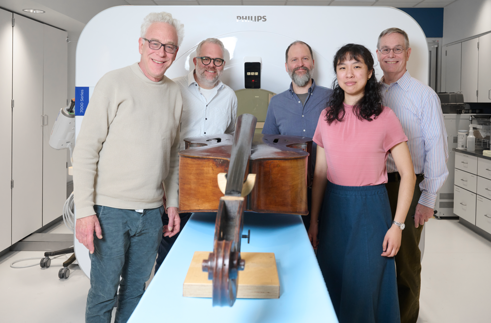
The instrument imaging team, from left: Philadelphia Orchestra bassist Duane Rosengard; Peter Noël, PhD, director of CT Research at the Perelman School of Medicine; luthier Zachary S. Martin; Leening Liu, a PhD student in Noël’s Laboratory of Advanced Computed Tomography Imaging; and Mark Kindig.
When you’re an expert in medical CT imaging, two things are bound to happen, says Peter Noël, PhD, associate professor of Radiology and director of CT Research at the Perelman School of Medicine. One: You develop an insatiable curiosity about the inner workings of all kinds of objects, including those unrelated to your research. And two: Both colleagues and complete strangers will ask for your help in imaging a wide variety of unexpected items.
Over the course of his career, in between managing his own research projects, Noël has imaged diverse objects ranging from animal skulls to tree samples from a German forest, all in the name of furthering scientific knowledge. But none has intrigued him as much as his current extracurricular project: the first known attempt to perform CT imaging of some of the world’s finest string basses.
The goal is to crack the code on what makes a world-class instrument. This knowledge could both increase the ability to better care for masterworks built between the 17th and 19th centuries, as well as providing insights into refining the building of new ones, including possibly shifting from older, scarcer European wood to the use of sustainably harvested U.S. wood.
That’s why Noël and Leening Liu, a PhD student in Noël’s Laboratory of Advanced Computed Tomography Imaging, have found themselves volunteering to run the basses through a Penn CT scanner occasionally, when they’re not developing next-generation CT technology.
“We always learn something out of projects like this … the more appealing part is that medical research can also be applied to non-medical things,” Noël said. “We have the opportunity to take what we learn in medicine and use it for something else—in this case, moving the arts forward.”
Leening Liu is a Ph.D. student in Bioengineering. She is a member of the Laboratory for Advanced Tomography Imaging (LACTI) with research interests including clinical applications of spectral CT and spectral CT thermometry.
We hope you will join us for the 2023 Herman P. Schwan Distinguished Lecture by Dr. Dorin Comaniciu, hosted by the Department of Bioengineering.
Wednesday, December 13, 2023 1:00 PM ET Location: Wu & Chen Auditorium (Levine 101) The lecture and Q&A will be followed by a light reception in Levine Lobby.
Speaker:Dorin Comaniciu, Ph.D. Senior Vice President Artificial Intelligence and Digital Innovations Siemens Healthineers
About Dorin Comaniciu:
Dr. Comaniciu serves as Senior Vice President for Artificial Intelligence and Digital Innovation at Siemens Healthineers. His scientific contributions to machine intelligence and computational imaging have translated to multiple clinical products focused on improving the quality of care, specifically in the fields of diagnostic imaging, image-guided therapy, and precision medicine.
Comaniciu is a member of the National Academy of Medicine, the Romanian Academy, and a Top Innovator of Siemens. He is a Fellow of the IEEE, ACM, MICCAI Society, and AIMBE, and a recipient of the IEEE Longuet-Higgins Prize for fundamental contributions to computer vision. Recent recognition of his work includes an honorary doctorate from Friedrich-Alexander University of Erlangen-Nuremberg.
He has co-authored 550 granted patents and 350 peer-reviewed publications that have received 61,000 citations, with an h-index of 102, in the areas of machine intelligence, medical imaging, and precision medicine.
A graduate of University of Pennsylvania’s Wharton School, Comaniciu received a doctorate in electrical and computer engineering from Rutgers University and a doctorate in electronics and telecommunications from Polytechnic University of Bucharest.
He is an advocate for technological innovations that save and enhance lives, addressing critical issues in global health.
About the Schwan Lecture:
The Herman P. Schwan Distinguished Lecture is in honor of one of the founding members of the Department of Bioengineering, who emigrated from Germany after World War II and helped create the field of bioengineering in the US. It recognizes people with a similar transformative impact on the field of bioengineering.
Congratulations to the members of the Penn Bioengineering community who were awarded 2023 Accelerating from Lab to Market Pre-Seed Grants from the University of Pennsylvania Office of the Vice Provost for Research (OVPR).
Andrew Tsourkas, Ph.D.
Three faculty affiliated with Bioengineering were included among the four winners. Andrew Tsourkas, Professor in Bioengineering and Co-Director of the Center for Targeted Therapeutics and Translational Nanomedicine (CT3N), was awarded for his project titled “Precise labeling of protein scaffolds with fluorescent dyes for use in biomedical applications.” Tsourkas’s team created protein scaffold that can better control the location and orientation of fluorescent dyes, commonly used for a variety of biomedical applications, such as labeling antibodies or fluorescence-guided surgery. The Tsourkas Lab specializes in “creating novel targeted imaging and therapeutic agents for the detection and/or treatment of diverse diseases.”
Also awarded were Penn Bioengineering Graduate Group members Mark Anthony Sellmeyer, Assistant Professor in Radiology in the Perelman School of Medicine, and Rahul M. Kohli, Associate Professor of Medicine in the Division of Infectious Diseases in the Perelman School of Medicine.
From the OVPR website:
“Penn makes significant commitments to academic research as one of its core missions, including investment in faculty research programs. In some disciplines, the path by which discovery makes an impact on society is through commercialization. Pre-seed grants are often the limiting step for new ideas to cross the ‘valley of death’ between federal research funding and commercial success. Accelerating from Lab to Market Pre-Seed Grant program aims to help to bridge this gap.”
Read the full list of winning projects and abstracts at the OVPR website.
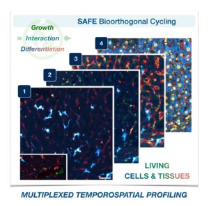
Cells in complex organisms undergo frequent changes, and researchers have struggled to monitor these changes and create a comprehensive profile for living cells and tissues. Historically researchers have been limited to only 3-5 markers due to spectral overlaps in fluorescence microscopy, an essential tool required for imaging cells. With only this small handful of markers, it is difficult to monitor protein expressions of live cells and a comprehensive profile of cellular dynamics cannot be created. However, a new study in Nature Biotechnology addresses these limitations by demonstrating a new method for comprehensive profiling of living cells.
Jina Ko, PhD
Jina Ko, Assistant Professor in Bioengineering in the School of Engineering and Applied Science and in Pathology and Laboratory Medicine in the Perelman School of Medicine, conducted postdoctoral research at Massachusetts General Hospital (MGH) and the Wyss Institute at Harvard University, and the work for this study was done under the supervision of Jonathan Carlson M.D., Ph.D. and Ralph Weissleder M.D., Ph.D. of MGH. Ko’s lab at Penn develops novel technologies using bioengineering, molecular biology, and chemistry to address diagnostic challenges for precision medicine.
To address these limitations in microscopy, the team developed a new chemistry tool which was highly gentle to cells. This “scission-accelerated fluorophore exchange (or SAFE)” method utilizes “click” chemistry, a type of chemistry that follows examples found in nature to create fast and simple reactions. This new SAFE method functions with non-toxic conditions to living cells and tissues, whereas previous methods have used harsh chemicals that would strip off fluorophores and consequently would not work with living cells and tissues.
With the development of SAFE, the authors demonstrated that researchers can now effectively perform multiple cycles of cell profiling and can monitor cellular changes over the course of their observations. Instead of the previous limitation of 3-5 markers total, SAFE allows for many more cycles and can keep track of almost as many markers as the researcher wants. One can now stain cells and quench/release fluorophores and repeat the cycle multiple times for multiplexing on living cells. Each cycle can profile 3 markers, and so someone interested in profiling 15 markers could easily perform 5 cycles to achieve this much more comprehensive cell profile. With this breakthrough in more detailed imaging of cells, SAFE demonstrates broad applicability for allowing researchers to better investigate the physiologic dynamics in living systems.
This study was supported by the Schmidt Science Fellows in Partnership with the Rhodes Trust and National Institutes of Health, National Cancer Institute (K99CA256353).
One of the reasons that cancer is notoriously difficult to treat is that it can look very different for each patient. As a result, most targeted therapies only work for a fraction of cancer patients. In many cases, patients will have tumors with no known markers that can be targeted, creating an incredible challenge in identifying effective treatments. A new study seeks to address this problem with the development of a simple methodology to help differentiate tumors from healthy, normal tissues.
This new study, published inScience Advances, was led by Andrew Tsourkas, Professor in Bioengineering and Co-Director of the Center for Targeted Therapeutics and Translational Nanomedicine (CT3N), who had what he describes as a “crazy idea” to use a patient’s antibodies to find and treat their own tumors, taking advantage of the immune system’s innate ability to identify tumors as foreign. This study, spearheaded by Burcin Altun, a former postdoctoral researcher in Tsourkas’s lab, and continued and completed by Fabiana Zappala, a former graduate student in Penn Bioengineering, details their new method for site-specifically labeling “off-the-shelf” and native serum autoantibodies with T cell–redirecting domains.
Researchers have known for some time that cancer patients will generate an antibody response to their own tumors. These anti-tumor antibodies are quite sophisticated in their ability to specifically identify cancer cells; however, they are not sufficiently potent to confer a therapeutic effect. In this study, Tsourkas’s team converted these antibodies into bispecific antibodies, thereby increasing their potency. T cell-redirecting bispecific antibodies are a new form of targeted therapeutic that forms a bridge between tumor cells and T cells which have been found to be as much as a thousand-times more potent than antibodies alone. By combining the specificity of a patient’s own antibodies with the potency of bispecific antibodies, researchers can effectively create a truly personalized therapeutic that is effective against tumors.
In order to test out this new targeted therapeutic approach, the Tsourkas lab had to develop an entirely new technology, allowing them to precisely label antibodies with T cell targeting domains, creating a highly homogeneous product. Previously it has not been possible to convert native antibodies into bispecific antibodies, but Tsourkas’s Targeted Imaging Therapeutics and Nanomedicine or TITAN lab specializes in the creation of novel targeted imaging and therapeutic agents for detection and treatment of various diseases. “Much is yet to be done before this could be considered a practical clinical approach,” says Tsourkas. “But I hope at the very least this works stimulates new ideas in the way we think about personalized medicine.”
In their next phase, Tsourkas’s team will be working to separate anti-tumor antibodies from other antibodies found in patients’ serum (which could potentially redirect the bispecific antibodies to other locations in the body), as well as examining possible adverse reactions or unintended effects and immunogenicity caused by the treatment. However, this study is just the beginning of a promising new targeted therapeutic approach to cancer treatment.
This work was supported by Emerson Collective and the National Institutes of Health, National Cancer Institute (R01 CA241661).
The Solomon R. Pollack Award for Excellence in Graduate Bioengineering Research is given annually to the most deserving Bioengineering graduate students who have successfully completed research that is original and recognized as being at the forefront of their field. This year Penn Bioengineering recognizes the outstanding work of two graduate students in Bioengineering: Erin Berlew and Rhea Chitalia.
Erin Berlew, Ph.D. candidate in Bioengineering
Erin Berlew is a Ph.D. candidate in the lab of Brian Chow, Associate Professor in Bioengineering. She successfully defended her thesis, titled “Single-component optogenetic tools for cytoskeletal rearrangements,” in December 2021. In her research, she used the BcLOV4 optogenetic platform discovered/developed in the Chow lab to control RhoGTPase signaling. Erin earned a B.S. in Chemistry from Haverford College in 2015 and was an Americorps member with City Year Philadelphia from 2015-2016. “Erin is a world-class bioengineering with an uncommon record of productivity gained through her complementary expertise in molecular, cellular, and computational biology,” says Chow. “She embodies everything wonderful, both academically and culturally, about our graduate program and its distinguished history.” Erin’s hobbies outside the lab include spending time with family, reading mystery novels, enjoying Philadelphia, and crossword puzzles. In the future, she hopes to continue to teach for the BE department (she has already taught ENGR 105 and served as a TA for undergraduate and graduate courses) and to conduct further research at Penn.
Rhea Chitalia, Ph.D. candidate in Bioengineering
Rhea Chitalia is a Ph.D. candidate in Bioengineering and a member of the Computational Biomarker Imaging Group (CBIG), advised by Despina Kontos, Matthew J. Wilson Associate Professor of Research Radiology II in the Perelman School of Medicine. Rhea completed her B.S.E. in Biomedical Engineering at Duke University in 2015. Her doctoral research concerns leveraging machine learning, bioinformatics, and computer vision to develop computational imaging biomarkers for improved precision cancer care. In December 2021 she successfully defended her thesis titled “Computational imaging biomarkers for precision medicine: characterizing intratumor heterogeneity in breast cancer.” “It has been such a privilege to mentor Rhea on her dissertation research,” says Kontos. “Rhea has been a star graduate student. Her work has made fundamental contributions in developing computational methods that will allow us to gain important insight into tumor heterogeneity by utilizing a multi-modality imaging approach.” David Mankoff, Matthew J. Wilson Professor of Research Radiology in the Perelman School of Medicine, served as Rhea’s second thesis advisor. “It was a true pleasure for me to work with Rhea and to Chair her BE Thesis Committee,” Mankoff adds. “Rhea’s Ph.D. thesis and thesis presentation was one of the best I have had the chance to be involved with in my graduate mentoring career.” After graduation, Rhea hopes to further precision medicine initiatives through the use of real world, multi-omic data in translational industry settings. She will be joining Invicro as an Imaging Scientist. In her spare time, Rhea enjoys trying new restaurants, reading, and spending time with friends and family.
To combat the COVID-19 pandemic caused by the SARS-CoV2 virus, Dr. Andrew Tsourkas’s Targeted Imaging Therapeutics and Nanomedicine (Titan) Lab in Penn Bioengineering, in collaboration with the Penn-based startup, AlphaThera, was recently awarded a $667,000 SBIR Phase II Grant Extension to support its efforts in commercializing COVID-19 detection technology. The grant supports work to address the growing need for anti-viral antibody testing. Specifically, the Tsourkas Lab and AlphaThera hope to leverage their expertise with antibody conjugation technologies to reduce the steps and complexity of existing detection assays to enable greater production and higher sensitivity tests. AlphaThera was founded in 2016 by Andrew Tsourkas, PhD, Professor of Bioengineering and James Hui, MD, PhD, a graduate of the Perelman School of Medicine and Penn Bioengineering’s doctoral program.
During this pandemic it is crucial to characterize disease prevalence among populations, understand immunity, test vaccine efficacy and monitor disease resurgence. Projections have indicated that millions of daily tests will be needed to effectively control the virus spread. One important testing method is the serological assay: These tests detect the presence of SARS-CoV2 antibodies in a person’s blood produced by the body’s immune system responding to infection. Serological tests not only diagnose active infections, but also establish prior infection in an individual, which can greatly aid in forecasting disease spread and contact tracing. To perform the serological assays for antibody detection, well-established immunoassay methods are used such as ELISA.
A variety of issues have slowed the distribution of these serological assays for antibody testing. The surge in demand for testing has caused shortages in materials and reagents that are crucial for the assays. Furthermore, complexity in some of the assay formats can slow both production and affect the sensitivity of test results. Recognizing these problems, AlphaThera is leveraging its novel conjugation technology to greatly improve upon traditional assay formats.
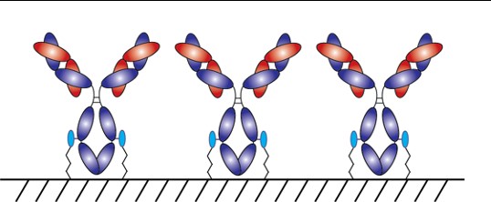
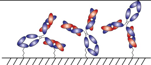
With AlphaThera’s conjugation technology, the orientation of antibodies can be precisely controlled so that they are aligned and uniformly immobilized on assay detection plates. This is crucial as traditional serological assays often bind antibodies to plates in a non-uniform manner, which increases variability of results and reduces sensitivity. See Fig 1 below. With AlphaThera’s uniform antibody immobilization, assay specificity could increase by as much as 1000- fold for detection of a patient’s SaRS-CoV2 antibodies.
Fig 1: Uniform vs Non-Uniform Immobilized Antibodies on Surface: Top is AlphaThera improvement, showing how antibodies would be uniformly immobilized and oriented on a plate for detection. Bottom is how many traditional serological assays immobilize antibodies, resulting in variability of results and lower specificity.
Furthermore, AlphaThera is addressing the shortage of assay reagents, specifically secondary antibody reagents, by removing certain steps from traditional serological assays. Rather than relying on secondary antibodies for detection of the patient antibodies, AlphaThera’s technology can label the patient SaRS-CoV2 primary antibodies directly in serum with a detection reagent. This eliminates several processing steps, reducing the time of the assay by as much as 50%, as well as the costs.
The Tsourkas Lab and AlphaThera have initiated their COVID-19 project, expanding into the Pennovation Center and onboarding new lab staff. Other antibody labeling products have also become available and are currently being prepared for commercialization. Check out the AlphaThera website to learn more about their technology at https://www.alphathera.com.
NIH SBIR Phase II Grant Extension— 5-R44-EB023750-03 (PI: Yu) — 10/07/2020 – 10/07/2021
DNA Microscopy Gives a Better Look at Cell and Tissue Organization
A new technique that researchers from the Broad Institute of MIT and Harvard University are calling DNA microscopy could help map cells for better understanding of genetic and molecular complexities. Joshua Weinstein, Ph.D., a postdoctoral associate at the Broad Institute, who is also an alumnus of Penn’s Physics and Biophysics department and former student in Penn Bioengineering Professor Ravi Radhakrishnan’s lab, is the first author of this paper on optics-free imaging published in Cell.
The primary goal of the study was to find a way of improving analysis of the spatial organization of cells and tissues in terms of their molecules like DNA and RNA. The DNA microscopy method that Weinstein and his team designed involves first tagging DNA, and allowing the DNA to replicate with those tags, which eventually creates a cloud of sorts that diffuses throughout the cell. The DNA tags subsequent interactions with molecules throughout the cell allowed Weinstein and his team to calculate the locations of those molecules within the cell using basic lab equipment. While the researchers on this project focused their application of DNA microscopy on tracking human cancer cells through RNA tags, this new method opens the door to future study of any condition in which the organization of cells is important.
If you’ve ever pressed a picture-hanging strip onto the wall only to realize it’s slightly off-center, you know the disappointment behind adhesion as we typically experience it: it may be strong, but it’s mostly irreversible. While you can un-stick the used strip from the wall, you can’t turn its stickiness back on to adjust its placement; you have to start over with a new strip or tolerate your mistake. Beyond its relevance to interior decorating, durable, reversible adhesion could allow for reusable envelopes, gravity-defying boots, and more heavy-duty industrial applications like car assembly.
Such adhesion has eluded scientists for years but is naturally found in snail slime. A snail’s epiphragm — a slimy layer of moisture that can harden to protect its body from dryness — allows the snail to cement itself in place for long periods of time, making it the ultimate model in adhesion that can be switched on and off as needed. In a new study, Penn Engineers demonstrate a strong, reversible adhesive that uses the same mechanisms that snails do.
Low-Dose Radiation CT Scans Could Be Improved by Machine Learning
Machine learning is a type of artificial intelligence growing more and more popular for applications in bioengineering and therapeutics. Based on learning from patterns in a way similar to the way we do as humans, machine learning is the study of statistical models that can perform specific tasks without explicit instructions. Now, researchers at Rensselaer Polytechnic Institute (RPI) want to use these kinds of models in computerized tomography (CT) scanning by lowering radiation dosage and improving imaging techniques.
A recent paper published in Nature Machine Intelligence details the use of modularized neural networks in low-dose CT scans by RPI bioengineering faculty member Ge Wang, Ph.D., and his lab. Since decreasing the amount of radiation used in a scan will also decrease the quality of the final image, Wang and his team focused on a more optimized approach of image reconstruction with machine learning, so that as little data as possible would be altered or lost in the reconstruction. When tested on CT scans from Massachusetts General Hospital and compared to current image reconstruction methods for the scans, Wang and his team’s method performed just as well if not better than scans performed without the use of machine learning, giving promise to future improvements in low-dose CT scans.
A Mind-Controlled Robotic Arm That Requires No Implants
A new mind-controlled robotic arm designed by researchers at Carnegie Mellon University is the first successful noninvasive brain-computer interface (BCI) of its kind. While BCIs have been around for a while now, this new design from the lab of Bin He, Ph.D., a Trustee Professor and the Department Head of Biomedical Engineering at CMU, hopes to eliminate the brain implant that most interfaces currently use. The key to doing this isn’t in trying to replace the implants with noninvasive sensors, but in improving noisy EEG signals through machine learning, neural decoding, and neural imaging. Paired with increased user engagement and training for the new device, He and his team demonstrated that their design enhanced continuous tracking of a target on a computer screen by 500% when compared to typical noninvasive BCIs. He and his team hope that their innovation will help make BCIs more accessible to the patients that need them by reducing the cost and risk of a surgical implant while also improving interface performance.
KIChE is an organization that aims “to promote constructive and mutually beneficial interactions among Korean Chemical Engineers in the U.S. and facilitate international collaboration between engineers in U.S. and Korea.”
We would also like to congratulate Natalia Trayanova, Ph.D., of the Department of Biomedical Engineering at Johns Hopkins University on being inducted into the Women in Tech International (WITI) Hall of Fame. Beginning in 1996, the Hall of Fame recognizes significant contributions to science and technology from women. Trayanova’s research specializes in computational cardiology with a focus on virtual heart models for the study of individualized heart irregularities in patients. Her research helps to improve treatment plans for patients with cardiac problems by creating virtual simulations that help reduce uncertainty in either diagnosis or courses of therapy.
Finally, we would like to congratulate Andre Churchwell, M.D., on being named Vanderbilt University’s Chief Diversity Officer and Interim Vice Chancellor for Equity, Diversity, and Inclusion. Churchwell is also a professor of medicine, biomedical engineering, and radiology and radiological sciences at Vanderbilt, with a long career focused in cardiology.


 A new technique that researchers from the Broad Institute of MIT and Harvard University are calling DNA microscopy could help map cells for better understanding of genetic and molecular complexities.
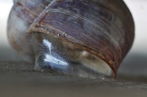