“QR Code for Cancer Cells” – Uncovering Why Some Cells Become Resistant to Anti-Cancer Therapies

by Win Reynolds

QR code
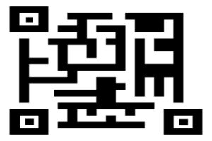
A research team led by engineers at the University of Pennsylvania and Northwestern University scientists has created a new synthetic biology approach, or a “QR code for cancer cells,” to follow tumor cells over time, finding there are meaningful differences in why a cancer cell dies or survives in response to anti-cancer therapies.

Remarkably, what fate cancer cells choose after months of therapy is “entirely predictable” based on seemingly small, yet important, differences that appear even before treatment begins. The researchers also discovered the reason is not genetics, contrary to beliefs held in the field.

The findings were recently published in Nature.

The study outlined the team’s new technology platform that developed a QR code for each of the millions of cells for scientists to find and use later — much like tagging swans in a pond. The QR code directs researchers to a genome-wide molecular makeup of these cells and provides information about how they’ve reacted to cancer treatment.

“We think this work stands to really change how we think about therapy resistance,” said Arjun Raj, co-senior author and Professor in Bioengineering in the School of Engineering and Applied Science at the University of Pennsylvania. “Rather than drug-resistant cells coming in just one flavor, we show that even in highly controlled conditions, different ‘flavors’ can emerge, raising the possibility that each of these flavors may need to be treated individually.”

In the study, the lab and collaborators sought to apply synthetic biology tools to answer a key question in cancer research: What makes certain tumors come back a few months or years after therapy? In other words, could the lab understand what causes some rare cells to develop therapeutic resistance to a drug?

“There are many ways cells become different from each other,” said Yogesh Goyal, the co-senior author at Northwestern University. “Our lab asks, how do individual cells make decisions? Understanding this in the context of cancer is all the more exciting because there’s a clinically relevant dichotomy: A cell dies or becomes resistant when faced with therapies.”

Using the interdisciplinary team, the scientists put the before-and-after cloned cells through a whole genome sequencing pipeline to compare the populations and found no systematic underlying genetic mutations to investigate the hypothesis. Raj and Goyal  helped develop the QR code framework, FateMap, that could identify each unique cell that seemed to develop resistance to drug therapy. “Fate” refers to whether a cell dies or survives (and if so, how), and the scientists “map” the cells across their lifespan, prior to and following anti-cancer therapy. FateMap is the result of work from several research institutions, and it applies an amalgamation of concepts spanning several disciplines, including synthetic biology, genome engineering, bioinformatics, machine learning and thermodynamics.

“Some are different by chance — just as not all leaves on a tree look the same — but we wanted to determine if that matters,” Goyal said. “The cell biology field has a hard time defining if differences have meaning.”

Read the full story in Penn Engineering Today.

Why New Cancer Treatments are Proliferating

by Karen L. Brooks

Doctors performing surgery.
Image: Penn Medicine News

In the five years since the FDA’s initial approval of chimeric antigen receptor (CAR) T cell therapy, Penn Medicine has gleaned 20 additional approvals related to drugs and techniques to treat or detect cancer.

Rather than being the single disease class many people refer to, “cancer” is a blanket term that covers more than 100 distinct diseases, many of which have little in common aside from originating with rapidly dividing cells. Since different cancers demand different treatments, it follows that any given new therapy emerging from any institution would be likely to be a new cancer treatment.

But why so many in just this five-year period?

The volume of new cancer treatments makes sense, says Abramson Cancer Center (ACC) director Robert Vonderheide, attributing the flurry of new cancer drug approvals to a recent “explosion” in knowledge about cancer biology.

“Much of that knowledge is about the immune system’s ability to attack cancer, which people seriously doubted until about 20 years ago. As soon as we had a clinical validation for this Achilles heel in cancer, the dam burst for ideas about other ways to exploit that vulnerability to come forward,” he says. “The first drug that came out to activate the immune system inspired the rest of the field to find the next drug, and the one after that. We as a field have moved from serendipity and empiricism to science-driven drug design.”

The first CAR T cell therapy approval invigorated Penn faculty interested in finding new ways to harness the immune system to fight cancer.

“An approval like that makes what you’re working on more of a reality,” says Avery Posey, an assistant professor of systems pharmacology and translational therapeutics in the Perelman School of Medicine, whose lab team spends much of its time trying to identify more specific antigens for solid tumors and also studies ways to optimize engineered donor T cells. “It brings a new perspective, showing that your work is more than basic research and can actually become drugs that impact patients’ lives. That’s a real motivator to keep pushing forward.”

Honing new immunotherapies is a priority among Penn researchers, but not every recently approved new cancer treatment or detection tool developed at the institution engages the immune system. Faculty have explored and introduced widely varying approaches to improving the standard of care for cancer patients.

Read the full story in Penn Medicine Magazine.

Avery Posey is a member of the Penn Bioengineering Graduate Group. Read more stories featuring Posey here.

RNA Nanoparticle Therapy Stops the Spread of Incurable Bone Marrow Cancer

by

Myeloma cells producing monoclonal proteins of varying types, created by Scientific Animations under the Creative Commons Attributions-Share Alike International 4.0 License

Multiple myeloma is an incurable bone marrow cancer that kills over 100,000 people every year. Known for its quick and deadly spread, this disease is one of the most challenging to address. As these cancer cells move through different parts of the body, they mutate, outpacing possible treatments. People diagnosed with severe multiple myeloma that is resistant to chemotherapy typically survive for only three to six months. Innovative therapies are desperately needed to prevent the spread of this disease and provide a fighting chance for those who suffer from it.

Michael Mitchell, J. Peter and Geri Skirkanich Assistant Professor of Innovation in Bioengineering (BE), and Christian Figueroa-Espada, doctoral student in BE at the University of Pennsylvania School of Engineering and Applied Science, created an RNA nanoparticle therapy that makes it impossible for multiple myeloma to move and mutate. The treatment, described in their study published in PNAS, turns off a cancer-attracting function in blood vessels, disabling the pathways through which multiple myeloma cells travel.

By shutting down this “chemical GPS” that induces the migration of cancer cells, the team’s therapy stops the spread of multiple myeloma, helping to eliminate it altogether.

Read the full story in Penn Engineering Today.

Penn Bioengineering Graduate Student on T Cell Therapy Improvements

Image: Courtesy of Penn Medicine News

 Neil Sheppard,  Adjunct Associate Professor of Pathology and Laboratory Medicine in the Perelman School of Medicine, and David Mai, a Bioengineering graduate student in the School of Engineering and Applied Science, explained the findings of their recent study, which offered a potential strategy to improve T cell therapy in solid tumors, to the European biotech news website Labiotech.

Mai is a graduate student in the lab of Carl H. June, the Richard W. Vague Professor in Immunotherapy in Penn Medicine, Director of the Center for Cellular Immunotherapies (CCI) at the Abramson Cancer Center, and member of the Penn Bioengineering Graduate Group.

Read “Immunotherapy in the fight against solid tumors” in Labiotech.

Read more about this collaborative study here.

Two Penn Bioengineering Students Win SFB STAR Awards

Congratulations to two Bioengineering graduate students who were awarded Student Travel Achievement Recognition (STAR) Awards from the Society for Biomaterials (SFB). The STAR Award recognizes research excellence and develops future leaders within SFB and comes with a certificate and a monetary award of $250. Penn Bioengineering graduate students Rebecca Haley and Alex Hamilton, both members of the lab of Michael J. Mitchell, Skirkanich Assistant Professor of Innovation in Bioengineering, received their awards and presented on their research in the SFB annual meeting in April 2023.

Rebecca Haley, Ph.D. student in Bioengineering

Rebecca Haley is a Ph.D. student in Bioengineering and a NSF Graduate Research Fellow. In the Mitchell Lab, she focuses on the use of ionizable lipid nanoparticles for the delivery of protein cargos. Supported by this STAR award, she presented her work delivering small protein RAS-inhibitors that reduce cancer cell proliferation. Rebecca is interested in expanding the applications of lipid nanoparticle technology, allowing currently limited therapeutics to achieve functional delivery and, hopefully, clinical success.

Alex Hamilton, Ph.D. student in Bioengineering

Alex Hamilton is a Ph.D. student in Bioengineering and an NSF Graduate Research Fellow. Alex’s work in the Mitchell lab focuses on non-viral nucleic acid delivery. His research interests include cancer immunotherapy, vaccines, and fetal-maternal medicine. He is currently engaged in using novel high-throughput screening techniques to accelerate the discovery process for lipid nanoparticle development for a variety of disease applications.

Two more Mitchell Lab members were likewise recognized with honorable mention inn the STAR Awards: Hannah Safford, a Ph.D. student in Bioengineering and NSF Fellow, and Rohan Palanki, a M.D.-Ph.D. student in Bioengineering and NIH Fellow

Learn more about the Mitchell Lab’s research in biomaterials science, drug delivery, and cellular and molecular bioengineering in the lab’s website.

Read more stories featuring Mitchell and his team here.

A Potential Strategy to Improve T Cell Therapy in Solid Tumors

A new Penn Medicine preclinical study demonstrates a simultaneous ‘knockout’ of two inflammatory regulators boosts T cell expansion to attack solid tumors.

by Meagan Raeke

Image: Courtesy of Penn Medicine News

A new approach that delivers a “one-two punch” to help T cells attack solid tumors is the focus of a preclinical study by researchers from the Perelman School of Medicine. The findings, published in the Proceedings of the National Academy of Sciences, show that targeting two regulators that control gene functions related to inflammation led to at least 10 times greater T cell expansion in models, resulting in increased anti-tumor immune activity and durability.

CAR T cell therapy was pioneered at Penn Medicine by Carl H. June, the Richard W. Vague Professor in Immunotherapy at Penn and director of the Center for Cellular Immunotherapies (CCI) at Abramson Cancer Center, whose work led to the first approved CAR T cell therapy for B-cell acute lymphoblastic leukemia in 2017. Since then, personalized cellular therapies have revolutionized blood cancer treatment, but remained stubbornly ineffective against solid tumors, such as lung cancer and breast cancer.

“We want to unlock CAR T cell therapy for patients with solid tumors, which include the most commonly diagnosed cancer types,” says June, the new study’s senior author. “Our study shows that immune inflammatory regulator targeting is worth additional investigation to enhance T cell potency.”

One of the challenges for CAR T cell therapy in solid tumors is a phenomenon known as T cell exhaustion, where the persistent antigen exposure from the solid mass of tumor cells wears out the T cells to the point that they aren’t able to mount an anti-tumor response. Engineering already exhausted T cells from patients for CAR T cell therapy results in a less effective product because the T cells don’t multiply enough or remember their task as well.

Previous observational studies hinted at the inflammatory regulator Regnase-1 as a potential target to indirectly overcome the effects of T cell exhaustion because it can cause hyperinflammation when disrupted in T cells—reviving them to produce an anti-tumor response. The research team, including lead author David Mai, a bioengineering graduate student in the School of Engineering and Applied Science, and co-corresponding author Neil Sheppard, head of the CCI T Cell Engineering Lab, hypothesized that targeting the related, but independent Roquin-1 regulator at the same time could boost responses further.

“Each of these two regulatory genes has been implicated in restricting T cell inflammatory responses, but we found that disrupting them together produced much greater anti-cancer effects than disrupting them individually,” Mai says. “By building on previous research, we are starting to get closer to strategies that seem to be promising in the solid tumor context.”

Read the full story in Penn Medicine News.

June is a member of the Penn Bioengineering Graduate Group. Read more stories featuring June’s research here.

Student Summer Research Spotlight: Dahin Song

Dahin Song
Dahin Song (BE 2024)

Dahin Song, a third year undergraduate student in Bioengineering, penned a guest blog post for Penn Career Services as part of their ongoing series of posts by recipients of the 2022 Career Services Summer Funding Grant. In this post, Song talks about her opportunity to conduct research in the SMART Lab of Daeyeon Lee, Professor and Evan C. Thompson Term Chair for Excellence in Teaching in the Department of Chemical and Biomolecular Engineering and member of the Penn Bioengineering Graduate Group. During her summer research, Song worked on increasing the stability of the monolayer in microbubbles, gas particles which have been put to therapeutic use. She writes:

“My project was on increasing the stability of the monolayer using cholesterol; theoretically, this would decrease the permeability while maintaining the fluidity of the monolayer. Being given my own project at the get-go was initially intimidating; initial learning curve was overwhelming – along with new wet lab techniques and protocols, I learned a whole new topic well enough to ask meaningful questions. But in retrospect, throwing myself headlong into a project was the best method to immerse me in the research environment, especially as a first-time researcher. I learned how to read papers efficiently, troubleshoot research problems, navigate in a laboratory environment, and be comfortable with working independently but more importantly, with others.”

Read “The Itsy Bitsy Bubble” in the Career Services blog.

New Insights into the Mechanisms of Tumor Growth

by

3d render of cells secreting exosomes
A team of researchers led by the School of Arts & Science’s Wei Guo offers new insights into a mechanism that promotes tumor growth. “This information could be used to help clinicians diagnose cancers earlier in the future,” says Guo.

In many instances, the physical manifestation of cancers and the ways they are subsequently diagnosed is via a tumor, tissue masses of mutated cells and structures that grow excessively. One of the major mysteries in understanding what goes awry in cancers relates to the environments within which these structures grow, commonly known as the tumor microenvironment.

These microenvironments play a role in facilitating tumor survival, growth, and spread. Tumors can help generate their own infrastructure in the form of vasculature, immune cells, signaling molecules, and extracellular matrices (ECMs), three-dimensional networks of collagen-rich support scaffolding for a cell. ECMs also help regulate cellular communications, and in the tumor microenvironment ECMs can be a key promoter of tumor growth by providing structural support for cancerous cells and in modulating signaling pathways that promote growth.

Now, new research led by the School of Arts & Science’s Wei Guo and published in the journal Nature Cell Biology has bridged the complex structural interactions within the tumor microenvironment to the signals that trigger tumor growth. The researchers studied cancerous liver cells grown on ECMs of varying stiffness and discovered that the stiffening associated with tumor growth can initiate a cascade that increases the production of small lipid-encapsulated vesicles known as exosomes.

“Think of these exosomes as packages that each cell couriers out, and, depending on the address, they get directed to other cells,” says Ravi Radhakrishnan, professor of bioengineering in the School of Engineering and Applied Science and a co-author of the paper.

“By recording the number of packages sent, the addresses on these packages, their contents, and most importantly, how they’re regulated and generated, we can better understand the relationship between a patient’s tumor microenvironment and their unique molecular signaling signatures, hinting at more robust personalized cancer therapies,” Radhakrishnan says.

While studying exosomes in relation to tumor growth and metastasis has been well-documented in recent years, researchers have mostly focused on cataloging their characteristics rather than investigating the many processes that govern the creation and shuttling of exosomes between cells. As members of Penn’s Physical Sciences Oncology Center (PSOC), Guo and Radhakrishnan have long collaborated on projects concerning tissue stiffness. For this paper, they sought to elucidate how stiffening promotes exosome trafficking in cancerous intracellular signaling.

“Our lab previously found that high stiffness promotes the secretion of exosomes,” says Di-Ao Liu, co-first author of the paper and a graduate student in the Guo Lab. “Now, we were able to model the stiffening processes through experiments and identify molecular pathways and protein networks that cause this, which better links ECM stiffening to cancerous signaling.”

Read the full story in Penn Today.

Carl June and Avery Posey Lead the Way in CAR T Cell Therapy

Perelman School of Medicine (PSOM) professors and Penn Bioengineering Graduate Group members Carl June and Avery Posey are leading the charge in T cell therapy and the fight against cancer.

Avery Posey, PhD
Carl June, MD

Advances in genome editing through processes such as CRISPR, and the ability to rewire cells through synthetic biology, have led to increasingly elaborate approaches for modifying and supercharging T cells for therapy. Avery Posey,  Assistant Professor of Pharmacology, and Carl June, the Richard W. Vague Professor in Immunotherapy, explain how new techniques are providing tools to counter some of the limitations of current CAR T cell therapies in a recent Nature feature.

The pair were also part of a team of researchers from PSOM, the Children’s Hospital of Philadelphia (CHOP), and the Corporal Michael J. Crescenz VA Medical Center to receive an inaugural $8 million Therapy ACceleration To Intercept CAncer Lethality (TACTICAL) Award from the Prostate Cancer Foundation. Their project will develop new clinic-ready CAR T cell therapies for Metastatic Castrate-Resistant Prostate Cancer (mCRPC).

Read “The race to supercharge cancer-fighting T cells” in Nature.

Read about the TACTICAL Award in the December 2022 Awards & Accolades section of Penn Medicine News.

Inside the Mitchell Lab: Crossing Biological Barriers

by

Black and white photo of Mike Mitchell working in the lab.
Mike Mitchell, Ph.D.

Engineers in the Center for Precision Engineering for Health (CPE4H) are focusing on innovations in diagnostics and delivery, cellular and tissue engineering, and the development of new devices that integrate novel materials with human tissues. Below is an excerpt from “Going Small to Win Big: Engineering Personalized Medicine,” featuring the research from the laboratory of Michael Mitchell, J. Peter and Geri Skirkanich Assistant Professor of Innovation in Bioengineering.

The Challenge

Solid tumors evade the immune system’s ability to attack them in part due to the tumors’ tough, fibrous biological barriers that circulating immune cells can’t cross. Researchers need to identify ways to deliver individualized treatments that can better target these tumors without causing damage to healthy tissues or affecting overall quality of life.

The Status Quo

Current cancer treatments typically involve surgery, radiation or chemo- therapy to eliminate solid tumors. These treatments are invasive and can cause numerous negative downstream effects. Newer treatments involve engineering a patient’s immune system to recognize and fight cancerous cells, but are so far only effective against certain “liquid” cancers, where the mutated cells circulate freely in the blood and bone marrow and are small enough to be picked off by the patient’s upgraded T cells. Additionally, existing methods can also require that the cell engineering take place in a lab rather than directly inside the body.

The Mitchell Lab’s Fix

Members of the lab of Michael Mitchell, J. Peter and Geri Skirkanich Assistant Professor of Innovation in Bioengineering, are looking to utilize nanoparticle delivery technology developed by their lab to engineer a different type of immune cell, the macrophage, in order to fight solid- tumor cancers from the inside.

The Mitchell lab is using lipid nanoparticles (LNPs) to carry mRNA and DNA sequences inside of macrophages, a type of immune cell that can consume tumor cells if engineered correctly. In theory, a patient would receive an injection carrying the LNP payload, and the macrophages, whose name literally means “big eaters,” would take up the genetic sequence, alter their function and be able to recognize a patient’s own unique tumor cells in the body.

Because of the way macrophages operate, they could cross the tumor’s biological barrier and attack the cells, destroying the tumor from the inside. An added benefit of the Mitchell Lab’s technology is that the destroyed tumor cells would then also allow other immune cells to present their antigens to circulating T cells, which could then learn to fight those same cancer cells in the future.

“One of the longstanding challenges that we face in the context of cancer and immunotherapies is that every tumor has unique antigens that are specific to patients,” says Mitchell. “This is why we’ve had a lot of trouble developing targeted therapies. Personalizing an approach by harnessing an individual’s immune system gives each patient a greater chance of a positive outcome.”

Read the full story in Penn Engineering magazine.