Secondary Cancers Following CAR T Cell Therapy Are Rare, Penn Medicine Analysis Shows

by Meagan Raeke

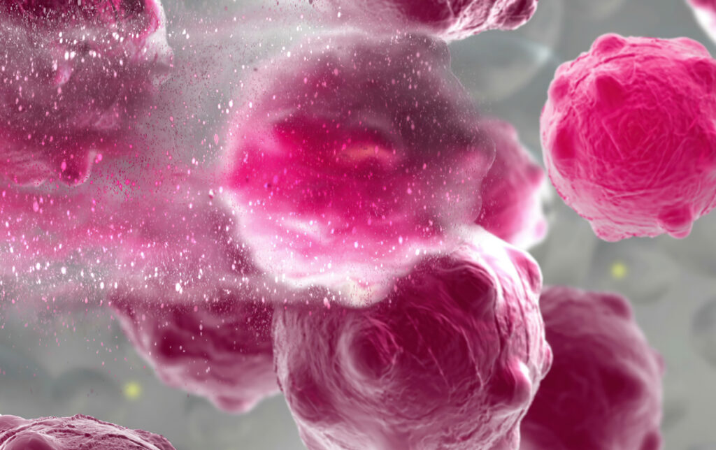
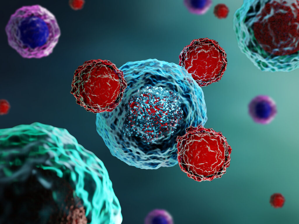
3d illustration of a damaged and disintegrating cancer cell. (Image: iStock/vitanovski)

The development of any type of second cancer following CAR T cell therapy is a rare occurrence, as found in an analysis of more than 400 patients treated at Penn Medicine, researchers from the Perelman School of Medicine at the University of Pennsylvania reported today in Nature Medicine. The team also described a single case of an incidental T cell lymphoma that did not express the CAR gene and was found in the lymph node of a patient who developed a secondary lung tumor following CAR T cell therapy.

CAR T cell therapy, a personalized form of immunotherapy in which each patient’s T cells are modified to target and kill their cancer cells, was pioneered at Penn. More than 30,000 patients with blood cancers in the United States—many of whom had few, if any, remaining treatment options available—have been treated with CAR T cell therapy since the first such therapy was approved in 2017. Some of the earliest patients treated in clinical trials have gone on to experience long-lasting remissions of a decade or more.

Secondary cancers, including T cell lymphomas, are a known, rare risk of several types of cancer treatment, including chemotherapy, radiation, and stem cell transplant. CAR T cell therapy is currently only approved to treat blood cancers that have relapsed or stopped responding to treatment, so patients who receive CAR T cell therapies have already received multiple other types of treatment and are facing dire prognoses.

In November 2023, the FDA announced an investigation into several reported cases of secondary T cell malignancies, including CAR-positive lymphoma, in patients who previously received CAR T cell therapy products. In January 2024, the FDA began requiring drugmakers to add a safety label warning to CAR T cell products. While the FDA review is still ongoing, it remains unclear whether the secondary T cell malignancies were caused by CAR T cell therapy.

As a leader in CAR T cell therapy, Penn has longstanding, clearly established protocols to monitor each patient both during and after treatment – including follow-up for 15 years after infusion – and participates in national reporting requirements and databases that track outcomes data from all cell therapy and bone marrow transplants.

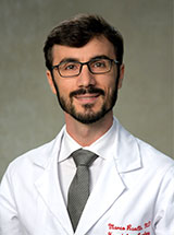
Marco Ruella, M.D.

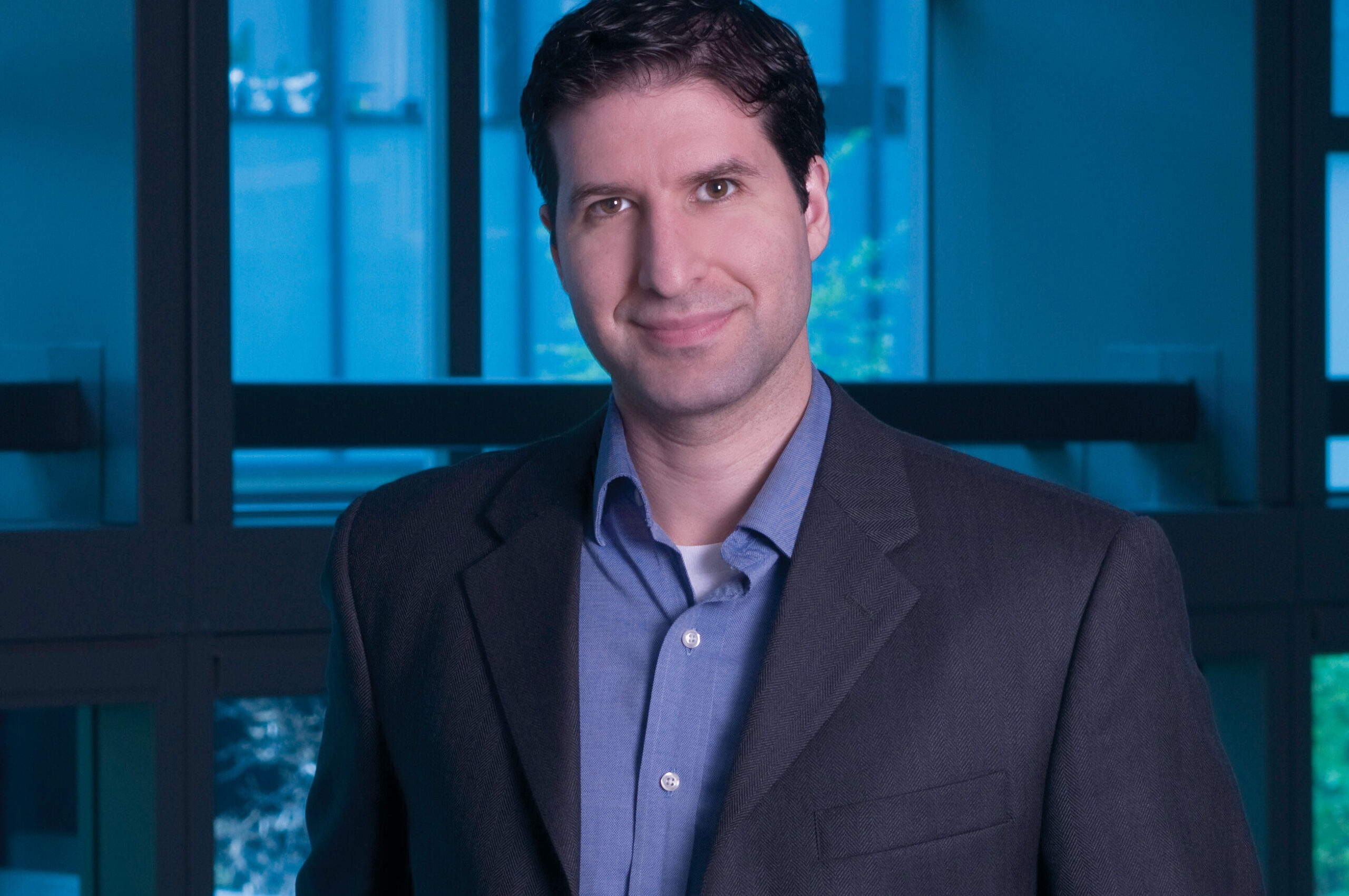
“When this case was identified, we did a detailed analysis and concluded the T cell lymphoma was not related to the CAR T cell therapy. As the news of other cases came to light, we knew we should go deeper, to comb through our own data to better understand and help define the risk of any type of secondary cancer in patients who have received CAR T cell products,” said senior author Marco Ruella, MD, an assistant professor of Hematology-Oncology and Scientific Director of the Lymphoma Program. “What we found was very encouraging and reinforces the overall safety profile for this type of personalized cell therapy.”

Read the full story in Penn Medicine News.

Marco Ruella is Assistant Professor of Medicine in the Perelman School of Medicine. He is a member of the Penn Bioengineering Graduate Group.

Sydney Shaffer Wins Christopher J. Marshall Award for Melanoma Research

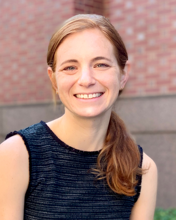
Sydney Shaffer, M.D., Ph.D.

Sydney Shaffer, Assistant Professor in Bioengineering in the School of Engineering and Applied Science and in Pathology and Laboratory Medicine in the Perelman School of Medicine, was named the 2023 Christopher J. Marshall Award winner by the Society for Melanoma Research (SMR). The award recognizes Shaffer’s contributions to melanoma research on oncogenic signalling and molecular pathogenesis of this disease, as well as her rapid development as a rising star and leader in the field, which have helped to further the SMR’s goal to eradicate melanoma. The award was presented at the SMR annual meeting in Philadelphia in November 2023. 

The Christopher J. Marshall Award was established in 2015 by the SMR in partnership with Melanoma Research Foundation Congress to recognize a student, postdoctoral fellow, or new independent PI who has published a substantial and original contribution to studies of signal transduction and melanoma.

Shaffer joined Penn as an Assistant Professor in 2019. She holds a M.D.-Ph.D. in Medicine and Bioengineering from the University of Pennsylvania and conducted postdoctoral research in cancer biology in the lab of Junwei Shi, Associate Professor in Penn Medicine. The Syd Shaffer Lab is an interdisciplinary team which focuses on “understanding how differences between single-cells generate phenotypes such as drug resistance, oncogenesis, differentiation, and invasion [using] a combination of imaging and sequencing technologies to investigate rare single-cell phenomena.” A recent paper in Nature Communications details the team’s method to quantify long-lived fluctuations in gene expression that are predictive of later resistance to targeted therapy for melanoma.

Read the award announcement and the full list of prior winners at the SMR website.

The NEMO Prize Goes to Research Improving Soft-Tissue Transplant Surgeries

by Melissa Pappas

Daeyeon Lee (left), Oren Friedman (center) and Sergei Vinogradov (right)

Each year, the Nemirovsky Engineering and Medicine Opportunity (NEMO) Prize, funded by Penn Health-Tech, awards $80,000 to a collaborative team of researchers from the University of Pennsylvania’s Perelman School of Medicine and the School of Engineering and Applied Science for early-stage, interdisciplinary ideas.

This year, the NEMO Prize has been awarded to Penn Engineering’s Daeyeon Lee, Russel Pearce and Elizabeth Crimian Heuer Professor in Chemical and Biomolecular Engineering, Oren Friedman, Associate Professor of Clinical Otorhinolaryngology in the Perelman School of Medicine, and Sergei Vinogradov, Professor in the Department of Biochemistry and Biophysics in the Perelman School of Medicine and the Department of Chemistry in the School of Arts & Sciences. Together, they are developing a new therapy that improves the survival and success of soft-tissue grafts used in reconstructive surgery.

More than one million people receive soft-tissue reconstructive surgery for reasons such as tissue trauma, cancer or birth defects. Autologous tissue transplants are those where cells and tissue such as fat, skin or cartilage are moved from one part of a patient’s body to another. As the tissue comes from the patient, there is little risk of transplant rejection. However, nearly one in four autologous transplants fail due to tissue hypoxia, or lack of oxygen. When transplants fail the only corrective option is more surgery. Many techniques have been proposed and even carried out to help oxygenate soft tissue before it is transplanted to avoid failures, but current solutions are time consuming and expensive. Some even have negative side effects. A new therapy to help oxygenate tissue quickly, safely and cost-effectively would not only increase successful outcomes of reconstructive surgery, but could be widely applied to other medical challenges. 

The therapy proposed by this year’s NEMO Prize recipients is a conglomerate or polymer of microparticles that can encapsulate oxygen and disperse it in sustainable and controlled doses to specific locations over periods of time up to 72 hours. This gradual release of oxygen into the tissue from the time it is transplanted to the time it functionally reconnects to the body’s vascular system is essential to keeping the tissue alive. 

“The microparticle design consists of an oxygenated core encapsulated in a polymer shell that enables the sustained release of oxygen from the particle,” says Lee. “The polymer composition and thickness can be controlled to optimize the release rate, making it adaptable to the needs of the hypoxic tissue.” 

These life-saving particles are designed to be integrated into the tissue before transplantation. However, because they exist on the microscale, they can also be applied as a topical cream or injected into tissue after transplantation. 

“Because the microparticles are applied directly into tissues topically or by interstitial injection (rather than being administered intravenously), they surpass the need for vascular channels to reach the hypoxic tissue,” says Friedman. “Their micron-scale size combined with their interstitial administration, minimizes the probability of diffusion away from the injury site or uptake into the circulatory system. The polymers we plan to use are FDA approved for sustained-release drug delivery, biocompatible and biodegrade within weeks in the body, presenting minimal risk of side effects.”

The research team is currently testing their technology in fat cells. Fat is an ideal first application because it is minimally invasive as an injectable filler, making it versatile in remodeling scars and healing injury sites. It is also the soft tissue type most prone to hypoxia during transplant surgeries, increasing the urgency for oxygenation therapy in this particular tissue type.

Read the full story in Penn Engineering Today.

Daeyeon Lee and Sergei Vinogradov are members of the Penn Bioengineering Graduate Group.

Penn Bioengineers Awarded 2023 “Accelerating from Lab to Market Pre-Seed” Grants

Congratulations to the members of the Penn Bioengineering community who were awarded 2023 Accelerating from Lab to Market Pre-Seed Grants from the University of Pennsylvania Office of the Vice Provost for Research (OVPR).

Andrew Tsourkas, Ph.D.

Three faculty affiliated with Bioengineering were included among the four winners. Andrew Tsourkas, Professor in Bioengineering and Co-Director of the Center for Targeted Therapeutics and Translational Nanomedicine (CT3N), was awarded for his project titled “Precise labeling of protein scaffolds with fluorescent dyes for use in biomedical applications.” Tsourkas’s team created protein scaffold that can better control the location and orientation of fluorescent dyes, commonly used for a variety of biomedical applications, such as labeling antibodies or fluorescence-guided surgery. The Tsourkas Lab specializes in “creating novel targeted imaging and therapeutic agents for the detection and/or treatment of diverse diseases.”

Also awarded were Penn Bioengineering Graduate Group members Mark Anthony Sellmeyer, Assistant Professor in Radiology in the Perelman School of Medicine, and Rahul M. Kohli, Associate Professor of Medicine in the Division of Infectious Diseases in the Perelman School of Medicine.

From the OVPR website:

“Penn makes significant commitments to academic research as one of its core missions, including investment in faculty research programs. In some disciplines, the path by which discovery makes an impact on society is through commercialization. Pre-seed grants are often the limiting step for new ideas to cross the ‘valley of death’ between federal research funding and commercial success. Accelerating from Lab to Market Pre-Seed Grant program aims to help to bridge this gap.”

Read the full list of winning projects and abstracts at the OVPR website.

Bioengineering Faculty Member Named ‘Young Innovator’ for Creation of Multiple Myeloma Therapy

by Abbey Porter

Michael Mitchell

Michael J. Mitchell, Associate Professor in Bioengineering at the University of Pennsylvania School of Engineering and Applied Science, has been named a “Young Innovator of Cellular and Molecular Bioengineering” by Cellular and Molecular Bioengineering, the journal of the Biomedical Engineering Society (BMES).

The award recognizes faculty who are conducting some of the most innovative and impactful studies in the field of biomedical engineering. Recipients will present their research and be officially recognized at the BMES Annual Meeting in October.

Mitchell is being honored for creating an RNA nanoparticle therapy that stops the spread of the deadly bone marrow cancer multiple myeloma and helps to eliminate it altogether. Known for being difficult to treat, the disease kills over 100,000 people every year.

“We urgently need innovative, effective therapies against this cancer,” Mitchell says. “The nanotechnology we developed can potentially serve as a platform to treat multiple myeloma and other bone marrow-based malignancies.”

Mitchell, along with Christian Figuerora-Espada, a doctoral student in Bioengineering, previously published a study in PNAS describing how their RNA nanoparticle therapy stops multiple myeloma from moving through the blood vessels and mutating. In their current paper in Cellular and Molecular Bioengineering, which expands upon this RNA nanoparticle platform, they show that inhibition of both multiple myeloma migration and adhesion to bone marrow blood vessels, combined with an FDA-approved multiple myeloma therapeutic, extends survival in a mouse model of multiple myeloma.

Read more in Penn Engineering Today.

A Suit of Armor for Cancer-fighting Cells

by Nathi Magubane

Chimeric antigen receptor T cell (CAR T) therapy has delivered promising results, transforming the fight against various forms of cancer, but for many, the therapy comes with severe and potentially lethal side effects. Now, a research team led by Michael Mitchell of the School of Engineering and Applied Science has found a solution that could help CAR T therapies reach their full potential while minimizing severe side effects. (Image: iStock / Meletios Verras)

In recent years, cancer researchers have hailed the arrival of chimeric antigen receptor T cell (CAR T) therapy, which has delivered promising results, transforming the fight against various forms of cancer. The process involves modifying patients’ T-cells to target cancer cells, resulting in remarkable success rates for previously intractable forms of cancer.

Six CAR T cell therapies have secured FDA approval, and several more are in the pipeline. However, these therapies come with severe and potentially lethal side effects, namely cytokine release syndrome (CRS) and neurotoxicity. These drawbacks manifest as a range of symptoms—from high fever and vomiting to multiple organ failure and patient death—posing significant challenges to broader clinical application.

Now, a research team led by Michael Mitchell, associate professor in the School of Engineering and Applied Science at the University of Pennsylvania, has found a solution that could help CAR T therapies reach their full potential while minimizing severe side effects. Their findings are published in the journal Nature Materials.

“Addressing CRS and neurotoxicity without compromising the therapeutic effectiveness of CAR T cells has been a complex challenge,” says Mitchell.

He says that unwanted interactions between CAR T and immune cells called macrophages drive the overactivation of macrophages, which in turn result in the release of toxic cytokines that lead to CRS and neurotoxicity.

“Controlling CAR T-macrophage interactions in vivo is difficult,” Mitchell says. “So, our study introduces a materials engineering-based strategy that involves incorporating a sugar molecule onto the surface of CAR T cells. These sugars are then used as a reactive handle to create a biomaterial coating around these cells directly in the body, which acts as a ‘suit of armor,’ preventing dangerous interactions with macrophages.”

First author Ningqiang Gong, a postdoctoral researcher in the Mitchell Lab, elaborates on the technique, “We attached this sugar molecule to the CAR T cells using metabolic labeling. This modification enables the CAR T cells to attack cancer cells without any hindrance.”

“When symptoms of CRS begin to manifest, we introduce another molecule—polyethylene glycol (PEG)—to create the suit of armor, which effectively blocks dangerous interactions between these engineered T cells, macrophages, and the tumor cells themselves,” Gong says.

Read the full story in Penn Today.

Carl June on the Boundless Potential of CAR T Cell Therapy

by Meagan Raeke

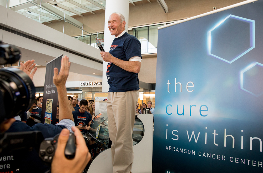
Carl June, at the flash mob celebration of the FDA approval of the CAR T cell therapy he developed, in August 2017. (Image: Courtesy of Penn Medicine Magazine)

For most of modern medicine, cancer drugs have been developed the same way: by designing molecules to treat diseased cells. With the advent of immunotherapy, that changed. For the first time, scientists engineered patients’ own immune systems to recognize and attack diseased cells.

One of the best examples of this pioneering type of medicine is CAR T cell therapy. Invented in the Perelman School of Medicine by Carl June, the Richard W. Vague Professor in Immunotherapy, CAR T cell therapy works by collecting T cells from a patient, modifying those cells in the lab so that they are designed to destroy cancerous cells, and reinfusing them into the patient. June’s research led to the first FDA approval for this type of therapy, in 2017. Six different CAR T cell therapies are now approved to treat various types of blood cancers. Carl June, at the flash mob celebration of the FDA approval of the CAR T cell therapy he developed, in August 2017. (Image: Courtesy of Penn Medicine Magazine)

CAR T cell therapy holds the potential to help millions more patients—if it can be successfully translated to other conditions. June and colleagues, including Daniel Baker, a fourth-year doctoral student in the Cell and Molecular Biology department, discuss this potential in a perspective published in Nature.

In the piece, June and Baker highlight other diseases that CAR T cell therapy could be effective.

“CAR T cell therapy has been remarkably successful for blood cancers like leukemias and lymphomas. There’s a lot of work happening here at Penn and elsewhere to push it to other blood cancers and to earlier stage disease, so patients don’t have to go through chemo first,” June says. “Another big priority is patients with solid tumors because they make up the vast majority of cancer patients. Beyond cancer, we’re seeing early signs that CAR T cell therapy could work in autoimmune diseases, like lupus.”

As for which diseases to pursue as for possible future treatment, June says, “essentially it boils down to two questions: Can we identify a population of cells that are bad? And can we target them specifically? Whether that’s asthma or chronic diseases or lupus, if you can find a bad population of cells and get rid of them, then CAR T cells could be therapeutic in that context.”

“What’s exciting is it’s not just theoretical at this point. There have been clinical reports in other autoimmune diseases, including myasthenia gravis and inflammatory myopathy,” Baker says. “But we are seeing early evidence that CAR T cell therapy will be successful beyond cancer. And it’s really opening the minds of people in the field to think about how else we could use CAR T. For example, there’s some pioneering work at Penn from the Epstein lab for heart failure. The idea is that you could use CAR T cells to get rid of fibrotic tissue after a cardiac injury, and potentially restore the damage following a heart attack.”

Baker adds, “there’s no question that over the last decade, CAR T cell therapy has revolutionized cancer. I’m hoping to play a role in bringing these next generation therapies to patients and make a real impact over the next decade. I think there’s potential for cell therapy to be a new pillar of medicine at large, and not just a new pillar of oncology.”

Read the full story at Penn Medicine Today.

SCALAR: A Microchip Designed to Transform the Production of mRNA Therapeutics and Vaccines

Led by Michael Mitchell and David Issadore of the School of Engineering and Applied Science, a team of researchers has developed a platform that could rapidly accelerate the development of mRNA-based lipid nanoparticle vaccines and therapeutics at both the small and large scale, SCALAR. (Image: iStock / Anatoly Morozov)

Following the global COVID-19 pandemic, the development and rapid deployment of mRNA vaccines highlighted the critical role of lipid nanoparticles (LNPs) in the context of pharmaceuticals. Used as the essential delivery vehicles for fragile RNA-based therapies and vaccines, LNPs protect the RNA from degradation and ensure effective delivery within the body.

Despite their critical importance, the large-scale manufacturing of these LNPs saw numerous bottlenecks during the pandemic, underscoring the need for scalable production techniques that could keep pace with global demand.

Now, in a paper published in the Proceedings of the National Academy of the Sciences, researchers at the University of Pennsylvania describe how the Silicon Scalable Lipid Nanoparticle Generation platform (SCALAR), a reusable silicon- and glass-based platform designed to transform the production landscape of LNPs for RNA therapeutics and vaccines, offers a scalable and efficient solution to the challenges exposed during the COVID-19 crisis.

“We’re excited to create a piece of technology platform that bridges the gap between small-scale discovery and large-scale manufacturing in the realm of RNA lipid nanoparticle vaccines and therapeutics,” says co-author Michael Mitchell, associate professor of bioengineering in the School of Engineering and Applied Science at Penn. “By doing so, we’ve effectively leapfrogged the clunky, time-consuming, and costly barriers that slow down the production ramp-up of promising new RNA medicines and vaccines.”

The intricacies of RNA-based therapies require the RNA to be encased in a delivery system capable of navigating the body’s biological obstacles. LNPs fulfill this role, allowing the RNA to reach the intended cells for maximum therapeutic impact. SCALAR aims to take this a step further, allowing for an unprecedented three orders of magnitude scalability in LNP production rates, addressing the speed and consistency bottlenecks that hinder existing methods.

Sarah Shepherd, the first author of the paper and a recent Ph.D. graduate who worked in the Mitchell Lab, says, “With SCALAR, we’re not just reacting to today’s challenges but proactively preparing for tomorrow’s opportunities and crises. This technology is flexible, uses mixing architectures well-documented in microfluidics, and is scalable enough to meet future demands in real time. That’s an enormous leap forward for the field.”

Shepherd says that SCALAR builds on prior work from the Mitchell lab and is based on a microfluidic chip platform. Akin to a computer chip, wherein a computer’s electrically integrated circuit has numerous little transistors transporting signals as ones or zeroes to produce an output, the SCALAR microchip precisely controls their two key reagents, lipids and RNA, to generate LNPs.

Read the full story in Penn Today.

“QR Code for Cancer Cells” – Uncovering Why Some Cells Become Resistant to Anti-Cancer Therapies

by Win Reynolds

QR codeA research team led by engineers at the University of Pennsylvania and Northwestern University scientists has created a new synthetic biology approach, or a “QR code for cancer cells,” to follow tumor cells over time, finding there are meaningful differences in why a cancer cell dies or survives in response to anti-cancer therapies.

Remarkably, what fate cancer cells choose after months of therapy is “entirely predictable” based on seemingly small, yet important, differences that appear even before treatment begins. The researchers also discovered the reason is not genetics, contrary to beliefs held in the field.

The findings were recently published in Nature.

The study outlined the team’s new technology platform that developed a QR code for each of the millions of cells for scientists to find and use later — much like tagging swans in a pond. The QR code directs researchers to a genome-wide molecular makeup of these cells and provides information about how they’ve reacted to cancer treatment.

“We think this work stands to really change how we think about therapy resistance,” said Arjun Raj, co-senior author and Professor in Bioengineering in the School of Engineering and Applied Science at the University of Pennsylvania. “Rather than drug-resistant cells coming in just one flavor, we show that even in highly controlled conditions, different ‘flavors’ can emerge, raising the possibility that each of these flavors may need to be treated individually.”

In the study, the lab and collaborators sought to apply synthetic biology tools to answer a key question in cancer research: What makes certain tumors come back a few months or years after therapy? In other words, could the lab understand what causes some rare cells to develop therapeutic resistance to a drug?

“There are many ways cells become different from each other,” said Yogesh Goyal, the co-senior author at Northwestern University. “Our lab asks, how do individual cells make decisions? Understanding this in the context of cancer is all the more exciting because there’s a clinically relevant dichotomy: A cell dies or becomes resistant when faced with therapies.”

Using the interdisciplinary team, the scientists put the before-and-after cloned cells through a whole genome sequencing pipeline to compare the populations and found no systematic underlying genetic mutations to investigate the hypothesis. Raj and Goyal  helped develop the QR code framework, FateMap, that could identify each unique cell that seemed to develop resistance to drug therapy. “Fate” refers to whether a cell dies or survives (and if so, how), and the scientists “map” the cells across their lifespan, prior to and following anti-cancer therapy. FateMap is the result of work from several research institutions, and it applies an amalgamation of concepts spanning several disciplines, including synthetic biology, genome engineering, bioinformatics, machine learning and thermodynamics.

“Some are different by chance — just as not all leaves on a tree look the same — but we wanted to determine if that matters,” Goyal said. “The cell biology field has a hard time defining if differences have meaning.”

Read the full story in Penn Engineering Today.

Why New Cancer Treatments are Proliferating

by Karen L. Brooks

Doctors performing surgery.
Image: Penn Medicine News

In the five years since the FDA’s initial approval of chimeric antigen receptor (CAR) T cell therapy, Penn Medicine has gleaned 20 additional approvals related to drugs and techniques to treat or detect cancer.

Rather than being the single disease class many people refer to, “cancer” is a blanket term that covers more than 100 distinct diseases, many of which have little in common aside from originating with rapidly dividing cells. Since different cancers demand different treatments, it follows that any given new therapy emerging from any institution would be likely to be a new cancer treatment.

But why so many in just this five-year period?

The volume of new cancer treatments makes sense, says Abramson Cancer Center (ACC) director Robert Vonderheide, attributing the flurry of new cancer drug approvals to a recent “explosion” in knowledge about cancer biology.

“Much of that knowledge is about the immune system’s ability to attack cancer, which people seriously doubted until about 20 years ago. As soon as we had a clinical validation for this Achilles heel in cancer, the dam burst for ideas about other ways to exploit that vulnerability to come forward,” he says. “The first drug that came out to activate the immune system inspired the rest of the field to find the next drug, and the one after that. We as a field have moved from serendipity and empiricism to science-driven drug design.”

The first CAR T cell therapy approval invigorated Penn faculty interested in finding new ways to harness the immune system to fight cancer.

“An approval like that makes what you’re working on more of a reality,” says Avery Posey, an assistant professor of systems pharmacology and translational therapeutics in the Perelman School of Medicine, whose lab team spends much of its time trying to identify more specific antigens for solid tumors and also studies ways to optimize engineered donor T cells. “It brings a new perspective, showing that your work is more than basic research and can actually become drugs that impact patients’ lives. That’s a real motivator to keep pushing forward.”

Honing new immunotherapies is a priority among Penn researchers, but not every recently approved new cancer treatment or detection tool developed at the institution engages the immune system. Faculty have explored and introduced widely varying approaches to improving the standard of care for cancer patients.

Read the full story in Penn Medicine Magazine.

Avery Posey is a member of the Penn Bioengineering Graduate Group. Read more stories featuring Posey here.