What originally drew me to this field was a “Women in Engineering Day” I attended at a local college while in high school. I had the opportunity to hear incredible women speak about their research regarding biomaterials and tissue engineering. This event showed me the impact this field can have on the world. This drove me to pursue an undergraduate degree in Biomedical Engineering, which only strengthened my passion. As I furthered my studies and began working full-time at a biotechnology company, I learned more about bioengineering. With encouragement from my coworkers and family, I decided to pursue my Master’s in Bioengineering and am delighted to have the opportunity to study at Penn.
What kind of research do you conduct, and what do you hope to focus on for your thesis?
I am actually a part-time student, who works full-time at a drug packaging and medical device company out in Exton, PA. Though I am not doing research on campus, my coursework has tied into previous research projects I have participated in at my job. My latest project entailed understanding different material properties used in container closure systems for mAb-based biologics and how they interact. This work was done to support an understanding of how to pick appropriate vial/syringe systems for various drug products in development.
What’s your favorite thing to do on Penn’s campus or in Philly?
My favorite thing to do is trying all the new restaurants and incredible foods this city has to offer. I think Philadelphia is so unique and has such rich cultural influences. With so many different neighborhoods and restaurant options you really can’t go wrong.
What did you study for your undergraduate degree, how does it pair with the work you’re doing now, and what advice would you give to your undergraduate self?
My undergraduate degree was in Biomedical Engineering. It has supported my graduate coursework very well and has given me a great opportunity to dive deeper into certain parts of my studies.
My advice to my younger self would be to take your time! It took me a little while to evaluate different graduate programs and choose which was right for me. Though it took some time, I ultimately decided what was best for me and couldn’t be happier with my choices.
What are you thinking about doing after graduate school?
Currently, I work full-time as an Associate Packaging Engineer at West Pharmaceutical Services in Exton, PA. I hope to take my degree to further my career and to help support my future aspirations at this company.
Growing up in Sri Lanka and being surrounded by relatives who were doctors, I have been fascinated by both modern and traditional medicine. However, during physician shadowing in high school, I came to the realization that I was far more fascinated with the technology doctors use rather than practicing medicine. Therefore, I made the decision to turn down studying medicine in the U.K. and come to Penn to study Bioengineering in the hopes of being more hands-on with medical technology.
Have you done research with a professor on campus? What did you like, and what didn’t you like about it?
I currently work in the Interventional Radiology Lab at the Hospital of the University of Pennsylvania (HUP) under Assistant Professor of Radiology Chamith Rajapakse. The best thing about research here is that I get to be hands-on with some of the most cutting edge technology in the world and help pioneer medical diagnostic techniques that aren’t traditionally being used anywhere else. The only downside is that the learning curve can be a little too steep.
What have been some of your favorite courses and/or projects in Bioengineering so far?
Without a doubt, my favorite BE class has to be BE 309 (Bioengineering Modeling, Analysis and Design Laboratory I) and especially the Computer-Cockroach Interface we have to develop for this lab.
What advice would you give to your freshman self?
There are way too many things happening at a given time at Penn. Take it easy and plan it out so you can do everything you want to! It’s totally possible. Who says you can’t work hard and play hard?!
What do you hope to pursue after obtaining your undergraduate degree?
My hope is to head my own health-tech startup and create technologies that will aid developing countries, starting out with my humble island of Sri Lanka first.
The blue circle is the global symbol for diabetes. Wikimedia Commons.
Diabetes is one of the more common diseases among Americans today, with the American Diabetes Association estimating that approximately 9.5 percent of the population battles the condition today. Though symptoms and causes may vary across types and patients, diabetes generally results from the body’s inability to produce enough insulin to keep blood sugar levels in check. A new experimental treatment from the lab of Sha Jin, Ph.D., a biomedical engineering professor at Binghamton University, aims to use about $1.2 million in recent federal grants to develop a method for pancreatic islet cell transplantation, as those are the cells responsible for producing insulin.
But the catch to this new approach is that relying on healthy donors of these islet cells won’t easily meet the vast need for them in diabetic patients. Sha Jin wants to use her grants to consider the molecular mechanisms that can lead pluripotent stem cells to become islet-like organoids. Because pluripotent stem cells have the capability to evolve into nearly any kind of cell in the human body, the key to Jin’s research is learning how to control their mechanisms and signaling pathways so that they only become islet cells. Jin also wants to improve the eventual culture of these islet cells into three-dimensional scaffolds by finding ways of circulating appropriate levels of oxygen to all parts of the scaffold, particularly those at the center, which are notoriously difficult to accommodate. If successful in her tissue engineering efforts, Jin will not only be able to help diabetic patients, but also open the door to new methods of evolving pluripotent stem cells into mini-organ models for clinical testing of other diseases as well.
A Treatment to Help Others See Better
Permanently crossed eyes, a medical condition called strabismus, affects almost 18 million people in the United States, and is particularly common among children. For a person with strabismus, the eyes don’t line up to look at the same place at the same time, which can cause blurriness, double vision, and eye strain, among other symptoms. Associate professor of bioengineering at George Mason University, Qi Wei, Ph.D., hopes to use almost $2 million in recent funding from the National Institute of Health to treat and diagnose strabismus with a data-driven computer model of the condition. Currently, the most common method of treating strabismus is through surgery on one of the extraocular muscles that contribute to it, but Wei wants her model to eventually offer a noninvasive approach. Using data from patient MRIs, current surgical procedures, and the outcomes of those procedures, Wei hopes to advance and innovate knowledge on treating strabismus.
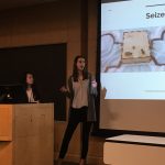
A Newly Analyzed Brain Mechanism Could be the Key to Stopping Seizures
Among neurological disorders, epilepsy is one of the most common. An umbrella term for a lot of different seizure-inducing conditions, many versions of epilepsy can be treated pharmaceutically. Some, however, are resistant to the drugs used for treatment, and require surgical intervention. Bin He, Ph. D., the Head of the Department of Biomedical Engineering at Carnegie Mellon University, recently published a paper in collaboration with researchers at Mayo Clinic that describes the way that seizures originating at a single point in the brain can be regulated by what he calls “push-pull” dynamics within the brain. This means that the propagation of a seizure through the brain relies on the impact of surrounding tissue. The “pull” he refers to is of the surrounding tissue towards the seizure onset zone, while the “push” is what propagates from the seizure onset zone. Thus, the strength of the “pull” largely dictates whether or not a seizure will spread. He and his lab looked at different speeds of brain rhythms to perform analysis of functional networks for each rhythm band. They found that this “push-pull” mechanism dictated the propagation of seizures in the brain, and suggest future pathways of treatment options for epilepsy focused on this mechanism.
Hyperspectral Imaging Might Provide New Ways of Finding Cancer
A new method of imaging called hyperspectral imaging could help improve the prediction of cancerous cells in tissue specimens. A recent study from a University of Texas Dallas team of researchers led by professor of bioengineering Baowei Fei, Ph.D., found that a combination of hyperspectral imaging and artificial intelligence led to an 80% to 90% level of accuracy in identifying the presence of cancer cells in a sample of 293 tissue specimens from 102 patients. With a $1.6 million grant from the Cancer Prevention and Research Institute of Texas, Fei wants to develop a smart surgical microscope that will help surgeons better detect cancer during surgery.
Fei’s use of hyperspectral imaging allows him to see the unique cellular reflections and absorptions of light across the electromagnetic spectrum, giving each cell its own specific marker and mode of identification. When paired with artificial intelligence algorithms, the microscope Fei has in mind can be trained to specifically recognize cancerous cells based on their hyperspectral imaging patterns. If successful, Fei’s innovations will speed the process of diagnosis, and potentially improve cancer treatments.
People and Places
A group of Penn engineering seniors won the Pioneer Award at the Rothberg Catalyzer Makerthon led be Penn Health-Tech that took place from October 19-20, 2019. SchistoSpot is a senior design project created by students Vishal Tien (BE ‘20), Justin Swirbul (CIS ‘20), Alec Bayliff (BE ‘20), and Bram Bruno (CIS ‘20) in which the group will design a low-cost microscopy dianostic tool that uses computer vision capabilities to automate the diagnosis of schistosomiasis, which is a common parasitic disease. Read about all the winners here.
Virginia Tech University will launch a new Cancer Research Initiative with the hope of creating an intellectual community across engineers, veterinarians, biomedical researchers, and other relevant scientists. The initiative will focus not only on building better connections throughout departments at the university, but also in working with local hospitals like the Carilion Clinic and the Children’s National Hospital in Washington, D.C. Through these new connections, people from all different areas of science and engineering and come together to share ideas.
Associate Professor of Penn Bioengineering Dani Bassett, Ph.D., recently sat down with the Penn Integrates Knowledge University Professor Duncan Watts, Ph.D., for an interview published in Penn Engineering. Bassett discusses the origins of network science, her research in small-world brain networks, academic teamwork, and the pedagogy of science and engineering. You can read the full interview here.
An all-female group of researchers from Northern Illinois University developed a device for use by occupational therapists that can capture three-dimensional images of a patient’s hand, helping to more accurately measure the hand or wrist’s range of motion. The group presented the abstract for their design at this year’s meeting of the Biomedical Engineering Society here in Philadelphia, where Penn students and researchers presented as well.
NB: Penn Bioengineering would like to congratulate one of its current Senior Design teams (Alec Bayliff, Bram Bruno, Justin Swirbul, and Vishal Then) which took home the $500 Pioneer Award at this year’s Rothberg Catalyzer competition this past weekend! Keep reading for more information on the competition, awards, and winners.
Penn Health-Tech’s Rothberg Catalyzer is a two-day makerthon that challenges interdisciplinary student teams to prototype and pitch medical devices that aim to address an unmet clinical need.
The Catalyzer’s third competition was held last weekend and was won by MAR Designs, a team of Mechanical Engineering and Applied Mechanics graduate students: Rebecca Li, Ariella Mansfield and Michael Sobrepera.
MAR Designs took home the top prize of $10,000 for their project, an orthotic device that children with cerebral palsy can more comfortably wear as they sleep.
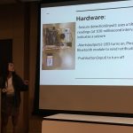
According to the team’s presentation, existing wrist orthoses “improve function and treat/prevent spasticity. However, patients report that these devices are uncomfortable which leads to lack of compliance and may also prevent patient’s eligibility for surgeries.” MAR Designs’ device initially allows full range of motion, but gradually straightens the wrist as the child is falling asleep.
In second place was Splash Throne. Team members Greg Chen, Nik Evitt, Jake Crawford and Meghan Lockwood proposed a toilet safety frame intended for elderly users. Embedded sensors track basic health information, like weight and heart-rate, as part of a preventative health routine.
Integrated Product Design students Jonah Arheim, Laura Ceccacci, Julia Lin and Alex Wan took third place with ONESCOPE, an untethered, hands-free laproscope designed to make minimally-invasive surgeries faster and safer.
Finally, SchistoSpot took home the Catalyzer’s Pioneer Award. Bioengineering and Computer and Information Science seniors Alec Bayliff, Bram Bruno, Justin Swirbul and Vishal Then designed a low-cost microscopy system that can aid in the diagnosis of the parasitic disease schistosomiasis by detecting eggs in urine samples, eliminating the need for a hospital visit.
The event was made possible by a three-year donation by scientist and entrepreneur Jonathan Rothberg, with the intent of inspiring the next generation of healthcare innovators.
The annual meeting of the Biomedical Engineering Society (BMES) will be held in our hometown of Philadelphia October 16-19, 2019. The professional society for bioengineers and biomedical engineers will be taking over the city of Brotherly Love, and lots of faculty and students from Penn’s Bioengineering will be attending and presenting their research.
As previously mentioned here, Jason Burdick, Ph.D., the Robert D. Bent Professor of Bioengineering, is one of three chairs of the 2019 annual meeting. He shares this position with two other local faculty: Alisa Morss Clyne, Ph.D., Associate Professor of Mechanical Engineering and Mechanics at Drexel University; and Ruth Ochia, Ph.D., Associate Professor of Instruction in Bioengineering at Temple University. They have worked together since their appointment in 2017 to plan and chair the Philadelphia conference. Check out the video below with details of what to expect from BMES in Philly.
For those of you who have never been to BMES, the event is comprised of a mixture of academic and networking events, including keynote talks from top researchers, thousands of oral and poster presentations, participants from around the world, and social receptions. To plan your itinerary, click here for the program and agenda and here for the schedule at a glance. With the meeting being held locally this year, there are far too many presentations by Penn Bioengineering faculty and staff to list here, so check out BMES’s searchable scientific program or our searchable schedule of Penn faculty student activities at this year’s meeting (separated by day).
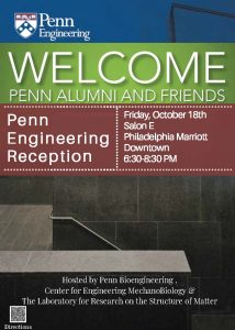
In addition to our academic participation, Penn Engineering and Bioengineering are also proud to sponsor this year’s meeting. Registered participants will have several venues to meet and mingle with Penn Engineering faculty, staff, and students and learn about its programs. Staff and volunteers will run a Penn Engineering booth (Booth #824) which will have literature on Penn departments and programs such as the Department of Bioengineering, the Center for Engineering MechanoBiology (CEMB), the Laboratory for Research on the Structure of Matter (LRSM), The Mahoney Institute for Neurosciences (MINS), and the Perelman School of Medicine’s Biomedical Graduate Studies group (BGS) and will be open 9:30am-5:00pm Thursday and Friday, and 9:30am-1:00pm during the conference.
For those interested in social events and networking, check out two back-to-back events on Friday night. From 6:30-8:30 pm, Penn’s Department of Bioengineering, CEMB, and LRSM will host a reception at the Philadelphia Marriott Downtown, Salon E. This will be followed by the meeting’s big BMES Dessert Bash at the Franklin Institute from 8:30-10:30 pm. (Please note: These events are open to registered conference participants only.) For those sticking around, there are no shortage of things to do in Philly, whether you are looking to site-see, shop, or dine.
We hope everyone has a wonderful time at the conference and enjoys Philadelphia! Let us know what activities you are enjoying most by tagging us on Twitter @pennbioeng or Instagram (pennbioengineering) and using the hashtag #pennbioengineering.
A New Sprayable Gel Can Help Prevent Surgical Adhesions
Adhesions are a common kind of scar tissue that can occur after surgery, and though usually not painful, they have the potential to result in complications like chronic pain or decreased heart efficiency, depending on where the scar tissue forms. Now, a sprayable gel developed by researchers at Stanford University will help to prevent adhesions from forming during surgical procedures. The gel, called PNP 1:10 in reference to its polymer-nanoparticle structure, has a similar stiffness to mayonnaise and achieves an ideal balance of slipperiness and stickiness that allows it to adhere easily to tissue of irregular shapes and surfaces. The flexible gel will actually dissolve in the body after two weeks, which is about how long most adhesions take to heal. Though lead author Lyndsay Stapleton, M.S., and senior authors Joseph Woo, M.D., and Eric Appel, Ph.D., have only tested the gel in rats and sheep so far, they hope that human applications are not too far in the future.
Learning to Understand Blood Clots in a New Model
Blood clots are the source of some of the deadliest human conditions and diseases. When a clot forms, blood flow can be interrupted, cutting off supply to the brain, heart, or other vital organs, resulting in potentially serious damage to the mind and body. For patients with certain bleeding disorders, clotting or the lack thereof can hold tremendous importance in surgery, and affect some of the typical procedures of a given operation. To help plan for such situations, researchers at the University of Buffalo created an in vitro model to help better illustrate the complex fluid mechanics of blood flow and clotting. Most importantly, this new model better demonstrates the role of shear stress in blood flow, and the way that it can affect the formation or destruction of blood clots – an aspect that current clinical devices often overlook. Led by Ruogang Zhao, Ph.D., the model can allow surgeons and hematologists to consider the way that certain chemical or physical treatments can affect clot formation on a patient-to-patient basis. The two key factors of the model are its incorporation of blood flow, and its relationship to shear stress, with clot stiffness through microfabrication technology using micropillars as force sensors for the stiffness. Going forward, Zhao and his research team hope to test the model on more patients, to help diversify the different bleeding disorders it can exhibit.
Training the Next Generation of Researchers
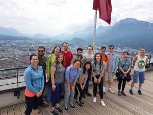
REACT 2019 students and Grenoble summer program interns, including undergraduate Rebecca Zappala (third from left, front), pose in front of the Chartreuse Mountains after completing a challenging ropes course. (Photo: Hermine Vincent)
Rebecca Zappala, a junior from Miami, Florida who is majoring in bioengineering, worked in Grenoble this summer on new ways to harvest water from fog. She describes her research project as a “futuristic” way to collect water and says that she’s thankful for the opportunity to work on her first independent research project through the Research and Education in Active Coatings Technology (REACT) program.
After learning the technical skills she needed for her project, Zappala spent her summer independently working on new ways to modify her material’s properties while working closely with her French PI and a post-doc in the lab. She was surprised to see how diverse the lab was, with researchers working on everything from biomolecular research to energy in the same space.
“I learned a lot,” she says about being in such an interdisciplinary setting. “I hadn’t been part of a research team before, and I got a lot of exposure to things that I wouldn’t have been exposed to otherwise.”
Virginia Tech Course Addresses the Needs of Wounded Veterans
A new course at Virginia Tech encourages students to apply engineering skills to real-life problems in the biomedical world by designing medical devices or other applications to assist veterans suffering from serious injuries or illnesses. Funded by the National Institute of Health, faculty from the Department of Biomedical Engineering and Mechanics hope that the course will allow students to see how theoretical knowledge from the classroom actually works in a clinical setting, and to understand how different stakeholder interests factor into designing a real device. What makes this new class unique from other traditional design-focused courses at other universities is its level of patient interaction. Students at Virginia Tech who choose to take this class will have the chance to gain input from field professionals like clinicians and engineers from the Salem Veterans Affairs Medical Center, while also being able to get direct feedback from the patients that the devices will actually help. Beginning in the spring of 2020, students can take the new course, and volunteer in the veterans clinics to gain even more experience with patients.
People and Places
Sevile Mannickarottu, the Director of the Educational Laboratories in Penn’s Department of Bioengineering and recent recipient of the Staff Recognition Award from the School of Engineering and Applied Sciences, presented a paper to highlight the Stephenson Foundation Bioengineering Educational Lab and Bio-Makerspace at the 126th annual conference of the American Society for Engineering Education. Thanks to the dedication of Mannickarottu and the lab staff to creating a space for simultaneous education and innovation, the Bioengineering Lab continues to be a hub for student community and projects of all kinds.
A week-long program for high school girls interested in STEM allows students to explore ideas and opportunities in the field through lab tours, guest speakers, and hands-on challenges. A collaboration across the University of Virginia Department of Biomedical Engineering, Charlottesville Women in Tech, and St. Anne’s Belfield School, the program gave this year’s students a chance to design therapies for children with disorders like hemiplegia and cerebral palsy, in the hopes that these interactive design challenges will inspire the girls to pursue future endeavors in engineering.
We would like to congratulate Nancy Albritton, Ph.D., on her appointment as the next Frank & Julie Jungers Dean of the College of Engineering at the University of Washington. Albritton brings both a deep knowledge of the research-to-marketplace pipeline and experience in the development of biomedical microdevices and pharmacoengineering to the new position.
We would also like to congratulate Jeffrey Brock, Ph.D., on his appointment as the dean of the Yale School of Engineering and Applied Science. Already both a professor of mathematics and a dean of science in the Faculty of Arts and Sciences at Yale, Brock’s new position will help him to foster collaborations across different departments of academia and research in science and engineering.
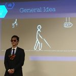
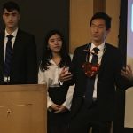
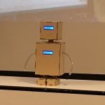
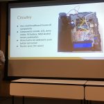
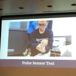
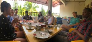
On May 8, 2019, first year Bioengineering students at the University of Pennsylvania gathered together for a marathon two-hour session in which no fewer than twenty-one groups presented the results of their final projects. These projects were the culmination of two semesters’ work in the courses BE 100 and 101, the department’s year-long introduction to Bioengineering. The topics were as diverse and creative as the students, ranging from medical devices and pediatric monitors to plant-care and diagnostic apps. They covered a variety of issues and needs, including tools to help the blind; lockboxes that incorporate breathalyzers (to stop you getting to your keys when intoxicated); mechanisms to sense epileptic seizures and monitor heart rate; and more. Each group had only four minutes to present the research, concept, and results of their project and give a brief demonstration. In the end, the entire class voted and two clear winners emerged. In first place was Group R7 with Heart Guide, a heart-shaped ultrasonic collision device for the blind. Group R3 came in second place with Pulsar the Robot, an adorable pediatric heart rate monitor. The course’s instructor, Dr. Michael Rizk, ended by saying that all of the students should be very proud of their work and that these final projects and the skills learned in year one are the foundation on which the rest of their BE curriculum will be based.
Congratulations to all of our first years on their amazing work. Check out some photos of their impressive work below! For more information on the Penn Bioengineering Undergraduate Curriculum, visit the department website. Most BE student projects are created in the George H. Stephenson Foundation Education Laboratory and “Bio-MakerSpace”, the department’s primary teaching lab.
In this series of posts, University of Pennsylvania students who took the spring 2019 APOC (Appropriate Point of Care Diagnostics) course write about their experience traveling to Ghana in May-June 2019.
by Allaire Morgan (Electrical Engineering, ’22)
Today, the team took various field trips related to water supply and public health. After being picked up on the bus, we drove for about an hour through the busy (and bumpy) streets of Kumasi to the Barakese Headworks of Ghana Water Company Limited. We spent the majority of the morning and early afternoon on a tour of the plant, following in chronological order the retrieval and treatment of water from the reservoir.
Upon walking up to the dam, the roar of the water was powerful. We went into a garage-like building which housed the four large pumps from the dam, pumping water uphill for further processing. After climbing approximately five stories on a shaky ladder, we reached the top of the dam to observe the reservoir from which the Ghana Water Company extracts its water. From there, our team ventured up the hill to observe the water treatment plant.
With the sun beating down, we slowly made our way through the facility, observing each stage of the treatment process in which millions of gallons of water were being treated at a time. The first stage, aeration, takes about six hours to complete. From there, water is pumped into giant drums for sedimentation, where water is stirred and polymers are added to force harmful chemicals to settle at the bottom. Clean water then slowly rises up in the million-gallon drum, with the clean water spilling over the edges to be collected and further processed in large sand filters. The sludge at the bottom of the drum is currently pumped back into the river, which may propose a serious public health problem in the future. The team then followed our guide into the labs, where we observed the various tests which are performed daily on the water after treatment to ensure proper sanitation. Lab technicians perform chemical experiments and culture the water to ensure that water-borne diseases cannot be carried by the filtered water.
After learning so much at the treatment plant, the team jumped on the bus to escape the heat and then traveled, after lunch, to the Komfo Akoye Teaching Hospital to observe the patient intake process in the hypertension clinic. We watched carefully in small groups from the corner of each doctor’s office to see how patients are treated and diagnosed. The doctors see around twenty to thirty patients per day, but on worse days they can see up to forty, with around two being new referrals from peripheral clinics. After speaking with the patient, the doctor makes a prescription recommendation on the patient’s paper file and gives it to the nurse for further processing. Each patient has a paper book which contains all of their medical data and history since coming to the hospital, and they retrieve it from a records room every time they visit. When asked about digitizing the process, the nurses were surprisingly resistant, arguing that they already were used to the paper filing system and they do not have the proper training to efficiently use a computer to file records.
After a long day of observations, the team traveled back to the guest house to eat dinner. Over our meal of pizza , spring rolls, and ambiguous but delicious juice, we discussed the events of the day and refocused our project, ironing out a specific plan for how we want to design our program and creating a vision for its implementation. We went to bed exhausted from a long day’s work but motivated for the project developments to come.
In this series of posts, University of Pennsylvania students who took the spring 2019 APOC (Appropriate Point of Care Diagnostics) course write about their experience traveling to Ghana in May-June 2019.
by Aime Bienfait Igiraneza (Computer Science, ’20)
Operation: Relaxation…and laundry
Itinerary:
Breakfast: Eggs, bread and tea. 8:30am
Laundry
Lunch at the Magnificent Foods
Running (or more of walking in my case) and swimming at the KNUST university
After what was a fun, informative, but busy week filled with hospital and clinic visits and walks through communities (and not forgetting the activity-filled day we had before, of course), this Sunday was supposed to be the time to relax…and do laundry.
Team meeting to talk about project.
Our breakfast started a little later than usual. Though breakfast was ready at 8:30 am, most of us had a lazy morning in and came out to eat at 9:00 am. When all the team members were assembled before the usual omelet and tea breakfast, we decided to do an impromptu recap of the week and brainstormed on how we could adapt our initial project to fit the clinics and hospitals we had visited during the week. This session, as spontaneous as it was, became a good way for us to build on our observations from the week not just for the applications of our project, but for identifying certain problems that can become future projects for future APOC teams.
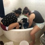
El and Laura doing laundry in the bathtub
Keeping the theme of lazy Sunday in mind, we did not start laundry until late morning, around 10:30 am. This didn’t prove to be a very wise idea, especially since we were supposed to do laundry the old-fashioned way with water, a bar of soap, and our good old hands. Furthermore, there proved to be a shortage of buckets for all of us to do laundry at the same time, which didn’t seem to leave enough time before the bus was supposed to pick us up at 1:00 pm for lunch. Nonetheless, we bonded as we shared our buckets (who knew that manual laundry-washing was such a social activity) and we made it in time for lunch; also nothing a little adrenaline and team work couldn’t fix.
Kyler and Bienfait doing laundry.
After laundry, we had lunch at the same restaurant we visited last Sunday: Magnificent Foods. The food was just as fantastic as we know it is in Ghana (the serving sizes are still too large for any of us to finish), but the most eventful thing was that the tailor came to take our measurements for the traditional clothes we are to wear to the King’s palace next Sunday. Everyone had their designs on their phones, their cloths bought from the market, and a bit of excitement on their faces as we saw our plans on the clothes being born. We anticipate receiving the clothes sometime this week. (Emotion check: beaming with excitement!!)
Once our long lunch meal was over, we decided to do a very un-lazy thing and went running on the KNUST campus before the sun set. This created another team bonding moment and we went swimming afterwards. This was the most memorable part for me because, though I can’t swim, I had my teammates with me and they taught me some basic swimming skills.
The rest of the night was very lazy. We played cards until late and each prepared for the week to come. (Emotion check: tired but excited to start the week.)
In this series of posts, University of Pennsylvania students who took the spring 2019 APOC (Appropriate Point of Care Diagnostics) course write about their experience traveling to Ghana in May-June 2019.
We started the day very early at 7:00 am with a drive to the Suntreso General Hospital where we were to visit Dr. Agyarko-Poku, a venerologist who would help us understand more about mother-to-child transmission of HIV/AIDS to aid in our study of noncommunicable diseases.
We arrived at the hospital, where the in-charge for the STI-OPD unit welcomed us and gave us a general overview of how events are run at the hospital’s HIV clinic which occurs on Wednesdays for adults and on Fridays for children. She gave us also an overview of how viral testing, data collection, and drug dispensary and treatment occur for the confirmed patients in the hospital. We got to see the relevant data points that were collected on each patient visit to help us better understand how well to modify our machine learning system.
We then went to the Disease Control Unit where the professionals there have the job of collating all hospital cases at the end of the week to identify diseases that are on the rise and have the potential to become public health concerns. We also got to understand more about how tuberculosis is diagnosed in the clinics and how treatment occurs for the disease. We found out also that drug-resistant tuberculosis has not been much of a problem for that particular hospital, contrary to what we thought. After this, we split into teams and went to visit the ART center, the counseling center, the dispensary, the data management room, as well as the consulting room where we got to interact more with the health professionals (i.e. nurses and pharmacists) in order to understand the processes that the patients have to go through from entrance into the hospital facility to diagnosis and the reception of their medication.
We left the Suntreso hospital and came back to KNUST where we had lunch before meeting with the civil engineers to discuss writing a report and making possible recommendations for the communities that we visited in order to improve their sanitation and water supply. We arrived at the Komfo Anokye Teaching Hospital’s Nursing Training School at about 2.30pm where we presented our Tuberculosis Triaging system to the students and received their questions which ranged from the usefulness of our algorithm and project to concerns about data contamination and invalidation. From KATH, we made a final run through KFC to grab dinner before making our way back to the guest house to enjoy our meal and conclude the day.