Researchers at Penn and colleagues have developed a tool to analyze single cells that assesses both the patterns of gene activation within a cell and which sibling cells shared a common progenitor.
Arjun Raj of the School of Engineering and Applied Science and the Perelman School of Medicine, former postdoc Lee Richman, now of Brigham and Women’s Hospital, and colleagues have developed a new analysis tool that combines a cell’s unique gene expression data with information about the cell’s origins. The method can be applied to identify new cell subsets throughout development and better understand drug resistance.
Recent advances in analyzing data at the single-cell level have helped biologists make great strides in uncovering new information about cells and their behaviors. One commonly used approach, known as clustering, allows scientists to group cells based on characteristics such as the unique patterns of active or inactive genes or by the progeny of duplicating cells, known as clones, over several generations.
Although single-cell clustering has led to many significant findings, for example, new cancer cell subsets or the way immature stem cells mature into “specialized” cells, researchers to this point had not been able to marry what they knew about gene-activation patterns with what they knew about clone lineages.
Now, research published in Cell Genomics led by University of Pennsylvania professor of bioengineering Arjun Raj has resulted in the development of ClonoCluster, an open-source tool that combines unique patterns of gene activation with clonal information. This produces hybrid cluster data that can quickly identify new cellular traits; that can then be used to better understand resistance to some cancer therapies.
“Before, these were independent modalities, where you would cluster the cells that express the same genes in one lot and cluster the others that share a common ancestor in another,” says Lee Richman, first paper author and a former postdoc in the Raj lab who is now at Brigham and Women’s Hospital in Boston. “What’s exciting is that this tool allows you to draw new lines around your clusters and explore their properties, which could help us identify new cell types, functions, and molecular pathways.”
Researchers in the Raj Lab use a technique known as barcoding to assign labels to cells they are interested in studying, particularly useful for tracking cells, clustering data based on cells’ offspring, and following lineages over time. Believing they could parse more valuable information out of this data by incorporating the cell’s unique patterns of gene activation, the researchers applied ClonoCluster to six experimental datasets that used barcoding to track dividing cells’ offspring. Specifically, they looked at the development of chemotherapy resistance and of stem cells into specialized tissue types.
Spencer Haws, Postdoctoral Research Fellow in the laboratory of Jennifer E. Phillips-Cremins, Associate Professor and Dean’s Faculty Fellow in Bioengineering and in Genetics, was awarded a 2022 Druckenmiller Fellowship from the New York Stem Cell Foundation Research Institute (NYSCF). This prestigious program is the largest dedicated stem cell fellowship program in the world and was developed to train and support young scientists working on groundbreaking research in the field of stem cell research. Haws is one of only five inductees into the 2022 class of fellows.
Haws earned his Ph.D. in Nutritional Sciences in 2021 from the University of Wisconsin-Madison, where he studied metabolism-chromatin connections under the mentorship of John Denu, Professor in Biomolecular Chemistry at the University of Wisconsin-Madison. As a NYSCF – Druckenmiller Fellow in the Cremins Laboratory for Genome Architecture and Spatial Neurobiology, Haws is using this previously developed expertise to frame his investigations into the underlying mechanisms driving the neurodegenerative disorder fragile X syndrome (FXS). “Ultimately, I hope that this work will help guide the development of future FXS-specific therapeutics of which none currently exist,” says Haws.
Jennifer E. Phillips-Cremins, Associate Professor and Dean’s Faculty Fellow in Bioengineering and Genetics, has been awarded the 2022 Dr. Susan Lim Award for Outstanding Young Investigator by the International Society for Stem Cell Research (ISSCR), the preeminent, global organization dedicated to stem cells research.
This award recognizes the exceptional achievements of an investigator in the early part of his or her independent career in stem cell research. Cremins works in the field of epigenetics, and is a pioneer in understanding how chromatin, the substance within a chromosome, works:
“Dr. Phillips-Cremins is a gifted researcher with diverse skills across cell, molecular, and computational biology. She is a shining star in the stem cell field who has already made landmark contributions in bringing long-range chromatin folding mechanisms to stem cell research. In addition to her skills as an outstanding researcher,” ISSCR President Melissa Little, Ph.D., said. “She has flourished as an independent investigator, providing the stem cell field with unique and creative approaches that have facilitated conceptual leaps in our understanding of long-range spatial regulation of stem cell fate. Congratulations, Jennifer, on this prestigious honor.”
Cremins was awarded a NIH Director’s Pioneer Award in 2021 and a Chan Zuckerberg Initiative (CZI) grant as part of the CZI Collaborative Pairs Pilot Project in 2020. The long-term goal of her lab is to understand the mechanisms by which chromatin architecture governs genome function. The ISSCR will recognize Cremins and her research in a plenary session during the ISSCR annual meeting on June 15.
A cross-disciplinary Penn team is pioneering a new approach to peripheral nerve repair.
In a new publication in the journal npjRegenerative Medicine, a team of Penn researchers from the School of Dental Medicine and the Perelman School of Medicine “coaxed human gingiva-derived mesenchymal stem cells (GMSCs) to grow Schwann-like cells, the pro-regenerative cells of the peripheral nervous system that make myelin and neural growth factors,” addressing the need for regrowing functional nerves involving commercially-available scaffolds to guide nerve growth. The study was led by Anh Le, Chair and Norman Vine Endowed Professor of Oral Rehabilitation in the Department of Oral and Maxillofacial Surgery/Pharmacology at the University of Pennsylvania School of Dental Medicine, and was co-authored by D. Kacy Cullen, Associate Professor in Neurosurgery at the Perelman School of Medicine at Penn and the Philadelphia Veterans Affairs Medical Center and member of the Bioengineering Graduate Group:
D. Kacy Cullen (Image: Eric Sucar)
“To get host Schwann cells all throughout a bioscaffold, you’re basically approximating natural nerve repair,” Cullen says. Indeed, when Le and Cullen’s groups collaborated to implant these grafts into rodents with a facial nerve injury and then tested the results, they saw evidence of a functional repair. The animals had less facial droop than those that received an “empty” graft and nerve conduction was restored. The implanted stem cells also survived in the animals for months following the transplant.
“The animals that received nerve conduits laden with the infused cells had a performance that matched the group that received an autograft for their repair,” he says. “When you’re able to match the performance of the gold-standard procedure without a second surgery to acquire the autograft, that is definitely a technology to pursue further.”
Read the full story and view the full list of collaborators in Penn Today.
We hope you will join us for the 2021 Grace Hopper Distinguished Lecture by Dr. Jennifer Lewis, presented by the Department of Bioengineering. For event links, email ksas@seas.upenn.edu.
Date: Thursday, March 25, 2021
Time: 3:00-4:00 PM EDT
Jennifer A. Lewis
Speaker: Jennifer A. Lewis, Sc.D.
Wyss Professor for Biologically Inspired Engineering
The Wyss Institue
Paulson School of Engineering and Applied Sciences
Harvard University
Title: “Biomanufacturing Vascularized Organoids and Functional Human Tissue”
Following the lecture, join us for a panel discussion “Horizon 2030: Engineering Life & Life in (Bio)Engineering” featuring Dr. Lewis and Penn faculty and moderated by Bioengineering students. Further details here.
Lecture Abstract:
Recent protocols in developmental biology are unlocking the potential for stem cells to undergo differentiation and self-assembly to form “mini-organs”, known as organoids. To bridge the gap from organoid building blocks (OBBs) to therapeutic functional tissues, integrative approaches that combine bottom-up organoid assembly with top-down bioprinting are needed. While it is difficult, if not impossible, to imagine how either organoids or bioprinting alone would fully replicate the complex multiscale features required for organ-specific function – their combination may provide an enabling foundation for de novo tissue manufacturing. My talk will begin by describing our recent efforts to generate organoids in vitro with perfusable microvascular networks that support their viability and maturation. Next, I will describe the generation of 3D vascularized organ-specific tissues by assembling OBBs into a living matrix that supports the embedded printing of macro-vessels by a process known as sacrificial writing in functional tissue (SWIFT). Though broadly applicable, I will highlight our recent work on kidney, cerebral, and cardiac tissue engineering.
Dr. Lewis Bio:
Jennifer A. Lewis is the Jianming Yu Professor of Arts and Sciences, the Wyss Professor for Biologically Inspired Engineering in the Paulson School of Engineering and Applied Sciences, and a core faculty member of the Wyss Institute at Harvard University. Her research focuses on 3D printing of functional, structural, and biological materials that emulate natural systems. Prior to joining Harvard, Lewis was a faculty member in the Materials Science and Engineering Department at the University of Illinois at Urbana-Champaign, where she served as the Director of the Materials Research Laboratory. Currently, she directs the Harvard Materials Research Science and Engineering Center (MRSEC) and serves the NSF Mathematical and Physical Sciences Advisory Committee.
Lewis has received numerous awards, including the Presidential Faculty Fellow Award, the American Chemical Society Langmuir Lecture Award, the Materials Research Society Medal Award, the American Ceramic Society Sosman and Roy Lecture Awards, and the Lush Science Prize. She is an elected member of the National Academy of Sciences, National Academy of Engineering, National Academy of Inventors, and the American Academy of Arts and Sciences. Her research has enjoyed broad coverage in the popular media. To date, she has co-founded two companies, Voxel8 Inc. and Electroninks, that are commercializing technology from her lab.
Information on the GraceHopper Lecture: In support of its educational mission of promoting the role of all engineers in society, the School of Engineering and Applied Science presents the GraceHopper Lecture Series. This series is intended to serve the dual purpose of recognizing successful women in engineering and of inspiring students to achieve at the highest level.
Rear Admiral GraceHopper was a mathematician, computer scientist, systems designer and the inventor of the compiler. Her outstanding contributions to computer science benefited academia, industry and the military. In 1928 she graduated from Vassar College with a B.A. in mathematics and physics and joined the Vassar faculty. While an instructor, she continued her studies in mathematics at Yale University where she earned an M.A. in 1930 and a Ph.D. in 1934. GraceHopper is known worldwide for her work with the first large-scale digital computer, the Navy’s Mark I. In 1949 she joined Philadelphia’s Eckert-Mauchly, founded by the builders of ENIAC, which was building UNIVAC I. Her work on compilers and on making machines understand ordinary language instructions lead ultimately to the development of the business language, COBOL. GraceHopper served on the faculty of the Moore School for 15 years, and in 1974 received an honorary degree from the University. In support of the accomplishments of women in engineering, each department within the School invites a prominent speaker for a one or two-day visit that incorporates a public lecture, various mini-talks and opportunities to interact with undergraduate and graduate students and faculty.
The first lecture in the Fall 2020 Penn Bioengineering Seminar Series will be held Thursday, September 10th. All seminars this semester will be held virtually on Zoom.
Quinton Smith, PhD
Speaker: Quinton Smith, Ph.D.
Postdoctoral Fellow
Laboratory for Multiscale Regenerative Technologies
Massachusetts Institute of Technology
Date: Thursday, September 10, 2020
Time: 3:00-4:00 pm
Zoom – check email for link or contact ksas@seas.upenn.edu
Title: “Stem Cell Fate is a Touchy Subject”
Abstract:
The success of regenerative cell therapy relies on the integration of a functional vascular system within the redeveloping tissue, to mediate the exchange of oxygen, nutrients and waste. Although the advent of human induced pluripotent stem cells (hiPSCs) has accelerated progress towards this goal, owing to their potential to generate clinically relevant scales of patient-specific cells, techniques to drive their specification mainly rely on chemical cues. In this seminar, I will discuss engineering strategies to control the complex stem cell extracellular milieu, emphasizing the importance of mechanical cues during hiPSC development, specification and downstream functionality as it relates to vascular differentiation.
Bio:
Quinton Smith received his PhD in Chemical and Biomolecular Engineering from Johns Hopkins University in 2017 after completing his bachelor’s degree in Chemical Engineering from the University of New Mexico. As a graduate student under the guidance of Dr. Sharon Gerecht, Quinton implemented various engineering tools to explore the roles of physical and chemical cues on stem cell lineage specification and downstream maturation. Dr. Smith is currently a postdoctoral fellow under the mentorship of Dr. Sangeeta Bhatia at MIT’s Koch Institute for Integrative Cancer Research, where he is investigating the role biliary epithelium in liver regeneration. Dr. Smith’s predoctoral work was supported by an NIH/NHLBI F-31 and NSF Graduate Research Fellowship. He is a recipient of the 2017 Siebel Scholar award, and most recently joined the class of 2018 HHMI Hanna Gray Fellows.
See the full list of upcoming Penn Bioengineering fall seminars here.
Our next Penn Bioengineering seminar will be held this Thursday. We hope to see you there!
Speaker: Tara L. Deans, Ph.D.
Assistant Professor
Biomedical Engineering
University of Utah
Date: Thursday, March 5, 2020
Time: 12:00-1:00 pm
Location: Room 337, Towne Building
Title: “Engineering Stem Cells to Create Novel Delivery Vehicles”
Abstract:
Synthetic biology has transformed how cells can be reprogrammed, providing a means to reliably and predictably control cell behavior with the assembly of genetic parts into more complex gene circuits. Using approaches and tools in synthetic biology, we are programming stem cells with novel genetic tools to control genes and pathways that result in changes in stem cell fate decisions, in addition to reprogramming terminally differentiated cells to function as unique therapeutic diagnostic and delivery vehicles.
Bio:
Dr. Tara Deans received her PhD from Boston University in Biomedical Engineering. Following her postdoctoral training at Johns Hopkins University, she became an Assistant Professor in Biomedical Engineering at the University of Utah. Currently, Dr. Deans runs an applied mammalian synthetic biology laboratory where her lab focuses on building novel genetic tools to study the mechanisms of stem cell differentiation for the purpose of directing cell fate decisions. Recently, Dr. Deans received four prestigious awards to support this area of research: the NSF CAREER Award, the Office of Naval Research (ONR) Young Investigator Award, the NIH Trailblazer Award and an NIH Director’s New Innovator Award. In addition to her research, Dr. Deans was recently named a STEM Ambassador in the STEM Ambassador Program (STEMAP) at the University of Utah to engage underrepresented groups in STEM fields.
Joel Boerckel, Ph.D, Assistant Professor of Orthopaedic Surgery and Bioengineering
To treat large gaps in long bones, like the femur, which result from bone tumor removal or a shattering trauma, researchers at Penn Medicine and the University of Illinois at Chicago developed a process that partially recreates the bone growth process that occurs before birth. A bone defect of more than two centimeters is considered substantial, and current successful healing rates stand at 50% or less, with failure often resulting in amputation. The team hopes that their method, which they’ve developed in rodent models to mimic the process of rapid fetal bone growth, can substantially improve success rates. Their findings are published in Science Translational Medicine.
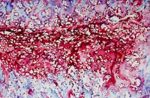
Watercolor- A watercolor image depicting the embryonic bone development process, endochondral ossification, featuring cartilage and bone. Credit: Joel Boerckel
“When bones are originally formed in the embryo, they’re first generated from cartilage, like a template,” says senior author Joel Boerckel, an assistant professor of orthopaedic surgery and bioengineering. “In order to regenerate bone within defects that otherwise won’t heal in grown people, we are seeking to recreate the embryonic bone development process.”
To do that, the researchers’ process begins with the delivery of specially engineered stem cells (called a condensation of mesenchymal cells) to the rodents’ bone defect, which sparks endochondral ossification, the specific term for embryonic bone development.
Vector Flow Imaging Helps Visualize Blood Flow in Pediatric Hearts
A group of biomedical engineers at the University of Arkansas used a new ultrasound-based imaging technique called vector flow imaging to help improve the diagnosis of congenital heart disease in pediatric patients. The study, led by associate professor of biomedical engineering Morten Jensen, Ph.D., collaborated with cardiologists at the local Children’s Hospital in Little Rock to produce images of the heart in infants to help potentially diagnose congenital heart defects. Though the use of vector flow imaging has yet to be developed for adult patients, this type of imaging could possibly provide more detail about the direction of blood flow through the heart than traditional techniques like echocardiography do. In the future, the use of both techniques could provide information about both the causes and larger effects of heart defects in patients.
Using Stem Cells to Improve Fertility in Leukemia Survivors
One of the more common side effects of leukemia treatment in female patients is infertility, but researchers at the University of Michigan want to change that. Led by associate professor of biomedical engineering Ariella Shikanov, Ph.D., researchers in her lab found ways of increasing ovarian follicle productivity in mice, which directly relates to the development of mature eggs. The project involves the use of adipose-derived stem cells, that can be found in human fat tissue, to surround the follicles in an ovary-like, three-dimensional scaffold. Because the radiation treatments for leukemia and some other cancers are harmful to follicles, increasing their survival rate with this stem cell method could reduce the rate of infertility in patients undergoing these treatments. Furthermore, this new approach is innovative in its use of a three-dimensional scaffold as opposed to a two-dimensional one, as it stimulates follicle growth in all directions and thus helps to increase the follicle survival rate.
Penn Engineers Look at How Stretching & Alignment of Collagen Fibers Help Cancer Cells Spread
Cancer has such a massive impact on people’s lives that it might be easy to forget that the disease originates at the cellular level. To spread and cause significant damage, individual cancer cells must navigate the fibrous extracellular environment that cells live in, an environment that Penn Engineer Vivek Shenoy has been investigating for years.
Shenoy’s most recent study on cancer’s mechanical environment was led by a postdoctoral researcher in his lab, Ehsan Ban. Paul Janmey, professor in Physiology and Bioengineering, and colleagues at Stanford University also contributed to the study. Shenoy also received the Heilmeier Award this March and delivered the Heilmeier Award Lecture in April.
Controlled Electrical Stimulation Can Prevent Joint Replacement Infections
Joint replacements are one of the most common kinds of surgery today, but they still require intense post-operative therapy and have a risk of infection from the replacement implant. These infections are usually due to the inflammatory response that the body has to any foreign object, and can become serious and life-threatening if left untreated. Researchers at the University of Buffalo Jacobs School of Medicine and Biomedical Sciences hope to offer a solution to preventing infections through the use of controlled electrical stimulation. Led by Mark Ehrensberger, Ph.D., Kenneth A. Krackow, M.D., and Anthony A. Campagnari, Ph.D., the treatment system uses the electrical signal to create an antibacterial environment at the interface of the body and the implant. While the signal does not prevent infections completely, these antibacterial properties will prevent infections from worsening to a more serious level. Patented as the Biofilm Disruption Device TM, the final product uses two electrode skin patches and a minimally invasive probe that delivers the electrical signal directly to the joint-body interface. The researchers behind the design hope that it can help create a more standard way of effectively treating joint replacement infections.
People and Places
TBx: Gabriel Koo, Ethan Zhao, Daphne Cheung, and Shelly Teng
For their senior design project, four bioengineering seniors — Gabriel Koo, Ethan Zhao, Daphne Cheung, and Shelly Teng — created a low-cost tuberculosis diagnostic that they called TBx. Using their knowledge of the photoacoustic effect of certain dyes, the platform the group created can detect the presence of lipoarabinomannan in patient urine. The four seniors presented TBx at the Rice360 Design Competition in Houston, Texas this spring, which annually features student-designed low-cost global health technologies.
New Vascularized Patches Could Help Patient Recovery from Heart Attacks
Heart attacks are the result of a stoppage of blood flow to the heart – an interruption to normal function that can result in severe tissue damage, or even tissue death. This loss of healthy tissue function is one of the biggest challenges in treating patients that undergo heart attacks, as the damaged tissue increases their risk of having future attacks. One of the main solutions to this issue right now is the creation of cardiac tissue scaffolds using stem cells to create a platform for new and healthy tissue to grow in vivo. A group of biomedical engineers at Michigan Technological University hopes to expand on this basis by focusing not just on cellular alignment in the scaffold but on that of microvessels too. Led by Feng Zhao, Ph.D., Associate Professor of Biomedical Engineering, the team hopes that this new attention on microvessel organization will improve the vasculature of the scaffolds, and thus improve the success of the scaffolds in vivo, allowing for a better recovery from heart attacks.
Some Stem Cells May Be More Fit Than Others
Stem cells are one of the hottest research areas in the field of bioengineering today. Widely known as the cells in the human embryo that have the ability to eventually transform into specific cells for the brain, lung, and every other organ, stem cells are also of recent interest because researchers found ways to reverse this process, transforming organ-specific cells back to the pluripotent stem cell level. This achievement however, is mostly applicable to individual stem cells, and doesn’t fully encapsulate the way this process might work on a larger population level. So Peter Zandstra, Ph. D., a bioengineering faculty member at the University of British Columbia, decided to research just that.
Using mouse embryonic fibroblasts (MEFs), Zandstra and his lab attempted to track the cells throughout their reprogramming, to more clearly trace each back to its respective parent population. Surprisingly, they found that after only one week of reprogramming, nearly 80% of the original cell population had been removed, meaning that most of the parent generation was not “fit” enough to undergo the process of reprogramming, indicating that perhaps some stem cells will have a better chance of survival in this process than others. This research may suggest that not all cells have the capacity to undergo reprogramming, as many researchers originally thought.
A New Microdevice Will Help Model Bronchial Spasms
The difficulty in breathing associated with asthma is the result of bronchial spasms, which are a kind of muscle contraction in the airways. But little was known about just how these spasms occurred in patients, so Andre Levchenko, Ph.D., Professor of Biomedical Engineering at Johns Hopkins, and his lab created a microdevice to model them. Calling the device a “bronchi on a chip,” Levchenko and his team used a microphysiological model to look at some of the biochemical and mechanical signals associated with these kinds of muscle contractions. They found that the contractions operate in a positive feedback system, so that those caused by disturbance from allergens will subsequently cause even more contractions to occur. But surprisingly, they also found that a second contraction, if triggered at the right time during the initial contraction, could actually stop the process and allow the muscles to relax. Because asthma is a notoriously difficult disease to translate from animal to human models, this new device opens the door to understanding different mechanisms of asthma before taking research to clinical trials.
New CHOP Research Center to Focus Research on Pediatric Airway Disorders
A new bioengineering lab at the Children’s Hospital of Philadelphia called the Center for Pediatric Airway Disorders will specialize in a variety of airway procedures for pediatric patients such as tracheal reconstruction and recurrent laryngeal nerve reinnervation. This new lab will be one of the first to give a unique focus to the application of bioengineering to pediatric laryngology. The interdisciplinary center brings together students and researchers from all different fields, including materials science and microbiology, to find new ways of repairing tissue and regenerating organs related to respiratory disorders. Specific areas of research will involve the modeling of children’s vocal cords, understanding the mechanisms of fibrosis, and improving surgical procedures.
Deeper Understanding of Sickle Cell Anemia Could Lead to New Treatments
Though sickle cell anemia is a common and well-known disease, a new study of its causes at the nanoscale level might reveal previously unknown information about the assembly of hemoglobin fibers. Using microscopes with the ability to visualize these molecules at such a small level, researchers at the University of Minnesota found that the beginning organizations that lead to sickle cell anemia are much less ordered than originally thought. Led by Associate Professor of Biomedical Engineering David Wood, Ph.D., the team of researchers used this higher level of microscopy to find that hemoglobin self-assembly process, which was originally thought to be 96% efficient, is actually only 4% efficient. Wood hopes that this new knowledge will help allow for the development of new and better treatments for patients with sickle cell anemia, as there are currently only two FDA-approved ones on the market.
People & Places
Penn Today asked five Penn researchers about the women in STEM who have been a source of inspiration and encouragement throughout their own careers. Their responses include active researchers who have paved the way for better inclusion in STEM and famous female scientists from the past who broke boundaries as they made strides with their research.
Joel Boerckel, Ph.D, Assistant Professor of Orthopaedic Surgery and Bioengineering
This week, we want to congratulate Joel Boerckel, Ph.D., Assistant Professor of Orthopaedic Surgery and Bioengineering, and his lab on receiving a second R01 Grant from the National Institute of Arthritis and and Musculoskeletal Skin Diseases for their work on defining the roles of YAP and TAZ in embryonic bone morphogenesis and mechanoregulation of fracture repair. Dr. Boerckel is a member of the McKay Orthopaedic Research Laboratory.
We would also like to congratulate Christopher Yip, Ph. D., on being appointed as the new dean of the University of Toronto’s Faculty of Applied Science and Engineering. A professor in both the Department of Chemical Engineering and Applied Chemistry the Institute of Biomaterials and Biomedical Engineering, Dr. Yip’s research involves the use of molecular imaging to understand the self-assembly of proteins.