Week in BioE (August 16, 2019)

by Sophie Burkholder

Electrode Arrays and Star Wars Help to Inspire a New Prosthetic Arm

Brain-controlled prosthetic arm, Wikimedia commons

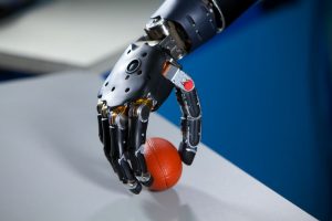
After nearly fifteen years of work, a new high-tech prosthetic arm from researchers at the University of Utah allows hand amputees to pluck grapes, pick up eggs without breaking them, and even put on their wedding rings. Named after Luke Skywalker’s robotic hand in the Star Wars saga, the LUKE Arm includes sensors that better mimic the way the human body sends information to the brain, allowing users to distinguish between soft and hard surfaces and to perform more complicated tasks. The arm relies heavily on an electrode array invented by University of Utah biomedical engineering professor Richard A. Normann, Ph.D., which is a bundle of microelectrodes that enable a computer to read signals from connected nerves in the user’s forearm.

But the biggest innovation in the use of these electrode arrays for the LUKE Arm is in the way they allow the prosthetic to mimic the sense of feeling on the surface of an object that indicates how much pressure should be applied when handling it. Gregory Clark, Ph.D., an associate professor of biomedical engineering at the University of Utah and the leader of the LUKE Arm project, says the key to improving these functions in the prosthetic was by more closely mimicking the path that this information takes to the brain, as opposed to merely what comprises that sensory information. In the future, Clark hopes to improve upon the LUKE Arm by including more inputs, like one for temperature data, and on making them more portable by eliminating the device’s need for computer connection.

Philly Voice Recognizes the Cremins Lab’s Innovations in Light-Activated Gene-Folding

While technological advancements over the past few decades have opened doors to understanding the topological structures of DNA, we still have far more to learn about how these structures impact and contribute to genome function. But here at Penn, the Cremins Laboratory in 3D Epigenomes and Systems Neurobiology hopes to fix that. Led by Jennifer E. Phillips-Cremins, Ph.D., members of the lab use light-activated dynamic looping (LADL) to better understand the way that genome topological properties and folding can affect protein translation. Cremins and her lab use this technique to force specific genome folds to interact with each other, and create temporary DNA loops that can then be bound together in the presence of blue light for certain proteins in the Arabidopsis plant. Using the data from these tests, researchers can better understand the genome structure-function relationships, and hopefully open the door to new treatments for diseases in which expression or mis-expression of certain genes is the cause.

Artificial Cells Can Deliver Molecules Better than the Real Thing

From pills to vaccines, ways to deliver drugs into the body have been constantly evolving since the early days of medicine.

Now, a new study from an interdisciplinary team led by researchers at the University of Pennsylvania provides a new platform for how drugs could be delivered to their targets in the future. Their work was published in the Proceedings of the National Academy of Sciences.

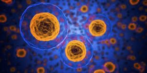
The research focuses on a dendrimersome, a compartment with a lamellar structure and size that mimic a living cell. It can be thought of as the shipping box of the cellular world that carries an assortment of molecules as cargo.

The scientists found that these dendrimersomes, which have a multilayered, onion-like structure, were able to “carry” high concentrations of molecules that don’t like water, which is common in pharmaceutical drugs. They were also able to carry these molecules more efficiently than other commercially available vessels. Additionally, the building block of the cell-like compartment, a janus dendrimer, is classified as an amphiphile, meaning it contains molecules that don’t like water and also molecules that are soluble in water, like lipids, that make up natural membranes.

“This is a different amphiphile that makes really cool self-assembled onions into which we were able to load a bunch of molecular cargos,” says co-author Matthew Good.

Read the rest of the story on Penn Today.

A Warm Evening Bath Could Improve Sleep Quality

In a recent review of over 5,000 sleep studies, biomedical engineering researchers at the University of Texas at Austin found a connection between water-based passive body heating and sleep onset latency, efficiency, and quality. Using meta-analytical tools to compare all of the studies and patient data, lead author and Ph.D. candidate Shahab Haghayegh and his team found that a warm bath in the temperature range of 104-109 degrees Fahrenheit taken 1-2 hours before bed has the ability to improve all three considered sleep categories. This makes sense considering that our body’s Circadian rhythms govern both our sleep cycles and temperature, bringing us to a higher temperature during the day and a lower one at night during sleep. In fact, this lowering of body temperature before sleep is what helps to trigger the onset of sleep, so taking a warm bath and allowing your body to cool down from it before going to sleep enhances the body’s own efforts of naturally cooling down before we go to bed. With this new and comprehensive review, those who suffer from poor sleep quality may soon find solace in temperature regulation therapy systems.

People & Places

With the recent 50th anniversary of the first moon landing by Americans Neil Armstrong, Buzz Aldrin, and Michael Collins in 1969, ABC News looked back at one of the women involved in the project. Judy Sullivan was a biomedical engineer at the time of the project, and served as the lead engineer of the biomedical system for Apollo 11. In this role, she led studies on the astronauts’ breathing rates and sensor capabilities for the devices being sent into space to help the astronauts monitor their health. For the Apollo 11 mission and a lot of Sullivan’s early work at NASA, she worked on teams of all men, as women were often encouraged to become teachers, secretaries, or homemakers over other professions. Today, Sullivan says she’s thrilled that women have more career options to choose from, and wants to continue seeing more women getting involved in math and science.

We would like to congratulate Sanjay Kumar, M.D., Ph.D., on his appointment as the new Department Chair of Bioengineering at the University of California, Berkeley. Since joining the faculty in 2005, Kumar has received several prestigious awards including the NSF Career Award, the NIH Director’s New Innovator Award, the Presidential Early Career Award for Scientists and Engineers, and the Berkeley student-voted Outstanding Teacher Award.


Week in BioE: March 29, 2019

by Sophie Burkholder

New Studies in Mechanobiology Could Open Doors for Cellular Disease Treatment

When we think of treatments at the cellular level, we most often think of biochemical applications. But what if we began to consider more biomechanical-oriented approaches in the regulation of cellular life and death? Under a grant from the National Science Foundation (NSF),Worcester Polytechnic Institute’s (WPI) Head of the Department of Biomedical Engineering Kristen Billiar, Ph.D., performs research that looks at the way mechanical stimuli can affect and trigger programmed cell death.

Billiar, who received his M.S.E. and Ph.D. from Penn, began his research by first noticing the way that cells typically respond to the mechanical stimuli in their everyday environment, such as pressure or stretching, with behaviors like migration, proliferation, or contraction. He and his research team hope to find a way to eventually predict and control cellular responses to their environment, which they hope could open doors to more forms of treatment for disorders like heart disease or cancer, where cellular behavior is directly linked to the cause of the disease.

Self-Learning Algorithm Could Help Improve Robotic Leg Functionality

Obviously, one of the biggest challenges in the field of prosthetics is the extreme difficulty in creating a device that perfectly mimics whatever the device replaces for its user. Particularly with more complex designs that involve user-controlled motion for joints in the limbs or hands, the electrical circuits implemented are by no means a perfect replacement of the neural connections in the human body from brain to muscle. But recently at the University of Southern California Viterbi School of Engineering, a team of researchers led by Francisco J. Valero-Cuevas, Ph. D.,  developed an algorithm with the ability to learn new walking tasks and adapt to others without any additional programming.

The algorithm will hopefully help to speed the progress of robotic interactions with the world, and thus allow for more adaptive technology in prosthetics, that responds to and learns with their users. The algorithm Valero-Cuevas and his team created takes inspiration from the cognition involved with babies and toddlers as they slowly learn how to walk, first through random free play and then from pulling on relevant prior experience. In a prosthetic leg, the algorithm could help the device adjust to its user’s habits and gait preferences, more closely mimicking the behavior of an actual human leg.

Neurofeedback Can Improve Behavioral Performance in High-Stress Situations

We’re all familiar with the concept of being “in the zone,” or the feeling of extraordinary focus that we can sometimes have in situations of high-stress. But how can we understand this shift in mindset on a neuroengineering level? Using the principal of the Yerkes-Dodson law, which says that there is a state of brain arousal that is optimal for behavioral performance, a team of biomedical engineering researchers at Columbia University hope to find ways of applying neurofeedback to improving this performance in demanding high-stress tasks.

Led by Paul Sajda, Ph.D., who received his doctoral degree from Penn, the researchers used a brain-computer interface to collect electroencephalography (EEG) signals from users immersed in virtual reality aerial navigation tasks of varying difficulty levels. In doing so, they were able to make connections between stressful situations and brain activity as transmitted through the EEG data, adding to the understanding of how the Yerkes-Dodson law actually operates in the human body and eventually demonstrating that the use of neurofeedback reduced the neural state of arousal in patients. The hope is that neurofeedback may be used in the future to help treat emotional conditions like post-traumatic stress disorder (PTSD).

Ultrasound Stimulation Could Lead to New Treatments for Inflammatory Arthritis

Arthritis, an autoimmune disease that causes painful inflammation in the joints, is one of the more common diseases among older patients, with more than 3 million diagnosed cases in the United States every year. Though extreme measures like joint replacement surgery are one solution, most patients simply treat the pain with nonsteroidal anti-inflammatory drugs or the adoption of gentle exercise routines like yoga. Recently however, researchers at the University of Minnesota led by Daniel Zachs, M.S.E., in the Sensory Optimization and Neural Implant Coding Lab used ultrasound stimulation treatment as a way to reduce arthritic pain in mice. In collaboration with Medtronic, Zachs and his team found that this noninvasive ultrasound stimulation greatly decreased joint swelling in mice who received the treatment as opposed to those that did not. They hope that in the future, similar methods of noninvasive treatment will be able to be used for arthritic patients, who otherwise have to rely on surgical remedies for serious pain.

People and Places

Leadership and Inspiration: EDAB’s Blueprint for Engineering Student Life

To undergraduates at a large university, the administration can seem like a mysterious, all-powerful entity, creating policy that affects their lives but doesn’t always take into account the reality of their day-to-day experience. The Engineering Deans’ Advisory Board (EDAB) was designed to bridge that gap and give students a platform to communicate with key decision makers.

The 13-member board meets once per week for 60 to 90 minutes. The executive board, comprised of four members, also meets weekly to plan out action items and brainstorm. Throughout his interactions with the group, board president Jonathan Chen, (ENG ‘19, W ‘19), has found a real kinship with his fellow board members, who he says work hard and enjoy one another’s company in equal measure.

Bioengineering major Daphne Cheung (ENG’19) joined the board as a first-year student because she saw an opportunity to develop professional skills outside of the classroom. “For me, it was about trying to build a different kind of aptitude in areas such as project management, and learning how to work with different kinds of people, including students and faculty, and of course, the deans,” she says.

Read the full story on Penn Engineering’s Medium Blog. Media contact Evan Lerner.

Purdue University College of Engineering and Indiana University School of Medicine Team Up in New Engineering-Medicine Partnership

The Purdue University College of Engineering and the Indiana University School of Medicine recently announced a new Engineering-Medicine partnership, that seeks to formalize ongoing and future collaborations in research between the two schools. One highlight of the partnership is the establishment of a new M.D./M.S. degree program in biomedical engineering that will allow medical students at Indiana University to receive M.S.-level training in engineering technologies as they apply to clinical practice. The goal of this new level of collaboration is to further involve Purdue’s engineering program in the medical field, and to exhibit the benefits that developing an engineering mindset can have for medical students. The leadership of this new partnership includes

Week in BioE (March 15, 2019)

by Sophie Burkholder

Synthetic Spinal Discs from a Penn Research Team Might Be the Solution to Chronic Back Pain

Spinal discs, the concentric circles of collagen fiber found between each vertebra of the spine, can be the source of immense back pain when ruptured. Especially for truck and bus drivers, veterans, and cigarette smokers, there is an increased risk in spinal disc rupture due to overuse or deterioration over time. But these patients aren’t alone. In fact, spinal discs erode over time for almost everyone, and are one of the sources of back pain in older patients, especially when the discs erode so much that they allow direct bone-to-bone contact between two or more vertebrae.

Robert Mauck, Ph.D.

Robert Mauck, Ph.D., who is the director of the McKay Orthopaedic Research Laboratory here at Penn and a member of the Bioengineering Graduate Group Faculty, led a research team in creating artificial spinal discs, with an outer layer made from biodegradable polymer and an inner layer made with a sugar-like gel. Their findings appear in Science Translational Medicine. These synthetic discs are also seeded with stem cells that produce collagen over time, meant to replace the polymer as it degrades in vivo over time. Though Mauck and his time are still far from human clinical trials for the discs, they’ve shown some success in goat models so far. If successful, these biodegradable discs could lead to a solution for back pain that integrates itself into the human body over time, potentially eliminating the need of multiple invasive procedures that current solutions require. Mauck’s work was recently featured in Philly.com.

An Untethered, Light-Activated Electrode for Innovations in Neurostimulation

Neurostimulation, a process by which nervous system activity can be purposefully modulated, is a common treatment for patients with some form of paralysis or neurological disorders like Parkinson’s disease. This procedure is typically invasive, and because of the brain’s extreme sensitivity, even the slightest involuntary movement of the cables, electrodes, and other components involved can lead to further brain damage through inflammation and scarring. In an effort to solve this common problem, researchers from the B.I.O.N.I.C. Lab run by Takashi D.Y. Kozai, Ph. D., at the University of Pittsburgh replaced long cables with long wavelength light and a formerly tethered electrode with a smaller, untethered one.

The research team, which includes Pitt senior bioengineering and computer engineering student Kaylene Stocking, centered the device on the principle of the photoelectric effect – a concept first described in a publication by Einstein as the local change in electric potential for an object when hit with a photon. Their design, which includes a 7-8 micron diameter carbon fiber implant, is now patent pending, and Kozai hopes that it will lead to safer and more precise advancements in neurostimulation for patients in the future.

A New Microfluidic Chip Can Detect Cancer in a Drop of Blood

Many forms of cancer cannot be detected until the disease has progressed past the point of optimum treatment time, increasing the risk for patients who receive late diagnoses of these kinds of cancer. But what if the diagnostic process could be simplified and made more efficient so that even a single drop of blood could be enough input to detect the presence of cancer in a patient? Yong Zeng, Ph.D., and his team of researchers at the University of Kansas in Lawrence sought to answer that question.

They designed a self-assembled 3D-nanopatterned microfluidic chip to increase typical microfluidic chip sensitivity so that it can now detect lower levels of tumor-associated exosomes in patient blood plasma. This is in large part due to the nanopatterns in the structure of the chip, which promote mass transfer and increase surface area, which in turn promotes surface-particle interactions in the device. The team applied the device to their studies of ovarian cancer, one of the notoriously more difficult kinds of cancer to detect early on in patients.

A Wearable Respiration Monitor Made from Shrinky Dinks

Michelle Khine, Ph. D., a professor of biomedical engineering at the University of California, Irvine incorporates Shrinky Dinks into her research. After using them once before in a medical device involving microfluidics, her lab recently worked to incorporate them into a wearable respiration monitor – a device that would be useful for patients with asthma, cystic fibrosis, and other chronic pulmonary diseases. The device has the capability to track the rate and volume of its user’s respiration based on measurements of the strain at the locations where the device makes contact with the user’s abdomen.

Paired with Bluetooth technology, this sensor can feed live readings to a smartphone app, giving constant updates to users and doctors, as opposed to the typical pulmonary function test, which only provides information from the time at which the test takes a reading. Though Khine and her team have only tested the device on healthy patients so far, they look forward to testing with patients who have pulmonary disorders, in hopes that the device will provide more comprehensive and accessible data on their respiration.

People and Places

Ashley Kimbel, a high school senior from Grissom High School in Huntsville, Alabama, designed a lightweight prosthetic leg for local Marine, Kendall Bane, after an attack in Afghanistan led him to amputate one of his legs below the knee. Bane, who likes to keep as active as possible, said the new lighter design is more ideal for activities like hiking and mountain biking, especially as any added weight makes balance during these activities more difficult. Kimbel used a CAD-modeling software produced by Siemens called Solid Edge, which the company hopes to continue improving in accessibility so that more students can start projects like Kimbel’s.

This week, we would like to congratulate Angela Belcher, Ph.D., on being named the new head of the Department of Biological Engineering at the Massachusetts Institute of Technology (MIT). With her appointment to this role, now half of the MIT engineering department heads are women. Belcher’s research is in the overlap of materials science and biological engineering, with a particular focus on creating nanostructures based on the evolution of ancient organisms for applications in medical diagnostics, batteries, solar cells, and more.

We would also like to congratulate Eva Dyer, Ph.D., and Chethan Pandarinath, Ph.D., both of whom are faculty members at the Walter H. Coulter Department of Biomedical Engineering at Georgia Tech and Emory University, on receiving research fellowships from the Alfred P. Sloan Foundation. Dr. Dyer, who formerly worked with Penn bioengineering faculty member Dr. Konrad Kording while he was at Northwestern University, leads research in the field of using data analysis methods to quantify neuroanatomy. Dr. Pandarinth leads the Emory and Georgia Tech Systems Neural Engineering Lab, where he works with a team of researchers to use properties of artificial intelligence and machine learning to better understand large neural networks in the brain.


Week in BioE (February 28, 2019)

by Sophie Burkholder

Louisiana Tech Sends First All-Female Team to RockOn

A team of faculty and students from Louisiana Tech University will participate in RockOn, a NASA-sponsored workshop on rocketry and engineering. Mechanical Engineering Lecturer Krystal Corbett, Ph.D., and Assistant Professor of bioengineering Mary Caldorera-Moore, Ph.D., will work together to lead the university’s first team of three all-female students at the event. At the program, they will have the chance to work on projects involving components of spacecraft systems, increasing students’ experience in hands-on activities and real-world engineering.

Refining Autism Treatments Using Big Data

Though treatments like therapy and medication exist for patients with autism, one of the biggest challenges that those caring for these patients face is in measuring their effects over time. Many of the markers of progress are qualitative, and based on a given professional’s opinion on a case-by-case basis. But now, a team of researchers from Rensselaer Polytechnic Institute (RPI) hopes to change that with the use of big data.

Juergen Hahn, Ph. D., and his lab recently published a paper in Frontiers in Cellular Neuroscience discussing their findings in connecting metabolic changes with behavioral improvements in autistic patients. Their analysis looks for multiple chemical and medical markers simultaneously in data from three distinct clinical trials involving metabolic treatment for patients. Being able to quantitatively describe the effects of current autism treatments would revolutionize clinical trials in the field, and lead to overall better patient care.

Penn Engineers Can Detect Ultra Rare Proteins in Blood Using a Cellphone Camera

One of the frontiers of medical diagnostics is the race for more sensitive blood tests. The ability to detect extremely rare proteins could make a life-saving difference for many conditions, such as the early detection of certain cancers or the diagnosis of traumatic brain injury, where the relevant biomarkers only appear in vanishingly small quantities. Commercial approaches to ultrasensitive protein detection are starting to become available, but they are based on expensive optics and fluid handlers, which make them relatively bulky and expensive and constrain their use to laboratory settings.

Knowing that having this sort of diagnostic system available as a point-of-care device would be critical for many conditions — especially traumatic brain injury — a team of engineers led by Assistant Professor in the Department of Bioengineering, David Issadore, Ph.D., at the University of Pennsylvania have developed a test that uses off-the-shelf components and can detect single proteins with results in a matter of minutes, compared to the traditional workflow, which can take days.

Read the full story on Penn Engineering’s Medium blog.

Treating Cerebral Palsy with Battery-Powered Exoskeletons

Cerebral palsy is one of the most common movement disorders in the United States. The disorder affects a patient’s control over even basic movements like walking, so treatments for cerebral palsy often involve the use of assistive devices in an effort to give patients better command over their muscles. Zach Lerner, Ph.D., is an Assistant Professor of Mechanical Engineering and faculty in Northern Arizona University’s Center for Bioengineering Innovation whose research looks to improve these kinds of assistive devices through the use of battery-powered exoskeletons.

Lerner and his lab recently received three grants, one each from the National Institute of Health (NIH), the National Science Foundation (NSF), and the Arabidopsis Biological Resource Center, to continue their research in developing these exoskeletons. Their goal is to create devices with powered assistance at joints like the ankle or knee to help improve patient gait patterns in rehabilitating the neuromuscular systems associated with walking. The team hopes that their work under these new grants will help further advance treatment for children with cerebral palsy, and improve overall patient care.

People & Places

David Aguilar, a 19-year-old bioengineering student at Universitat Internacional de Catalunya made headlines recently for a robotic prosthetic arm that he built for himself using Lego pieces. Due to a rare genetic condition, Aguilar was born without a right forearm, a disability that inspired him to play with the idea of creating his own prosthetic arm from age nine. His design includes a working elbow joint and grabber that functions like a hand. In the future, Aguilar hopes to continue improving his own prosthetic designs, and to help create similar versions of affordable devices for other patients who need them.

This week, we would like to congratulate two recipients of the National Science Foundation’s Career Awards, given to junior faculty that exemplify the role of teacher-scholars in their research. The first recipient we’d like to acknowledge is the University of Arkansas’ Kyle Quinn, Ph.D., who received the award for his work in developing new image analysis methods and models using the fluorescence of two metabolic cofactors. Dr. Quinn completed his Ph.D. here at Penn in Dr. Beth Winkelstein’s lab, and received the Solomon R. Pollack Award for Excellence in Graduate Bioengineering Dissertation Research for his work.

The second recipient of the award we wish to congratulate is Reuben Kraft, Ph.D., who is an Assistant Professor in Mechanical and Biomedical Engineering at Penn State. Dr. Kraft’s research centers around developing computational models of the brain through linking neuroimaging and biomechanical assessments. Dr. Kraft also collaborates with Kacy Cullen, Ph.D., who is a secondary faculty member in Penn’s bioengineering department and a member of the BE Graduate Group faculty.

Finally, we’d like to congratulate Dawn Elliott, Ph.D., on being awarded the Orthopaedic Research Society’s Adele L. Boskey, PhD Award, awarded annually to a member of the Society with a commitment to both mentorship and innovative research. Dr. Elliott’s spent 12 years here at Penn as a member of the orthopaedic surgery and bioengineering faculty before joining the University of Delaware in 2011 to become the founding director of the bioengineering department there. Her research focuses primarily on the biomechanics of fibrous tissue in tendons and the spine.

Week in BioE (August 9, 2018)

Converting Fat to Fight Obesity

White fat stories calories and provides the body with insulation.

There are two types of fat in the human body: brown and white. Brown fat, the “good” fat, is rich in mitochondria, which gives it its brown appearance. Whereas white fat stores calories and acts as an insulator, mitochondria-rich brown fat burns energy to produce heat throughout the body and maintains body temperature. White fat, conversely, uses its stored energy to insulate the body and keep its temperature level. While all fat serves a purpose in the body, an excess of white fat cells causes obesity, a condition affecting one in three adults in the U.S. and the root cause of many potential health problems. Finding ways to convert white fat to brown opens a possibility of treating this problem naturally.

A new study in Scientific Reports proposes a clever way to convert fat types. Professor of Biomedical Engineering Samuel Sia, PhD, of the Columbia University School of Engineering and Applied Science, led a team which developed a method of converting white fat into brown using a tissue-grafting technique. After extracting and converting the fat, it can then be transplanted back into the patient. White fat is hard-wired to convert to brown under certain conditions, such as exposure to cold temperatures, so the trick for Dr. Sia’s team was finding a way to make the conversion last for long periods. The studies conducted with mice suggested that using these methods, newly-converted fat stayed brown for a period of two months.

Dr. Sia’s team will proceed to conduct further tests, especially on the subjects’ metabolism and overall weight after undergoing the procedure, and they hope that eventual clinical trials will result in new methods to treat or even prevent obesity in humans.

Cremins Lab Student Appointed Blavatnik Fellow

Linda Zhou is currently pursuing her MD/PhD in Genomics and Computational Biology under the supervision of Dr. Jennifer Phillips-Cremins.

The Perelman School of Medicine named Linda Zhou, a student in BE’s Cremins Laboratory, a Blavatnik Fellow for the 2018-2019 academic year. The selection process for this award is highly competitive, and Linda’s selection speaks to the excellent quality of her scholarship and academic performance. The fellows will be honored in a special ceremony at the Museum of Natural History in New York City.

Linda received her B.S. in Biophysics and Biochemistry from Yale University and is currently pursuing her M.D./Ph.D. in the Genomics and Computational Biology Program at Penn. “I am honored to be named a Blavatnik Fellow and am extremely excited to continue my graduate studies investigating neurological disorders and the 3D genome,” she said. “This support will be integral to achieving my long term goal of driving scientific discovery that will help treat human disease.”

Linda’s research is overseen by Penn Bioengineering Assistant Professor Jennifer Phillips-Cremins, PhD. “Linda is an outstanding graduate student,” said Dr. Cremins. “It is a true delight to work with her. She is hard working, intelligent, kind, and has extraordinary leadership ability. Her unrelenting search for ground-state truth makes her a shining star.”

The Blavatnik Family Fellowship in Biomedical Research is a new award announced by the Perelman School of Medicine in May of this year. This generous gift from the Blavatnik Family Foundation awards $2 million to six recipients in the Biomedical Graduate Studies Program at Penn for each of the next four years.

Growing Lungs in a Lab

As the demand for lung transplants continues to rise, so does the need for safe and effective transplanted lungs. Bioengineered lungs grown or created in labs are one way of meeting this demand. The problem – as is ever the case with transplants – is the high rate of rejection. The results of success are always better when cells from the patient herself (or autologous cells) are used in the transplanted organ.

Recently Joan Nichols, PhD, Professor of Internal Medicine, and Microbiology and Immunology, at the University of Texas Medical Branch at Galveston, successfully bioengineered the first human lung. Her latest study published in Science Translational Medicine describes the next milestone for Dr. Nichols’ lab: successfully transplanting a bioengineered lung into a pig.

These advances are possible due to Dr. Nichols’ work with autologous cells, continuing the trend of “on demand” medicine (i.e. medicine tailor for a specific patient) which we track on this blog. Dr. Nichols’ particular method is to build the structure of a lung (using the harvested organs of dead pigs in this case), de-cellularize the tissue, and then repopulate it with autologous cells from the intended recipient. This way, the host body recognizes the cells as friendly and the likelihood of acceptance increases. While further study is needed before clinical trials can begin, Dr. Nichols and her team see the results as extremely promising and believe that we are on the way to bioengineered human lungs.

Nanoparticles Combat Dental Plaque

Combine a diet high in sugar with poor oral hygiene habits and dental cavities likely result. The sugar triggers the formation of an acidic biofilm (plaque) on the teeth, eroding the surface. Early childhood dental cavities affect one in every four children in the United States and hundreds of millions more globally. It’s a particularly severe problem in underprivileged populations.

In a study published in Nature Communications this week, researchers led by Hyun (Michel) Koo of the University of Pennsylvania School of Dental Medicine in collaboration with David Cormode of Penn’s Perelman School of Medicine and School of Engineering and Applied Science used FDA-approved nanoparticles to effectively disrupt biofilms and prevent tooth decay in both an experimental human-plaque-like biofilm and in an animal model that mimics early-childhood caries.

Dr. David Cormode is Assistant Professor of Radiology and Secondary Faculty in Bioengineering at Penn. His research includes Bioengineering Therapeutics, Devices and Drug Delivery and Biomaterials.

Read the full story at Penn Today. Media contact Katherine Unger Baillie.

Stopping the Flu from Catching On

The flu virus is notoriously contagious, but there may be a way to stop it before it starts. In order for the influenza virus to successfully transport itself into the cells of a human host, it needs a certain protein called hemagglutinin which mediates its entry. By interfering with this vital ingredient, researchers can effectively kill the virus.

A new study in the Proceedings of the National Academy of Sciences discusses a method of disrupting the process by which this protein causes the virus to infect its host cells. This discovery could lead to more effective flu vaccines that target the flu virus at its root, rather than current ones which have to keep up with the ongoing changes and mutations of the virus itself. Indeed, the need for different vaccines to address various “strains” of the flu is moot if a vaccine can stop the virus from infecting people in the first place.

This breakthrough results from grants provided by the NSF, the Welch Foundation, and the NIH to Rice University and Baylor College of Medicine. Lead researchers José Onuchic, PhD, Harry C. and Olga K. Wiess Chair of Physics and Professor of Chemistry and BioSciences at Rice University; Jianpeng Ma, PhD, Professor of Bioengineering at Rice University and Lodwick T. Bolin Professor of Biochemistry at Baylor College of Medicine; and Qinghua Wang, PhD, Assistant Professor of Biochemistry at Baylor College of Medicine. Their team will continue to study the important role proteins play in how the flu virus operates.

People and Places

This week, we congratulate a few new leadership appointments in bioengineering. First, the Georgia Institute of Technology appointed Penn BE alumnus Andréas García, PhD, the new Executive Director of the Parker H. Petit Institute for Bioengineering and Bioscience. In addition to his new role, Dr. García is also the George W. Woodruff School of Mechanical Engineering Regents Professor. He conducts research in biomolecular, cellular, and tissue engineering and collaborates with a number of research centers across Georgia Tech. Dr. García graduated with both his M.S.E. and Ph.D. from the University of Pennsylvania’s Department of Bioengineering.

Secondly, the University of Minnesota Institute for Engineering in Medicine (IEM) named the Distinguished McKnight University Professor John Bischof, PhD, their new director. This follows Dr. Bischof’s recent position as interim director for the IEM. Dr. Bischof earned his Ph.D. in Mechanical Engineering at the University of California at Berkeley, and is currently a faculty member in both the Mechanical Engineering and Biomedical Engineering Departments at the University of Minnesota. Dr. Bischof holds the Carl and Janet Kuhrmeyer Chair in Mechanical Engineering.

At an earlier, but no less impressive, point in his academic career, Tanishq Abraham became the youngest person to graduate with a degree in biomedical engineering. The fifteen year old recently graduated summa cum laude from the University of California, Davis. As part of his graduating research, Abraham – a first-generation Indian-American – designed a device to measure the heart rates of burn victims. Abraham has already been accepted by U.C. Davis for his Ph.D. and plans to continue on to his M.D.

Finally, the work continues to create affordable and well-fitted prosthetics, especially for remote, rural, and underfunded areas both in the U.S. and abroad. Unfortunately, recent studies published by the Centre for Biomedical Engineering at the India Institute of Technology Delhi (IIT) demonstrate the uphill nature of this battle; stating that India alone contains over half a million upper limb amputees. To address this explosive population, researchers and entrepreneurs are using new bioengineering technologies such as digital manufacturing, 3D scanning and printing, and more. The best innovations are those that save time, resources, and money, without sacrificing quality in the prosthetic or patient comfort. Penn Engineering’s Global Biomedical Service (GBS) program similarly responds to this need, as each year students follow an academically rigorous course with a two-week immersive trip to China, where they learn how to create and fit prosthetic limbs for local children in conjunction with Hong Kong Polytechnic University.

Week in BioE (July 25, 2018)

Advances in Stem Cell Research

Stem cell therapy has been used to treat a variety of conditions.

A paper published this month in Scientific Reports announced a new a strategy for the treatment of segmental bone defects. The new technique, called Segmental Additive Tissue Engineering (or SATE) comes from a team of researchers with the New York Stem Cell Foundation Research Institute (NYSCF). A press release from the NYSCF and an accompanying short video (below) describe the breakthrough technique, which will “[allow] researchers to combine segments of bone engineered from stem cells to create large scale, personalized grafts that will enhance treatment for those suffering from bone disease or injury through regenerative medicine.”

Segmental Additive Tissue Engineering from NYSCF on Vimeo.

Ralph Lauren Senior Investigator Guiseppe Maria de Peppo, PhD, and first author Martina Sladkova, PhD, express their hope that this new procedure will help address some of the limitations of bone grafts, such as immune system rejection, the need for growing bones in pediatric patients, and the difficulty of creating larger bone grafts made from patient stem cells.

Elsewhere in stem cell research, the Spanish Agency for Medicines and Medical Devices has given the company Viscofan BioEngineering approval to start clinical trials for stem cell therapy to treat heart failure. Already a world leader in the market for medical collagen, Viscofan is now turning its research toward using collagen (a protein found in the connective tissue of mammals) to strengthen the weakened heart muscle of those with ischemic cardiomyopathy, a type of heart failure and the leading cause of death in the world. This new “Cardiomesh” project includes collaborators from industry, academia, and hospitals to create this elastic and biodegradable product. Viscofan expects to start clinical trials after the summer of this year, and the full details can be found in Viscofan’s press release.

Federal Grant Supports International Bioengineering Research

The Canadian government awarded a $1.65 million federal grant to two top Canadian universities to develop a center based on engineering RNA. The University of Lethbridge and the Université de Sherbrooke will team up with international collaborators from the United States, Germany, France, Australia, and more and to found and develop the Ribonucleic Acid Bioengineering and Innovation Network Collaborative Research and Training Experience over the next six years. This comes as part of the Canadian government’s CREATE initiative, which awards grants to research teams across the country to support research, innovation, and jobs-creation in the sciences. These two universities are national leaders in the field of RNA research, a diverse and interdisciplinary field. This new network will focus on training of both young academics transitioning to industry and entrepreneurs looking to develop new technologies. This project is led by Hans-Joachim Wieden, PhD, Chemistry and Biochemistry faculty at the University of Lethbridge and an Alberta Innovates Strategic Chair in RNA Bioengineering.

Lehigh University Awarded Grant in Ebola Research

Close to Philadelphia in Allentown, PA, researchers at Lehigh University received a National Science Foundation (NSF) grant to support their research into one of the deadliest of modern diseases, the Ebola virus, which is highly infectious and currently without vaccine or cure. Entitled “TIM Protein-Mediated Ebola Virus-Host Cell Adhesion: Experiments and Models,” the goal of this research is to create a “predictive and quantitative model of the Ebola Virus (EBOV)-host cell interactions at the molecular through single-virus levels.” Building on past research, the investigators ultimately hope to provide the first quantitative study of this type of cell interaction. By studying how EBOV enters the body through healthy cells, the aim is to understand how it works and ultimately develop a technique to stop its entry. The lead investigator, Anand Jagota, PhD, is the current Professor and Founding Chair of Lehigh University’s Bioengineering program.

New Research in Brain Tumor Removal

The National Institute of Biomedical Imaging and Bioengineering (NIBIB) awarded a grant to Fake (Frank) Lu, PhD, Assistant Professor of Biomedical Engineering at the State University of New York (SUNY) at Binghamton in support of his research to design more accurate techniques for the removal of brain tumors. His technique, called Stimulated Raman Scattering or SRS, is a mode of identifying molecules during surgery which can be used to create a highly detailed and accurate image. Dr. Lu’s SRS techniques will improve both the speed of the surgery and the accuracy of the tissue removal. With this grant support, Dr. Lu’s team will collaborate with local universities and hospitals on collecting more data as their next step before making the technology more widely available.

People and Places

Undergraduate students at our neighbor Drexel University received the Robert Noyce Scholarship, an NSF program that supports students seeking their teacher certification in science and math at the middle school level. The co-investigators and undergraduates are from a variety of disciplines and programs across the university, including co-investigator Donald L. McEachron, PhD, Teaching Professor of Biomedical Engineering, Science and Health Systems. The students’ curriculum in the DragonsTeach Middle Years program will combine rigorous preparation for teaching STEM subjects and the foundational knowledge to work with under-served schools.

Another group of students, this time from California State University, Long Beach, used their victory in the university’s annual Innovation Challenge as an opportunity to launch a startup called Artemus Labs. Their first product, “Python,” uses body heat other physical sensations to regulate a prosthetic liner, useful in making sure prosthetic limbs are comfortable for the wearer. The students explained that their idea was driven by need, as few prosthetic manufacturers prioritize such factors as temperature or sweat regulation in the creation of their products.

Finally, the University of Southern California Viterbi School of Engineering has a new Chair of Biomedical Engineering: Professor K. Kirk Shung, PhD. Dr. Shung obtained his doctorate from the University of Washington and joined USC in 2002. With a background in electrical engineering, Dr. Shung’s research focuses on high frequency ultrasonic imaging and transducer development, and has been supported by a NIH grant as well as won multiple awards from the American Institute of Ultrasound in Medicine and the Institute of Electrical and Electronics Engineers (IEEE), among others.

Week in BioE (October 30, 2017)

Using Stem Cells to Repair Damaged Tissue

CCND2
Induced pluripotent stem cells

Repairing heart tissue after a heart attack is a major focus of tissue engineering. A key challenge here is keeping grafted cardiomyocytes in place within the tissue to promote repair. As we reported a couple of weeks ago, using tissue spheroids and nanowires is one approach to overcome this challenge. Another approach involves manipulating the cell cycle — the process by which normal cells reproduce, grow, and eventually die.

In the latest advance in cellular engineering for this purpose, Jianyi Zhang, M.D., Ph.D., chair of the Department of Biomedical Engineering at the University of Alabama, Birmingham (UAB) and T. Michael and Gillian Goodrich Endowed Chair of Engineering Leadership, published an article in Circulation Research showing how to control key cell-cycle activators to improve the success rate of cardiomyocyte transplants. Dr. Zhang and his coauthors, using a mouse model of myocardial infarction, engineered the transplanted cells so that they expressed much higher levels of cyclin d2, a protein that plays a key role in cell division. Cardiac function improved significantly, and infarct size decreased in mice receiving these engineered the cells. The authors plan to test their discovery next in larger animal models.

Use of stem cells in tissue regeneration isn’t limited to the heart, of course. Stephanie Willerth, Ph.D., Canada Research Chair in Biomedical Engineering at the University of Victoria in Canada, is one of two recipients from that school of an Ignite Award from the British Columbia Innovation Council. Dr. Willerth will use her award to create “bioink” for three-dimensional printers. The bioink will convert skin cells into pluripotent stem cells using technology developed by Aspect Biosystems, a biotech company in Vancouver. Once induced, the pluripotent stem cells can be converted again into a number of different cell types. Dr. Willerth’s specific focus is building brain tissue with this technology.

Making Music

Prosthetic limbs have been a standard of care for amputees and people with underdeveloped arms or legs. Many current prostheses are designed to resemble actual limbs and use myoelectrical interfaces to re-create normal movements. Alternatively, other prostheses designed for specific purposes, such as the Flex-Foot Cheetah prosthetic foot for running, do not resemble the human limb but are optimized for a specific prosthetic function.

Now, a group of undergraduate bioengineering students at George Mason University (GMU) produced a prosthetic arm to play the violin. The students, who were instructed by Laurence Bray, Ph.D., associate chair of the Department of Bioengineering at GMU, were connected with a local fifth grader from nearby Alexandria, Va., named Isabella Nicola. Nicola was born without a left hand and only part of her left arm, and she had been learning violin using a prosthesis designed for her by her music teacher. The teacher, a GMU alumnus, reached out the department for help.

The design team used a three-dimensional printer to create a prosthetic arm for Isabella. The prosthesis is made of durable, lightweight plastic and includes a built-in bow, which Isabella can use to play her instrument. The prosthesis is hot pink — the color of Isabella’s choosing. She can now play the violin much more easily than before. Whether a symphony chair is in her future is up to her.

People and Places

The University of New Hampshire will use a five-year Center of Biomedical Research Excellence grant by the National Institutes of Health to create the Center of Integrated Biomedical and Bioengineering Research. The center will unite several colleges under the rubric of bioengineering and biomedical engineering. Similarly, the University of Iowa will use a $1.4 million grant from the Roy J. Carver Charitable Trust, an Iowa-based charity, to add a biomedical engineering laboratory for its College of Engineering.

Finally, congratulations to University of Minnesota Ph.D. BME student Lizzy Crist, who has been named the NCAA’s Woman of the Year, for her undergraduate record as a scholar-athlete (soccer) at Washington University in St. Louis.  She joins last year’s winner, MIT biological engineering student Margaret Guo, a swimmer who is now an M.D./Ph.D. student at Stanford.