Decoding How the Brain Accurately Depicts Ever-changing Visual Landscapes

A collaborative study finds that deeper regions of the brain encode visual information more slowly, enabling the brain to identify fast-moving objects and images more accurately and persistently.

by Erica K. Brockmeier

Busy pedestrian crossing at Hong Kong

New research from the University of Pennsylvania, the Scuola Internazionale Superiore de Studi Avanzati (SISSA), and KU Leuven details the time scales of visual information processing across different regions of the brain. Using state-of-the-art experimental and analytical techniques, the researchers found that deeper regions of the brain encode visual information slowly and persistently, which provides a mechanism for explaining how the brain accurately identifies fast-moving objects and images. The findings were published in Nature Communications.

Understanding how the brain works is a major research challenge, with many theories and models developed to explain how complex information is processed and represented. One area of particular interest is vision, a major component of neural activity. In humans, for example, there is evidence that around half of the neurons in the cortex are related to vision.

Researchers are eager to understand how the visual cortex can process and retain information about objects in motion in a way that allows people to take in dynamic scenes while still retaining information about and recognizing the objects around them.

“One of the biggest challenges of all the sensory systems is to maintain a consistent representation of our surroundings, despite the constant changes taking place around us. The same holds true for the visual system,” says Davide Zoccolan, director of SISSA’s Visual Neuroscience Laboratory. “Just look around us: objects, animals, people, all on the move. We ourselves are moving. This triggers rapid fluctuations in the signals acquired by the retina, and until now it was unclear whether the same type of variations apply to the deeper layers of the visual cortex, where information is integrated and processed. If this was the case, we would live in tremendous confusion.”

Experiments using static stimuli, such as photographs, have found that information from the sensory periphery are processed in the visual cortex according to a finely tuned hierarchy. Deeper regions of the brain then translate this information about visual scenes into more complex shapes, objects, and concepts. But how this process works in more dynamic, real-world settings is not well understood.

To shed light on this, the researchers analyzed neural activity patterns in multiple visual cortical areas in rodents while they were being shown dynamic visual stimuli. “We used three distinct datasets: one from SISSA, one from a group in KU Leuven led by Hans Op de Beeck and one from the Allen Institute for Brain Science in Seattle,” says Zoccolan. “The visual stimuli used in each were of different types. In SISSA, we created dedicated video clips showing objects moving at different speeds. The other datasets were acquired using various kinds of clips, including from films.”

Next, the researchers analyzed the signals registered in different areas of the visual cortex through a combination of sophisticated algorithms and models developed by Penn’s Eugenio Pasini and Vijay Balasubramanian. To do this, the researchers developed a theoretical framework to help connect the images in the movies to the activity of specific neurons in order to determine how neural signals evolve over different time scales.

“The art in this science was figuring out an analysis method to show that the processing of visual images is getting slower as you go deeper and deeper in the brain,” says Balasubramanian. “Different levels of the brain process information over different time scales; some things could be more stable, some quicker. It’s very hard to tell if the time scales across the brain are changing, so our contribution was to devise a method for doing this.”

Read the full story in Penn Today.

Vijay Balasubramanian is the Cathy and Marc Lasry Professor in the Department of Physics and Astronomy in the School of Arts & Sciences and a member of the Penn Bioengineering Graduate Group at the University of Pennsylvania.

New Grant Aims to Broaden Participation in Cutting-Edge Materials Research

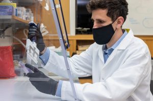
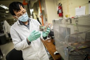
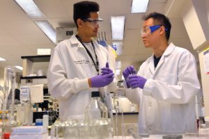
University of Puerto Rico’s Edgardo Sánchez (left) and Penn graduate Zhiwei Liao working in the lab of Daeyeon Lee. Via the Advancing Device Innovation through Inclusive Research and Education program, researchers from Penn and the University of Puerto Rico will continue their materials science collaboration while supporting STEM career pathways for underrepresented groups. (Image credit: Felice Macera).

The National Science Foundation (NSF) has awarded grants to eight research teams to support partnerships that will increase diversity in cutting-edge materials research, education, and career development. One of those teams is Penn’s Laboratory for Research on the Structure of Matter (LRSM) and the University of Puerto Rico (UPR), whose long-running collaboration has now received an additional six years of support.

With the goal of supporting partnerships between minority-serving educational institutions and leading materials science research centers, NSF’s Partnership for Research & Education in Materials (PREM) program funds innovative research programs and provides institutional support to increase recruitment, retention, and graduation by underrepresented groups as well as providing underserved communities access to materials research and education.

‘Research at the frontier’

With this PREM award, known as the Advancing Device Innovation through Inclusive Research and Education (ADIIR) program, researchers from Penn and UPR’s Humacao and Cayey campuses will conduct research on the properties of novel carbon-based materials with unique properties, and will study the effects of surface modification in new classes of sensors, detectors, and purification devices.

Thanks to this collaboration of more than 20 years, both institutions have made significant scientific and educational progress aided by biannual symposia and regular pre-pandemic travel between both institutions before the pandemic, resulting in a rich portfolio of publications, conference presentations, patents, students trained, and outreach programs.

“Together we have been publishing good papers that have impact, and we’ve really cultivated a culture of collaboration and friendship between our institutions,” says Penn’s Arjun Yodh, former director of the LRSM. “Our goal is to carry out research at the frontier and, in the process, nurture promising students from Puerto Rico and Penn.”

Ivan Dmochowski, a chemistry professor at Penn who has been involved with PREM for several years, says that this program has helped his group connect with experts in Puerto Rico whose skills complement his group’s interests in protein engineering. Dmochowski has also hosted UPR faculty members and students in his lab and also travelled to Puerto Rico before the pandemic to participate in research symposia, seminars, and outreach events.

“I’ve had students who have benefitted from being a co-author on a paper or having a chance to mentor students, and the faculty we’ve interacted with are exceptional,” Dmochowski says. “There’s a lot of benefit for both me and my students, and I’ve enjoyed our interactions both personally and scientifically.”

Penn’s Daeyeon Lee, a chemical and biomolecular engineering professor who has been involved with PREM for several years, regularly hosts students and faculty from UPR while working on nanocarbon-based composite films for sensor applications. The success of this collaboration relies on unique materials made by researchers at UPR combined with a method for processing them into composite structures developed in Lee’s lab.

“What I really admire about people at PREM, both faculty and students, is their passion,” says Lee. “I think that’s had a really positive impact on my students and postdocs who got to interact with them because they got to see the passion that the students brought.”

Read the full story in Penn Today.

Daeyeon Lee is a professor and the Evan C Thompson Term Chair for Excellence in Teaching in the Department of Chemical and Biomolecular Engineering and a member of the Bioengineering Graduate Group in Penn’s School of Engineering and Applied Science.

Arjun Yodh is the James M. Skinner Professor of Science in the Department of Physics & Astronomy in Penn’s School of Arts & Sciences and a member of the Bioengineering Graduate Group in Penn’s School of Engineering and Applied Science.

“’Electronic Nose’ Accurately Sniffs Out Hard-to-Detect Cancers”

A.T. Charlie Johnson, Ph.D.

A.T. Charlie Johnson, Rebecca W. Bushnell Professor of Physics and Astronomy at the Penn School of Arts & Sciences, and member of the Penn Bioengineering Graduate Group has been working with a team of researchers on a new “electronic nose” that could help track the spread of COVID-19 based on the disease’s unique odor profile. Now, similar research shows that vapors emanating from blood samples can be tested to distinguish between benign and cancerous pancreatic and ovarian cells. Johnson presented the results at the annual American Society of Clinical Oncology meeting on June 4 (Abstract # 5544):

“It’s an early study but the results are very promising,” Johnson said. “The data shows we can identify these tumors at both advanced and the earliest stages, which is exciting. If developed appropriately for the clinical setting, this could potentially be a test that’s done on a standard blood draw that may be part of your annual physical.”

Read the full story in Penn Medicine News.

Bioengineering Contributes to “New COVID-19 Testing Technology at Penn”

César de la Fuente, Ph.D., a Presidential Assistant Professor in Psychiatry, Microbiology, and Bioengineering, is leading a team to develop an electrode that can be easily printed at low cost to provide COVID-19 test results from your smart phone.

A recent Penn Medicine blog post surveys the efforts across Penn and the Perelman School of Medicine to develop novel says to detect SARS-CoV-2 and features several Department of Bioengineering faculty and Graduate Group members, including César de la Fuente, Presidential Assistant Professor in Psychiatry, Microbiology, and Bioengineering; Arupa Ganguly, Professor in Genetics; A.T. Charlie Johnson, Rebecca W. Bushnell Professor in Physics and Astronomy; Lyle Ungar, Professor in Computer and Information Science; and Ping Wang, Associate Professor in Pathology and Laboratory Medicine.

Read “We’ll Need More than Vaccines to Vanquish the Virus: New COVID-19 Testing Technology at Penn” by Melissa Moody in Penn Medicine News.

An ‘Electronic Nose’ to Sniff Out COVID-19

by Erica K. Brockmeier

Postdoc Scott Zhang at work in the Johnson lab. (Photo: Eric Sucar, University Communications)

Even as COVID-19 vaccines are being rolled out across the country, the numerous challenges posed by the pandemic won’t all be solved immediately. Because herd immunity will take some time to reach and the vaccine has not yet been approved for some groups, such as children under 16 years of age, the coming months will see a continued need for tools to rapidly track the disease using real-time community monitoring.

A team of Penn researchers is working on a new “electronic nose” that could help track the spread of COVID-19. Led by physicist Charlie Johnson, the project, which was recently awarded a $2 million grant from the NIH, aims to develop rapid and scalable handheld devices that could spot people with COVID-19 based on the disease’s unique odor profile.

Dogs and devices that can detect diseases

Long before “coronavirus” entered into the vernacular, Johnson was collaborating with Cynthia Otto, director of the Penn Vet Working Dog Center, and Monell Chemical Senses Center’s George Preti to diagnose diseases using odor. Diseases are known to alter a number of physical processes, including body odors, and the goal of the collaboration was to develop new ways to detect the volatile organic compounds (VOCs) that were unique to ovarian cancer.

The next step is to scale down the current device, and the researchers are aiming to develop a prototype for testing on patients within the next year.

Since 2012, the researchers have been developing new ways to diagnose early-stage ovarian cancer. Otto trained dogs to recognize blood plasma samples from patients with ovarian cancer using their acute sense of smell. Preti, who passed away last March, was looking for the specific VOCs that gave ovarian cancer a unique odor. Johnson developed a sensor array, an electronic version of the dog’s nose, made of carbon nanotubes interwoven with single-stranded DNA. This device binds to VOCs and can determine samples that came from patients with ovarian cancer.

Last spring, as the pandemic’s threat became increasingly apparent, Johnson and Otto shifted their efforts to see if they could train their disease-detecting devices and dogs to spot patients with COVID-19.

Continue reading at Penn Today.

N.B.: A.T. Charlie Johnson, Rebecca W. Bushnell Professor of Physics and Astronomy at the Penn School of Arts & Sciences, and Lyle Ungar, Professor in Computer and Information Science at Penn Engineering and Psychology at the School of Arts & Sciences, are both members of the Penn Bioengineering Graduate Group.

Arjun Yodh Named 2021 Michael S. Feld Biophotonics Award Recipient by The Optical Society

Arjun Yodh, Ph.D.

The Department of Phsyics in the Penn School of Arts & Sciences has announced that Arjun Yodh, Professor in Physics and Astronomy and member of the Bioengineering Graduate Group, was awarded the 2021 Michael S. Biophotonics Award by the Optical Society (OSA):

“He was selected for his ‘pioneering research on optical sensing in scattering media, especially diffuse optical and correlation spectroscopy and tomography, and for advancing the field of biophotonics through mentorship.’

The award ‘recognizes innovative and influential contributions to the field of biophotonics, regardless of career stage.'”

Nader Engheta Awarded Isaac Newton Medal and Prize


Nader Engheta, PhD

Nader Engheta, H. Nedwill Ramsey Professor in Electrical and Systems Engineering, Bioengineering and Materials Science and Engineering, has been awarded the 2020 Isaac Newton Medal and Prize by the Institute of Physics (IOP). The IOP is the professional body and scholarly society for physics in the UK and Ireland.

Engheta has been recognized for ” groundbreaking innovation and transformative contributions to electromagnetic complex materials and nanoscale optics, and for pioneering development of the fields of near-zero-index metamaterials, and material-inspired analogue computation and optical nanocircuitry.”

Read the full story in Penn Engineering Today.

Neuroengineering/Bioengineering Seminar: “Photovoltaic Restoration of Sight in Age-related Macular Degeneration” (Daniel Palanker)

Daniel Palanker, PhD

The Center for Neuroengineering and Therapeutics and the Department of Bioengineering present:

Speaker: Daniel Palanker, Ph.D.
Director of the Hansen Experimental Physics Laboratory and Professor of Ophthalmology
Stanford University

Date: Wednesday, November 18, 2020
Time: 1:00-2:00 PM EST
Zoom – check email for link or contact eprince@seas.upenn.edu

Title: “Photovoltaic Restoration of Sight in Age-related Macular Degeneration”

Abstract:

Retinal degenerative diseases lead to blindness due to loss of the “image capturing” photoreceptors, while neurons in the “image-processing” inner retinal layers are relatively well preserved. Information can be reintroduced into the visual system using electrical stimulation of the surviving inner retinal neurons. We developed a photovoltaic substitute of photoreceptors which convert light into pulsed electric current, stimulating the secondary retinal neurons. Visual information captured by a camera is projected onto the retina from augmented-reality glasses using pulsed near-infrared (~880nm) light. This design avoids the use of bulky electronics and wiring, thereby greatly reducing the surgical complexity. Optical activation of the photovoltaic pixels allows scaling the number of electrodes to thousands. In preclinical studies, we found that prosthetic vision with subretinal implants preserves many features of natural vision, including flicker fusion at high frequencies (>30 Hz), adaptation to static images, antagonistic center-surround organization and non-linear summation of subunits in receptive fields, providing high spatial resolution. Results of the clinical trial with our implants (PRIMA, Pixium Vision) having 100μm pixels, as well as preclinical measurements with 75 and 55μm pixels, confirm that spatial resolution of prosthetic vision can reach the pixel pitch. Remarkably, central prosthetic vision in AMD patients can be perceived simultaneously with peripheral natural vision. For broader acceptance of this technology by patients who lost central vision due to agerelated macular degeneration, visual acuity should exceed 20/100, which requires pixels smaller than 25μm. I will describe the fundamental limitations in electro-neural interfaces and 3-dimensional configurations which should enable such a high spatial resolution. Ease of implantation of these wireless arrays, combined with high resolution opens the door to highly functional restoration of sight.

Bio:

Daniel Palanker is a Professor of Ophthalmology and Director of the Hansen Experimental Physics Laboratory at Stanford University. He received MSc in Physics in 1984 from the State University of Armenia in Yerevan, and PhD in Applied Physics in 1994 from the Hebrew University of Jerusalem, Israel. Dr. Palanker studies interactions of electrical field with biological cells and tissues, and develops optical and electronic technologies for diagnostic, therapeutic, surgical and prosthetic applications, primarily in ophthalmology. In the range of optical frequencies, his studies include laser-tissue interactions with applications to ocular therapy and surgery, and interferometric imaging of neural signals. In the field of electro-neural interfaces, he is developing highresolution photovoltaic retinal prosthesis for restoration of sight and implants for electronic control of organs. Several of his developments are in clinical practice world-wide: Pulsed Electron Avalanche Knife (PEAK PlasmaBlade, Medtronic), Patterened Scanning Laser Photocoagulator (PASCAL, Topcon), Femtosecond Laser-assisted Cataract Surgery (Catalys, J&J), and Neural Stimulator for enhancement of tear secretion (TrueTear, Allergan). Photovoltaic retinal prosthesis for restoration of sight (PRIMA, Pixium Vision) is in clinical trials.

See the full list of upcoming Penn Bioengineering events here.

What do ‘Bohemian Rhapsody,’ ‘Macbeth,’ and a list of Facebook Friends All Have in Common?

New research finds that works of literature, musical pieces, and social networks have a similar underlying structure that allows them to share large amounts of information efficiently.

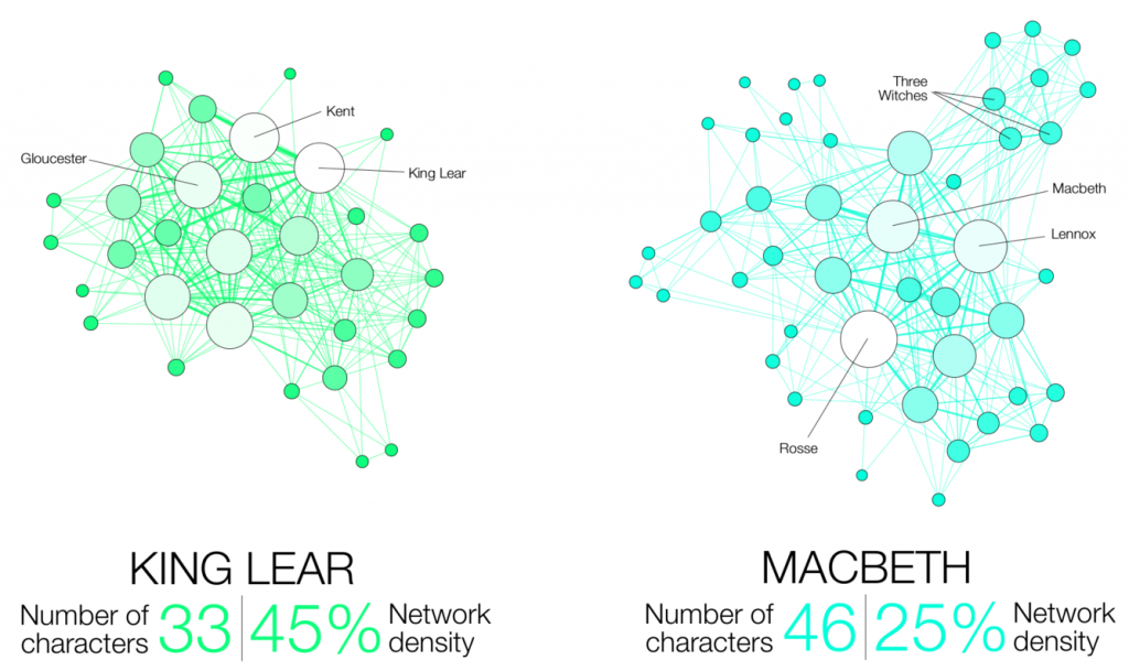
Examples of statistical network analysis of characters in two of Shakespeare’s tragedies. Two characters are connected by a line, or edge, if they appear in the same scene. The size of the circles that represent these characters, called nodes, indicate how many other characters one is connected to. The network’s density relates to how complete the graph is, with 100% density meaning that it has all of the characters are connected. (Image: Martin Grandjean)


By Erica K. Brockmeier

To an English scholar or avid reader, the Shakespeare Canon represents some of the greatest literary works of the English language. To a network scientist, Shakespeare’s 37 plays and the 884,421 words they contain also represent a massively complex communication network. Network scientists, who employ math, physics, and computer science to study vast and interconnected systems, are tasked with using statistically rigorous approaches to understand how complex networks, like all of Shakespeare, convey information to the human brain.

New research published in Nature Physics uses tools from network science to explain how complex communication networks can efficiently convey large amounts of information to the human brain. Conducted by postdoc Christopher Lynn, graduate students Ari Kahn and Lia Papadopoulos, and professor Danielle S. Bassett, the study found that different types of networks, including those found in works of literature, musical pieces, and social connections, have a similar underlying structure that allows them to share information rapidly and efficiently.

Technically speaking, a network is simply a statistical and graphical representation of connections, known as edges, between different endpoints, called nodes. In pieces of literature, for example, a node can be a word, and an edge can connect words when they appear next to each other (“my” — “kingdom” — “for” — “a” — “horse”) or when they convey similar ideas or concepts (“yellow” — “orange” — “red”).

The advantage of using network science to study things like languages, says Lynn, is that once relationships are defined on a small scale, researchers can use those connections to make inferences about a network’s structure on a much larger scale. “Once you define the nodes and edges, you can zoom out and start to ask about what the structure of this whole object looks like and why it has that specific structure,” says Lynn.

Building on the group’s recent study that models how the brain processes complex information, the researchers developed a new analytical framework for determining how much information a network conveys and how efficient it is in conveying that information. “In order to calculate the efficiency of the communication, you need a model of how humans receive the information,” he says.

Continue reading at Penn Today.

The Optimal Immune Repertoire for Bacteria

by Erica K. Brockmeier

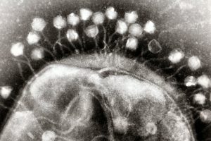
Transmission electron micrograph of multiple bacteriophages, viruses that infect bacteria, attached to a cell wall. New research describes how bacteria can optimize their “memory” of past viral infections in order to launch an effective immune response against a new invader. (Image: Graham Beards)

Before CRISPR became a household name as a tool for gene editing, researchers had been studying this unique family of DNA sequences and its role in the bacterial immune response to viruses. The region of the bacterial genome known as the CRISPR cassette contains pieces of viral genomes, a genomic “memory” of previous infections. But what was surprising to researchers is that rather than storing remnants of every single virus encountered, bacteria only keep a small portion of what they could hold within their relatively large genomes.

Work published in the Proceedings of the National Academy of Sciences provides a new physical model that explains this phenomenon as a tradeoff between how much memory bacteria can keep versus how efficiently they can respond to new viral infections. Conducted by researchers at the American Physical Society, Max Planck Institute, University of Pennsylvania, and University of Toronto, the model found an optimal size for a bacteria’s immune repertoire and provides fundamental theoretical insights into how CRISPR works.

In recent years, CRISPR has become the go-to biotechnology platform, with the potential to transform medicine and bioengineering. In bacteria, CRISPR is a heritable and adaptive immune system that allows cells to fight viral infections: As bacteria come into contact with viruses, they acquire chunks of viral DNA called spacers that are incorporated into the bacteria’s genome. When the bacteria are attacked by a new virus, spacers are copied from the genome and linked onto molecular machines known as Cas proteins. If the attached sequence matches that of the viral invader, the Cas proteins will destroy the virus.

Bacteria have a different type of immune system than vertebrates, explains senior author Vijay Balasubramanian, but studying bacteria is an opportunity for researchers to learn more about the fundamentals of adaptive immunity. “Bacteria are simpler, so if you want to understand the logic of immune systems, the way to do that would be in bacteria,” he says. “We may be able to understand the statistical principles of effective immunity within the broader question of how to organize an immune system.”

Read more on Penn Today.

Vijay Balasubramanian is the Cathy and Marc Lasry Professor in the Department of Physics and Astronomy in the School of Arts & Sciences at the University of Pennsylvania and a member of the Department of Bioengineering Graduate Group

This research was supported by the Simons Foundation (Grant 400425) and National Science Foundation Center for the Physics of Biological Function (Grant PHY-1734030).