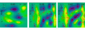
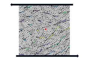
Developing new soft materials requires new data-driven research techniques, such as autonomous experimentation. Data regarding nanometer-scale material structure, taken by X-ray measurements at a synchrotron, can be fed into an algorithm that identifies the most relevant features, represented here as red dots. The algorithm then determines the optimum conditions for the next set of measurements and directs their execution without human intervention. Brookhaven National Laboratory’s Kevin Yager, who helped develop this technique, will co-teach a course on it as part of a new Penn project on Data Driven Soft Materials Research.
The National Science Foundation’s Research Traineeship Program aims to support graduate students, educate the STEM leaders of tomorrow and strengthen the national research infrastructure. The program’s latest series of grants are going toward university programs focused on artificial intelligence and quantum information science and engineering – two areas of high priority in academia, industry and government.
Chinedum Osuji, Eduardo D. Glandt Presidential Professor and Chair of the Department of Chemical and Biomolecular Engineering (CBE), has received one of these grants to apply data science and machine learning to the field of soft materials. The grant will provide five years of support and a total of $3 million for a new Penn project on Data Driven Soft Materials Research.
Osuji will work with co-PIs Russell Composto, Professor and Howell Family Faculty Fellow in Materials Science and Engineering, Bioengineering, and in CBE, Zahra Fakhraai, Associate Professor of Chemistry in Penn’s School of Arts & Sciences (SAS) with a secondary appointment in CBE, Paris Perdikaris, Assistant Professor in Mechanical Engineering and Applied Mechanics, and Andrea Liu, Hepburn Professor of Physics and Astronomy in SAS, all of whom will help run the program and provide the connections between the multiple fields of study where its students will train.
These and other affiliated faculty members will work closely with co-PI Kristin Field, who will serve as Program Coordinator and Director of Education.
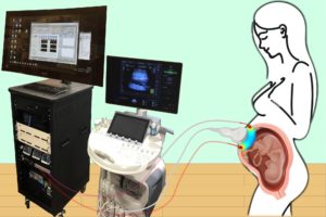
Combining optical measurements with ultrasound, an interdisciplinary team from the School of Arts & Sciences, Perelman School of Medicine, and CHOP developed a device to better measure blood flow and oxygenation in the placenta. (Image: Lin Wang)
A study published in Nature Biomedical Engineering details a novel method for imaging the placenta in pregnant patients as well as the results of a pilot clinical study. By combining optical measurements with ultrasound, the findings show how oxygen levels can be monitored noninvasively and provides a new way to generate a better understanding of this complex, crucial organ. This research was the result of a collaboration of the groups of the University of Pennsylvania’s Arjun Yodh and Nadav Schwartz with colleagues from the Children’s Hospital of Philadelphia (CHOP) and was led by postdoc Lin Wang.
Schwartz describes the placenta as the “engine” of pregnancy, an organ that plays a crucial role in delivering nutrients and oxygen to the fetus. Placental dysfunction can lead to complications such as fetal growth restriction, preeclampsia, and stillbirth. To increase knowledge about this crucial organ, the National Institute of Child Health and Human Development launched the Human Placenta Project in 2014. One focus of the program is to develop tools to assess human placental structure and function in real time, including optical devices.
For three years, the researchers optimized the design of their instrument and tested it in preclinical settings. The process involved integrating optical fibers with ultrasound probes, exploring various ultrasound transducers, and improving the multimodal technology so that measurements were stable, accurate, and reproducible while collecting data at the bedside. The resulting instrumentation now enables researchers to study the anatomy of the placenta while also collecting detailed functional information about placenta blood flow and oxygenation, capabilities that existing commercially devices do not have, the researchers say.
Because the placenta is located far below the body’s surface, one of the key technical challenges addressed by Wang, a postdoc in Yodh’s lab, was reducing background noise in the opto-electronic system. Light is scattered and absorbed when it travels through thick tissues, Yodh says, and the key for success was to reduce background interference so that the small amount of light that penetrates deep into the placenta and then returns is still large enough for a high-quality measurement.
“We’re sending a light signal that goes through the same deep tissues as the ultrasound. The extremely small amount of light that returns to the surface probe is then used to accurately assess tissue properties, which is only possible with very stable lasers, optics, and detectors,” says Yodh. “Lin had to overcome many barriers to improve the signal-to-noise ratio to the point where we trusted our data.”
The authors are Lin Wang, Jeffrey M. Cochran, Kenneth Abramson, Lian He, Venki Kavuri, Samuel Parry, Arjun G. Yodh, and Nadav Schwartz from Penn; Tiffany Ko, Wesley B. Baker, and Rebecca L. Linn from the Children’s Hospital of Philadelphia, and David R. Busch, previously a research associate at Penn and now at the University of Texas Southwestern Medical School.
This research was supported by National Institutes of Health grants F31HD085731, R01NS113945, R01NS060653, P41EB015893, P41EB015893, T32HL007915, and U01HD087180.
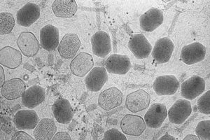
A group of bacteriophages, viruses that infect bacteria, imaged using transmission electron microscopy. New research sheds light on how bacteria fight off these invaders without triggering an autoimmune response. (Image: ZEISS Microscopy, CC BY-NC-ND 2.0)
During the last few years, CRISPR has grabbed headlines for helping treat patients with conditions as varied as blindness and sickle cell disease. However, long before humans co-opted CRISPR to fight genetic disorders, bacteria were using CRISPR as an immune system to fight off viruses.
In bacteria, CRISPR (Clustered Regularly Interspaced Short Palindromic Repeats) works by stealing small pieces of DNA from infecting viruses and storing those chunks in the genes of the bacteria. These chunks of DNA, called spacers, are then copied to form little tags, which attach to proteins that float around until they find a matching piece of DNA. When they find a match, they recognize it as a virus and cut it up.
Now, a paper published in Current Biology by researchers from the University of Pennsylvania Department of Physics and Astronomy shows that the risk of autoimmunity plays a key role in shaping how CRISPR stores viral information, guiding how many spacers bacteria keep in their genes, and how long those spacers are.
Ideally, spacers should only match DNA belonging to the virus, but there is a small statistical chance that the spacer matches another chunk of DNA in the bacteria itself. That could spell death from an autoimmune response.
“The adaptive immune system in vertebrates can produce autoimmune disorders. They’re very serious and dangerous, but people hadn’t really considered that carefully for bacteria,” says Vijay Balasubramanian, principal investigator for the paper and the Cathy and Marc Lasry Professor of Physics in the School of Arts & Sciences.
Balancing this risk can put the bacteria in something of an evolutionary bind. Having more spacers means they can store more information and fend off more types of viruses, but it also increases the likelihood that one of the spacers might match the DNA in the bacteria and trigger an autoimmune response.
Vijay Balasubramanian is the Cathy and Marc Lasry Professor of Physics at the Department of Physics and Astronomy of the University of Pennsylvania, a visiting professor at Vrije Universiteit Brussel, and a member of the Penn Bioengineering Graduate Group.
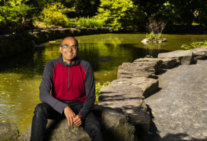
Cathy and Marc Lasry Professor Vijay Balasubramanian at Penn’s BioPond.
In an interview with Quanta Magazine, Vijay Balasubramanian discusses his work as a theoretical physicist, noting his study of the foundations of physics and the fundamentals of space and time. He speaks of the importance of interdisciplinary study and about how literature and the humanities can contextualize scientific exploration in the study of physics, computer science, and neuroscience.
Balasubramanian is Cathy and Marc Lasry Professor in the Department of Physics and Astronomy in the Penn School of Arts and Sciences and a member of the Penn Bioengineering Graduate Group.
A suspension of particles of different sizes during shearing experiments conducted in the lab of Paulo Arratia, with arrows indicating particle “flow” and trajectories. In a new study published in Nature Physics, researchers detail the relationship between a disordered material’s individual particle arrangement and how it reacts to external stressors. The study also found that these materials have “memory” that can be used to predict how and when they will flow. (Image: Arratia lab)
New research published in Nature Physics details the relationship between a disordered material’s individual particle arrangement and how it reacts to external stressors. The study also found that these materials have “memory” that can be used to predict how and when they will flow. The study was led by Larry Galloway, a Ph.D. student in the lab of Paulo Arratia, and Xiaoguang Ma, a former postdoc in the lab of Arjun Yodh, in collaboration with researchers in the labs of Douglas Jerolmack and Celia Reina.
A disordered material is randomly arranged at the particle-scale, e.g. atoms or grains, instead of being systematically distributed—think of a pile of sand instead of a neatly stacked brick wall. Researchers in the Arratia lab are studying this class of materials as part of Penn’s Materials Research Science & Engineering Center, where one of the program’s focuses is on understanding the organization and proliferation of particle-scale rearrangements in disordered, amorphous materials.
The key question in this study was whether one could observe the structure of a disordered material and have some indication as to how stable it is or when it might begin to break apart. This is known as the yield point, or when the material “flows” and begins to move in response to external forces. “For example, if you look at the grains of a sand castle and how they are arranged, can I tell you whether the wind can blow it over or if it has to be hit hard to fall over?” says Arratia. “We want to know, just by looking at the way the particles are arranged, if we can say anything about the way they’re going to flow or if they are going to flow at all.”
While it has been known that individual particle distribution influences yield point, or flow, in disordered materials, it has been challenging to study this phenomenon since the field lacks ways to “quantify” disorder in such materials. To address this challenge, the researchers collaborated with colleagues from across campus to combine expertise across the fields of experimentation, theory, and simulations.
The authors are Larry Galloway, Erin Teich, Christoph Kammer, Ian Graham, Celia Reina, Douglas Jerolmack, Arjun Yodh, and Paulo Arratia from Penn; Xiaoguang Ma, previously a postdoc at Penn and now at the Southern University of Science and Technology in Shenzhen, China; and Nathan Keim, previously a postdoc at Penn and now at Pennsylvania State University.
A processed image representative of the types of images used in this study. Natural landscapes were transformed into binary images, ones made of black and white pixels, that were decomposed into different textures defined by specific statistics. (Image: Eugenio Piasini)
The human brain uses more energy than any other organ in the body, requiring as much as 20% of the body’s total energy. While this may sound like a lot, the amount of energy would be even higher if the brain were not equipped with an efficient way to represent only the most essential information within the vast, constant stream of stimuli taken in by the five senses. The hypothesis for how this works, known as efficient coding, was first proposed in the 1960s by vision scientist Horace Barlow.
Now, new research from the Scuola Internazionale Superiore di Studi Avanzati (SISSA) and the University of Pennsylvania provides evidence of efficient visual information coding in the rodent brain, adding support to this theory and its role in sensory perception. Published in eLife, these results also pave the way for experiments that can help understand how the brain works and can aid in developing novel artificial intelligence (AI) systems based on similar principles.
According to information theory—the study of how information is quantified, stored, and communicated—an efficient sensory system should only allocate resources to how it represents, or encodes, the features of the environment that are the most informative. For visual information, this means encoding only the most useful features that our eyes detect while surveying the world around us.
Vijay Balasubramanian, a computational neuroscientist at Penn, has been working on this topic for the past decade. “We analyzed thousands of images of natural landscapes by transforming them into binary images, made up of black and white pixels, and decomposing them into different textures defined by specific statistics,” he says. “We noticed that different kinds of textures have different variability in nature, and human subjects are better at recognizing those which vary the most. It is as if our brains assign resources where they are most necessary.”
Cathy and Marc Lasry Professor Vijay Balasubramanian at Penn’s BioPond.
A study published in PLOS Computational Biology describes a new model for how the olfactory system discerns unique odors. Researchers from the University of Pennsylvania found that a simplified, statistics-based model can explain how individual odors can be perceived as more or less similar from others depending on the context. This model provides a starting point for generating new hypotheses and conducting experiments that can help researchers better understand the olfactory system, a complex, crucial part of the brain.
The sense of smell, while crucial for things like taste and hazard avoidance, is not as well studied as other senses. Study co-author Vijay Balasubramanian, a theoretical physicist with an interest in how living systems process information, says that olfaction is a prime example of a complex information-processing system found in nature, as there are far more types of volatile molecules—on the scale of tens or hundreds of thousands—than there are receptor types in the nose to detect them, on the scale of tens to hundreds depending on the species.
“Every molecule can bind to many receptors, and every receptor can bind to many molecules, so you get this combinatorial mishmash, with the nose encoding smells in a way that involves many receptor types to collectively tell you what a smell is,” says Balasubramanian. “And because there are many fewer receptor types than molecular species, you basically have to compress a very high dimensional olfactory space into a much lower dimensional space of neural responses.”
The prestigious APS Fellowship Program signifies recognition by one’s professional peers. Each year, no more than one half of one percent of the APS membership is recognized with this distinct honor. Bassett’s election and groundbreaking work in biological physics and network science will be recognized through presentation of a certificate at the APS March Meeting.
Bassett is a pioneer in the field of network neuroscience, an emerging subfield which incorporates elements of mathematics, physics, biology and systems engineering to better understand how the overall shape of connections between individual neurons influences cognitive traits. They lead the Complex Systems lab which tackles problems at the intersection of science, engineering, and medicine using systems-level approaches, exploring fields such as curiosity, dynamic networks in neuroscience, and psychiatric disease.
Bassett recently collaborated with Penn artist-in-residence Rebecca Kamen and other scholars on an interdisciplinary art exhibit on the creative process in art and science at the Katzen Art Center at American University. They have also published research modeling different types of curiosity and exploring gender-based citation bias in neuroscience publishing.
“I’m thrilled and humbled to receive this honor from the American Physical Society,” says Bassett. “I am indebted to the many fantastic mentees, colleagues, and mentors that have made my time in science such an exciting adventure. Thank you.”
New collaborative research describes how electrons move through two different configurations of bilayer graphene, the atomically-thin form of carbon. These results provide insights that researchers could use to design more powerful and secure quantum computing platforms in the future.
“Today’s computer chips are based on our knowledge of how electrons move in semiconductors, specifically silicon,” says first and co-corresponding author Zhongwei Dai, a postdoc at Brookhaven. “But the physical properties of silicon are reaching a physical limit in terms of how small transistors can be made and how many can fit on a chip. If we can understand how electrons move at the small scale of a few nanometers in the reduced dimensions of 2-D materials, we may be able to unlock another way to utilize electrons for quantum information science.”
When a material is designed at these small scales, to the size of a few nanometers, it confines the electrons to a space with dimensions that are the same as its own wavelength, causing the material’s overall electronic and optical properties to change in a process called quantum confinement. In this study, the researchers used graphene to study these confinement effects in both electrons and photons, or particles of light.
The work relied upon two advances developed independently at Penn and Brookhaven. Researchers at Penn, including Zhaoli Gao, a former postdoc in the lab of Charlie Johnson who is now at The Chinese University of Hong Kong, used a unique gradient-alloy growth substrate to grow graphene with three different domain structures: single layer, Bernal stacked bilayer, and twisted bilayer. The graphene material was then transferred onto a special substrate developed at Brookhaven that allowed the researchers to probe both electronic and optical resonances of the system.
“This is a very nice piece of collaborative work,” says Johnson. “It brings together exceptional capabilities from Brookhaven and Penn that allow us to make important measurements and discoveries that none of us could do on our own.”
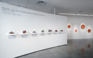
Rebecca Kamen, Penn artist-in-residence and visiting scholar, has a new exhibition titled “Reveal: The Art of Reimagining Scientific Discovery” at American University Museum at the Katzen Arts Center that explores curiosity and the creative process across art and science. (Image: Greg Staley)
Rebecca Kamen, Penn artist-in-residence and visiting scholar, has long been interested in science and the natural world. As a Philadelphia native and an artist with a 40-plus-year career, her intersectional work sheds light on the process of scientific discovery and its connections to art, with previous exhibitions that celebrate Apollo 11’s “spirit of exploration and discovery” to new representations of the periodic table of elements.
Now, in her latest exhibition, Kamen has created a series of pieces that highlight how the creative processes in art and science are interconnected. In “Reveal: The Art of Reimagining Scientific Discovery,” Kamen chronicles her own artistic process while providing a space for self-reflection that enables viewers to see the relationship between science, art, and their own creativity.
The exhibit, on display at the Katzen Art Center at American University, was inspired by the work of Penn professor Dani Bassett and American University professor Perry Zurn, the exhibit’s faculty sponsor. The culmination of three years of work, “Reveal” features collaborations with a wide range of scientists, including philosophers at American University, microscopists at the National Institutes of Health studying SARS-CoV-2 , and researchers in Penn’s Complex Systems Lab and the Addiction, Health, and Adolescence (AHA!) Lab.
“Reveal: The Art of Reimagining Scientific Discovery,” presented by the Alper Initiative for Washington Art and curated by Sarah Tanguy, is on display at the American University Museum in Washington, D.C., until Dec. 12.
The exhbition catalog, which includes an essay on “Radicle Curiosity” by Perry Zurn and Dani S. Bassett, can be viewed online.