César de la Fuente, PhD, Presidential Assistant Professor in Bioengineering, Chemical and Biomolecular Engineering, Psychiatry, and Microbiology, was featured in the Philadelphia Business Journal’s Class of 2021 “40 Under 40” list. Currently focused on antibiotic discovery, creating tools for microbiome engineering, and low-cost diagnostics, de le Fuente pioneered the world’s first computer-designed antibiotic with efficacy in animal models.
De la Fuente was previously included in the AIChE’s “35 Under 35” list in 2020 and most recently published his work demonstrating a rapid COVID-19 diagnostic test which delivers highly accurate results within four minutes.
Read “40 Under 40: Philadelphia Business Journal’s complete Class of 2021” here.
Read other BE blog posts featuring Dr. de la Fuente here.
Even as COVID-19 vaccinations are being rolled out, testing for active infections remains a critical tool in fighting the pandemic. Existing rapid tests that can directly detect the virus rely on reverse transcription polymerase chain reaction (RT-PCR), a common genetic assay that nevertheless requires trained technicians and lab space to conduct.
Alternative testing methods that can be scaled up and deployed in places where those are in short supply are therefore in high demand.
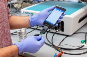
Penn researchers have now demonstrated such a method, which senses the virus by measuring the change in an electrical signal when a piece of the SARS-CoV-2 virus binds to a biosensor in their device, which they call RAPID 1.0.
The work, published in the journal Matter, was led by César de la Fuente, a Presidential Assistant Professor who has appointments in Engineering’s departments of Chemical and Biomolecular Engineering, and Bioengineering, as well as in Psychiatry and Microbiology in the Perelman School of Medicine.
“Prior to the pandemic, our lab was working on diagnostics for bacterial infections. But then, COVID-19 hit. We felt a responsibility to use our expertise to help—and the diagnostic space was ripe for improvements,” de la Fuente said. “We feel strongly about the health inequities witnessed during the pandemic, with testing access and the vaccine rollout, for example. We believe inexpensive diagnostic tests like RAPID could help bridge some of those gaps.”
The RAPID technology uses electrochemical impedance spectroscopy (EIS), which transforms the binding event between the SARS-CoV-2 viral spike protein and its receptor in the human body, the protein ACE2 (which provides the entry point for the coronavirus to hook into and infect human cells), into an electrical signal that clinicians and technicians can detect. That signal allows the test to discriminate between infected and healthy human samples. The signal can be read through a desktop instrument or a smartphone.
Carl June, MD, the Richard W. Vague Professor in Immunotherapy in the department of Pathology and Laboratory Medicine in the Perelman School of Medicine at the University of Pennsylvania, director of the Center for Cellular Immunotherapies at Penn’s Abramson Cancer Center, and member of the Penn Bioengineering Graduate Group, received the $1 million Sanford Lorraine Cross Award for his groundbreaking work in developing chimeric antigen receptor (CAR) T cell therapy. June is a world renowned cancer cell therapy pioneer.
“Sanford Health, the only health system in the country to award a $1 million prize for achievements in the medical sciences, announced the award on April 13 at a special ceremony in Sioux Falls, South Dakota. The biennial award recognizes life-changing breakthroughs and bringing emerging transformative medical innovations to patients.
‘This is a well-deserved and exciting award for one of Penn’s most distinguished faculty members, whose pioneering research has reshaped the fight against cancer and brought fresh hope for both adults and children with the disease,’ said J. Larry Jameson, MD, PhD, Executive Vice President of the University of Pennsylvania for the Health System and Dean of the Perelman School of Medicine. ‘His contributions truly have been transformative for patients across the globe and taken the field of oncology in new and powerful directions.'”
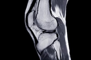
As we age, the cushioning cartilage between our joints begins to wear down, making it harder and more painful to move. Known as osteoathritis, this extremely common condition has no known cure; if the symptoms can’t be managed, the affected joints must be surgically replaced.
Now, researchers are exploring whether their specially designed nanoparticles can deliver a new inflammation inhibitor to joints, targeting a previously overlooked enzyme called sPLA2.
The normal function of sPLA2 is to provide lipids (fats) that promote a variety of inflammation processes. The enzyme is always present in cartilage tissue, but typically in low levels. However, when the researchers examined mouse and human cartilage taken from those with osteoarthritis, disproportionately high levels of the enzyme were discovered within the tissue’s structure and cells.
“This marked increase strongly suggests that sPLA2 plays a role in the development of osteoarthritis,” said the study’s corresponding author, Zhiliang Cheng, PhD, a research associate professor of Bioengineering. “Being able to demonstrate this showed that we were on the right track for what could be a potent target for the disease.”
The next step was for the study team – which included lead author Yulong Wei, MD, a researcher in Penn Medicine’s McKay Orthopaedic Research Laboratory – to put together a nanoparticle loaded with an sPLA2 inhibitor. This would block the activity of sPLA2 enzyme and, they believed, inflammation. These nanoparticles were mixed with animal knee cartilage in a lab, then observed as they diffused deeply into the dense cartilage tissue. As time progressed, the team saw that the nanoparticles stayed there and did not degrade significantly or disappear. This was important for the type of treatment the team envisioned.
Popular accounts of the human genome often depict it as a long string of DNA base pairs, but in reality the genome is separated into chromosomes that are tightly twisted and coiled into complex three-dimensional structures. These structures create a myriad of connections between sites on the genome that would be distant from one another if stretched out end-to-end. These “long range interactions” are not incidental — they regulate the activity of our genes during development and can cause disease when disrupted.
Now two teams of researchers at the Perelman School of Medicine at the University of Pennsylvania, each led by Jennifer E. Phillips-Cremins, associate professor and Dean’s Faculty Fellow in the Department of Bioengineering at the School of Engineering and Applied Science and of Genetics at the Perelman School of Medicine have been awarded grants totaling $9 million from the National Institutes of Health (NIH), as part of a major NIH Common Fund initiative to understand such 3D-genomic interactions.
The initiative, known as the 4D Nucleome Program, broadly aims to map higher-order genome structures across space and time, as well as to understand how the twists and loops of the DNA sequence govern genome function and cellular phenotype in health and disease.
N.B.: In addition to Phillips-Cremins, collaborators include Arjun Raj, Professor in Bioengineering and Genetics, and Bioengineering Graduate Group Members Melike Lakadamyali, Associate Professor in Physiology, and Bomyi Lim, Assistant Professor in Chemical and Biomolecular Engineering.
Penn Medicine researchers have developed a unifying definition of ‘cytokine storm’ to provide a framework to assess and treat patients whose immune systems have gone rogue.
One of the most elusive aspects for clinicians treating COVID-19 is the body’s immune response to the virus. In the most severe cases of COVID-19, the immune system goes into overdrive, resulting in a fever, multiorgan system damage, and often death—a cytokine storm. But how to detect and treat a cytokine storm requires that clinicians can identify it as such.
Two Penn Medicine researchers have developed a unifying definition of “cytokine storm” to provide physicians with a framework to assess and treat severely-ill patients whose immune systems have gone rogue. Cytokine storms can be triggered by different pathogens, disorders, or treatments, from COVID-19 to Castleman disease to CAR T cell therapy.
In a paper published in the New England Journal of Medicine, David Fajgenbaum,an assistant professor of translational medicine & human genetics and director of the Center for Cytokine Storm Treatment & Laboratory (CSTL), and Carl June,a professor of pathology and laboratory medicine and director of the Center for Cellular Immunotherapies in the Abramson Cancer Center, and the Parker Institute for Cancer Immunotherapies define a cytokine storm as requiring elevated circulating cytokine levels, acute systemic inflammatory symptoms, and secondary organ dysfunction beyond what could be attributed to a normal response to a pathogen, if a pathogen is present.
“There has never been a defining central review of what a cytokine storm is and how to treat one, and now with COVID-19, that is a major issue,” says Fajgenbaum, a Castleman disease patient who has previously experienced five cytokine storms himself. “I’ve spent the last 10 years of my life as a cytokine storm patient and researcher, so I know the importance of having a comprehensive unified definition to find therapies that work across the various types of cytokine storms.”
There is widespread recognition that the immune response to a pathogen, but not the pathogen itself, can contribute to multiorgan dysfunction and other symptoms. Additionally, similar cytokine storm syndromes can occur with no obvious infection.
Chlorine gas is a commonly used industrial chemical. It is also highly toxic and potentially deadly; it was used as a chemical weapon in both World War I and the Syrian Civil War and has led to multiple deaths from industrial accidents. Mixing certain household cleaners can also produce the toxic gas, leading to lasting lung injuries for which there are currently no effective treatments.
Now, researchers at Penn Engineering and Penn’s Perelman School of Medicine are collaborating with BARDA, the U.S. Office of Health and Human Services’ Biomedical Advanced Research and Development Authority, to address this need using their lung-on-a-chip technology.
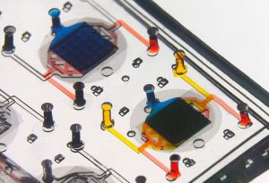
The laboratory of Dan Huh, associate professor in the Department of Bioengineering, has developed a series of organ-on-a-chip platforms. These devices incorporate human cells into precisely engineered microfluidic channels that mimic an organ’s natural environment, providing a way to conduct experiments that would not otherwise be feasible.
Huh’s previous research has involved using a placenta-on-a-chip to study which drugs are able to reach a developing fetus; investigating microgravity’s effect on the immune system by sending one of his chips to the International Space Station; and testing treatments for dry eye disease using an eye-on-a-chip, complete with a mechanical blinking eyelid.
The Perelman School of Medicine has announced the winners of the 2020 Penn Medicine Awards of Excellence. The Office of the Dean says:
“These awardees exemplify our profession’s highest values of scholarship, teaching, innovation, commitment to service, leadership, professionalism and dedication to patient care. They epitomize the preeminence and impact we all strive to achieve. The awardees range from those at the beginning of their highly promising careers to those whose distinguished work has spanned decades.
Each recipient was chosen by a committee of distinguished faculty from the Perelman School of Medicine or the University of Pennsylvania. The contributions of these clinicians and scientists exemplify the outstanding quality of patient care, mentoring, research, and teaching of our world-class faculty.”
Two faculty members affiliated with Penn Bioengineering are among this year’s recipients.
Yale Cohen, PhD, Professor of Otorhinolaryngology with secondary appointments in Neuroscience and Bioengineering, is the recipient of the Jane M. Glick Graduate Student Teaching Award. Cohen is an alumnus of the Penn Bioengineering doctoral program and is currently the department’s Graduate Chair.
“Dr. Cohen’s commitment to educating and training the next generation of scientists exemplifies the type of scientist and educator that Jane Glick represented. His students value his highly engaging and supportive approach to teaching, praising his enthusiasm, energy, honesty, and compassion.”
Douglas H. Smith, MD, Robert A. Groff Endowed Professor of Research and Teaching in Neurosurgery and member of the Penn Bioengineering Graduate Group, is the recipient of this year’s William Osler Patient Oriented Research Award:
“Dr. Smith is the foremost authority on diffuse axonal injury (DAI) as the unifying hypothesis behind the short- and long-term consequences of concussion. After realizing early in his career that concussion, or mild traumatic brain injury (TBI), was a much more serious event than broadly appreciated, Dr. Smith and his team have used computer biomechanical modeling, in vitro and in vivo testing in parallel with seminal human studies to elucidate mechanisms of concussion.”
Using a magnetic field and hydrogels, a team of researchers in the Perelman School of Medicine have demonstrated a new possible way to rebuild complex body tissues, which could result in more lasting fixes to common injuries, such as cartilage degeneration. This research was published in Advanced Materials.
“We found that we were able to arrange objects, such as cells, in ways that could generate new, complex tissues without having to alter the cells themselves,” says the study’s first author, Hannah Zlotnick, a graduate student in bioengineering who works in the McKay Orthopaedic Research Laboratory at Penn Medicine. “Others have had to add magnetic particles to the cells so that they respond to a magnetic field, but that approach can have unwanted long-term effects on cell health. Instead, we manipulated the magnetic character of the environment surrounding the cells, allowing us to arrange the objects with magnets.”
In humans, tissues like cartilage can often break down, causing joint instability or pain. Often, the breakdown isn’t in total, but covers an area, forming a hole. Current fixes are to fill those holes in with synthetic or biologic materials, which can work but often wear away because they are not the same exact material as what was there before. It’s similar to fixing a pothole in a road by filling it with gravel and making a tar patch: The hole will be smoothed out but eventually wear away with use because it’s not the same material and can’t bond the same way.
What complicates fixing cartilage or other similar tissues is that their makeup is complex.
“There is a natural gradient from the top of cartilage to the bottom, where it contacts the bone,” Zlotnick explains. “Superficially, or at the surface, cartilage has a high cellularity, meaning there is a higher number of cells. But where cartilage attaches to the bone, deeper inside, its cellularity is low.”
So the researchers, which included senior author Robert Mauck, PhD, director of the McKay Lab and a professor of Orthopaedic Surgery and Bioengineering, sought to find a way to fix the potholes by repaving them instead of filling them in. With that in mind, the research team found that if they added a magnetic liquid to a three-dimensional hydrogel solution, cells, and other non-magnetic objects including drug delivery microcapsules, could be arranged into specific patterns that mimicked natural tissue through the use of an external magnetic field.
And while innovation in health care usually brings to mind new treatments and medicines, the efforts of clinicians, engineers, and IT specialists demonstrate the importance technological infrastructure for rapidly deployable, tech-based solutions so clinicians can provide the best care to patients amid social distancing and coronavirus restrictions.
The telemedicine revolution
In late March, telemedicine was key for allowing Penn Medicine clinicians to deliver care while avoiding potentially risky in-person interactions. Chief Medical Information Officer C. William Hanson III and his team helped set up the IT infrastructure for scaling up telemedicine capabilities and provided guidance to clinicians. Thanks to the quick pivot, Penn Medicine went from 300 telemedicine visits in February to more than 7,500 visits per day in a matter of weeks.
But far from seeing telemedicine as a temporary solution during the pandemic, Hanson has been a long-time advocate for this approach to health care. In his role as liaison between clinicians and the IT community in the past 10 years Hanson, helped establish remote ICU monitoring protocols and broadened opportunities for televisits with specialists. Now, with the pandemic removing many of the previous barriers to entry, be they technical, insurance-based, or simply a lack of familiarity, Hanson believes that telemedicine is here to stay.
“As the pandemic evolved, people were aware that telemedicine could help the health care system, as well as doctors and patients, during this crisis,” he says. “Now, there are definitely places where telemedicine makes good sense, and we will continue to use that as part of our way of handling a problem.” Other benefits include removing geographic barriers to entry for new patients, reduced appointment times, increased patient satisfaction, and reduced health care provider burnout.
Simple solutions for COVID-19 challenges
As the director of Penn’s Telestroke Program, neurologist Michael Mullen has experience diagnosing from a distance. This spring, telemedicine carts his group uses were repurposed in COVID ICUs. At the same time, Mullen and group wanted to expand their ability to assess stroke patients remotely, so he reached out to Brian Litt, faculty director of Penn Health-Tech, to see how he could collaborate to create an analogous telemedicine station using readily available, cost-effective components.
Rapid and simple solutions are at the heart of Penn’s ModLab, a subgroup of the GRASP lab focused on robots made of configurable individual components. As part of a COVID-19 rapid response initiative, engineers worked with Mullen to figure out a viable solution in record time. “The idea was to make it as simple and as fast as possible,” says graduate student Caio Mucchiani. “With robotics, usually you want to make things more sophisticated, however, given the situation, we needed to know how we could use off-the-shelf components to make something.”
Fellow graduate student Ken Chaney, postdoc Bernd Pfrommer, and Mucchiani came up with a plan that replicated the required specs of the existing telemedicine carts, including state-of-the-art cameras for detailed imaging as well as a reliable, easily rechargeable battery. The team then put together 10 telemedicine carts, assembling the prototypes with social distancing and masks at the GRASP lab in early April.
While changes to treatment approaches mean that these carts still require additional field testing, Mullen is still eager to expand the program, be it for diagnosing patients safely or educating medical students in an era of social distancing. “In the setting of COVID, when everything was getting crazy, it was remarkable to see the energy that GRASP brought to help,” adds Mullen. “Everyone was really busy, and it was amazing to see this group of people who wanted to use their expertise to help.”