Carl June Highlighted for Success in Gene Therapy

Carl June, MD

Scientific American recently featured two gene therapies that were invented at Penn, including research from Carl June, MD, the Richard W. Vague Professor in Immunotherapy in Pathology and Laboratory Medicine, director of the Center for Cellular Immunotherapies, and member of the Penn Bioengineering Graduate Group, which led to the FDA approval for the CAR T therapy (sold by Novartis as Kymriah) for treating acute lymphoblastic leukemia (ALL), one of the most common childhood cancers.

Read “Four Success Stories in Gene Therapy” in Scientific American.

Nerve Repair, With Help From Stem Cells

A cross-disciplinary Penn team is pioneering a new approach to peripheral nerve repair.

In a new publication in the journal npj Regenerative Medicine, a team of Penn researchers from the School of Dental Medicine and the Perelman School of Medicine “coaxed human gingiva-derived mesenchymal stem cells (GMSCs) to grow Schwann-like cells, the pro-regenerative cells of the peripheral nervous system that make myelin and neural growth factors,” addressing the need for regrowing functional nerves involving commercially-available scaffolds to guide nerve growth. The study was led by Anh Le, Chair and Norman Vine Endowed Professor of Oral Rehabilitation in the Department of Oral and Maxillofacial Surgery/Pharmacology at the University of Pennsylvania School of Dental Medicine, and was co-authored by D. Kacy Cullen, Associate Professor in Neurosurgery at the Perelman School of Medicine at Penn and the Philadelphia Veterans Affairs Medical Center and member of the Bioengineering Graduate Group:

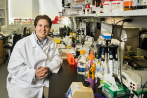
D. Kacy Cullen (Image: Eric Sucar)

“To get host Schwann cells all throughout a bioscaffold, you’re basically approximating natural nerve repair,” Cullen says. Indeed, when Le and Cullen’s groups collaborated to implant these grafts into rodents with a facial nerve injury and then tested the results, they saw evidence of a functional repair. The animals had less facial droop than those that received an “empty” graft and nerve conduction was restored. The implanted stem cells also survived in the animals for months following the transplant.

“The animals that received nerve conduits laden with the infused cells had a performance that matched the group that received an autograft for their repair,” he says. “When you’re able to match the performance of the gold-standard procedure without a second surgery to acquire the autograft, that is definitely a technology to pursue further.”

Read the full story and view the full list of collaborators in Penn Today.

Penn Anti-Cancer Engineering Center Will Delve Into the Disease’s Physical Fundamentals

by Evan Lerner

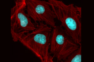
A colorized microscope image of an osteosarcoma shows how cellular fibers can transfer physical force between neighboring nuclei, influencing genes. The Penn Anti-Cancer Engineering Center will study such forces, looking for mechanisms that could lead to new treatments or preventative therapies.

Advances in cell and molecular technologies are revolutionizing the treatment of cancer, with faster detection, targeted therapies and, in some cases, the ability to permanently retrain a patient’s own immune system to destroy malignant cells.

However, there are fundamental forces and associated challenges that determine how cancer grows and spreads. The pathological genes that give rise to tumors are regulated in part by a cell’s microenvironment, meaning that the physical push and pull of neighboring cells play a role alongside the chemical signals passed within and between them.

The Penn Anti-Cancer Engineering Center (PACE) will bring diverse research groups from the School of Engineering and Applied Science together with labs in the School of Arts & Sciences and the Perelman School of Medicine to understand these physical forces, leveraging their insights to develop new types of treatments and preventative therapies.

Supported by a series of grants from the NIH’s National Cancer Institute, the PACE Center is Penn’s new hub within the Physical Sciences in Oncology Network. It will draw upon Penn’s ecosystem of related research, including faculty members from the Abramson Cancer Center, Center for Targeted Therapeutics and Translational Nanomedicine, Center for Soft and Living Matter, Institute for Regenerative Medicine, Institute for Immunology and Center for Genome Integrity.

Dennis Discher and Ravi Radhakrishnan

The Center’s founding members are Dennis Discher, Robert D. Bent Professor with appointments in the Departments of Chemical and Biomolecular Engineering (CBE), Bioengineering (BE) and Mechanical Engineering and Applied Mechanics (MEAM), and Ravi Radhakrishnan, Professor and chair of BE with an appointment in CBE.

Discher, an expert in mechanobiology and in delivery of cells and nanoparticles to solid tumors, and Radhakrishnan, an expert on modeling physical forces that influence binding events, have long collaborated within the Physical Sciences in Oncology Network. This large network of physical scientists and engineers focuses on cancer mechanisms and develops new tools and trainee opportunities shared across the U.S. and around the world.

Lukasz Bugaj, Alex Hughes, Jenny Jiang, Bomyi Lim, Jennifer Lukes and Vivek Shenoy (Clockwise from upper left).

Additional Engineering faculty with growing efforts in the new Center include Lukasz Bugaj, Alex Hughes and Jenny Jiang (BE), Bomyi Lim (CBE), Jennifer Lukes (MEAM) and Vivek Shenoy (Materials Science and Engineering).

Among the PACE Center’s initial research efforts are studies of the genetic and immune mechanisms associated with whether a tumor is solid or liquid and investigations into how physical stresses influence cell signaling.

Originally posted in Penn Engineering Today.

With NIH Pioneer Award, Jennifer E. Phillips-Cremins Will Study Genome Folding’s Role in Long-term Memory

by Evan Lerner

Jennifer E. Phillips-Cremins (upper left) and members of her lab.

Each year, the National Institutes of Health (NIH) recognizes exceptionally creative scientists through its High-Risk, High-Reward Research Program. The four awards granted by this program are designed to support researchers whose “out of the box” and “trailblazing” ideas have the potential for broad impact.

Jennifer E. Phillips-Cremins, Associate Professor and Dean’s Faculty Fellow in Penn Engineering’s Department of Bioengineering and the Perelman School of Medicine’s Department of Genetics, is one such researcher. As a recipient of an NIH Director’s Pioneer Award, she will receive $3.5 million over five years to support her work on the role that the physical folding of chromatin plays in the encoding of neural circuit and synapse properties contributing to long-term memory.

Phillips-Cremins’ award is one of 106 grants made through the High-Risk, High-Reward program this year, though she is only one of 10 to receive the Pioneer Award, which is the program’s largest funding opportunity.

“The science put forward by this cohort is exceptionally novel and creative and is sure to push at the boundaries of what is known,” said NIH Director Francis S. Collins.

Phillips-Cremins’ research is in the general field of epigenetics, the molecular and structural modifications that allow the genome — an identical copy of which is found in each cell — to express genes differently at different times and in different parts of the body. Within this field, her lab focuses on higher-order folding patterns of the DNA sequence, which bring distant sets of genes and regulatory elements into close proximity with one another as they are compressed inside the cell’s nucleus.

Previous work from the Cremins lab has investigated severe genome misfolding patterns common across a class of genetic neurological disorders, including fragile X syndrome, Huntington’s disease, ALS and Friedreich’s ataxia.

With the support of the Pioneer Award, she and the members of her lab will extend that research to a more fundamental question of neuroscience: how memory is encoded over decades, despite the rapid turnover of the relevant proteins and RNA sequences within the brain’s synapses.

“Our long-term goals are to understand how, when and why pathologic genome misfolding leads to synaptic dysfunction by way of disrupted gene expression,” said Phillips-Cremins, “as well as to engineer the genome’s structure-function relationship to reverse pathologic synaptic defects in debilitating neurological diseases.”

Originally posted in Penn Engineering Today.

How a Diversity Program Enabled a Childhood Orthopaedics Patient’s Research Dreams

by Julie Wood

As a child, Sonal Mahindroo would go to her orthopaedics appointments with her family, slowly becoming more and more fascinated by the workings and conditions of the musculoskeletal system. While being treated for scoliosis, she would receive children’s books from her doctor that helped provide clear and simplified explanations of orthopaedic topics, which supported her interest.

Nearly a decade later, Mahindroo is still interested in expanding her orthopaedic knowledge, and a Penn Medicine program is helping fuel that expansion. Now a senior at St. Bonaventure University in New York, Mahindroo spends her time at the university’s lab. But in addition to that, this year, she was able to take part in more learning opportunities with Penn Medicine’s support, via the McKay Orthopaedic Research Lab’s Diversity, Equity, and Inclusion (DEI) committee’s conference grant program.

McKay’s DEI committee — consisting of faculty, post-docs, graduate students, and staff — offers a welcoming environment and resources that support people of all identities, empowering them to bring forward unique perspectives to orthopaedic research.

“Our goal is to improve diversity and culture both within McKay and in the orthopaedic research community outside of Penn,” said Sarah Gullbrand, PhD, a research assistant professor at the McKay Lab. “We wanted to provide an opportunity for students to attend a conference and make connections to help them pursue their interest in orthopaedic research.”

The McKay conference grant supports undergraduate students who have been unable to get hands-on research experience. Participants are provided with the opportunity to network with leaders in the field of orthopaedic research, listen to cutting-edge research presentations, and learn about ways to get involved in orthopaedic research themselves.

“When launching the conference grant program earlier this year, I was motivated by my own experience attending a conference as an undergraduate. That experience really increased my interest in attending graduate school and taught me a lot about the breadth of research in orthopaedics,” said Hannah Zlotnick, a PhD student at the McKay Lab and member of the DEI committee. Through the McKay Conference Grants, the committee has supported two cohorts of students. “So far, we’ve been able to fund 11 undergraduate students from around the country to virtually attend orthopaedics conferences and receive early exposure to careers in STEM.”

Along with the conference grant, the McKay Lab holds workshops, book clubs, and other programs focused on DEI-related topics. As part of their efforts for promoting gender diversity in the field, the McKay Lab has previously partnered with the Perry Initiative to offer direct orthopaedic experiences for girls in high school, where they can learn how to suture, and perform mock fracture fixation surgeries on sawbones.

As a primarily male-populated field, orthopaedics could benefit greatly from diversity efforts. While women comprise approximately 50 percent of medical school graduates in the United States, they represent only 14 percent of orthopaedic surgery residents.

“The only women on staff at my orthopaedist’s office were receptionists. There were no female physicians or engineers to make my scoliosis brace,” Mahindroo said. “It was really cool coming to the McKay Lab and seeing how much the field has progressed since then.”

Read more at Penn Medicine News.

N.B. Hannah Zlotnick is a PhD student in Bioengineering studying in the lab of Robert Mauck, Mary Black Ralston Professor in Bioengineering and Orthopaedic Surgery.

Penn Engineers Create Faster and Cheaper COVID-19 Testing With Pencil Lead

by Melissa Pappas

César de la Fuente, PhD

Testing is key to understanding and controlling the spread of COVID-19, which has already taken more than four million lives around the world. However, current tests are limited by the tradeoff between accuracy and the time it takes to analyze a sample.

Another challenge of current COVID-19 tests is cost. Most tests are expensive to produce and require trained personnel to administer and analyze them. Testing in low-and middle-income communities has therefore been largely inaccessible, leaving individuals at greater risk of viral spread.

To address cost, time and accuracy, a new electrochemical test developed by Penn researchers uses electrodes made from graphite — the same material found in pencil lead. Developed by César de la Fuente, Presidential Assistant Professor in Bioengineering,  Microbiology and Psychiatry with a secondary appointment in Chemical and Biomolecular Engineering, these electrodes reduce the cost to $1.50 per test and require only 6.5 minutes to deliver 100-pecent-accurate results from saliva samples and up to 88 percent accuracy in nasal samples.

While his previous research highlights the invention of RAPID (Real-time Accurate Portable Impedimetric Detection prototype 1.0), a COVID-19 testing kit which uses screen-printed electrodes, this new research published in PNAS presents LEAD (Low-cost Electrochemical Advanced Diagnostic), using the same concept as RAPID but with less expensive materials. De la Fuente’s current test reduces costs from $4.67 per test (RAPID) to $1.50 per test (LEAD) just by changing the building material of the electrodes.

“Both RAPID and LEAD work on the same principle of electrochemistry,” says de la Fuente. “However, LEAD is easier to assemble, it can be used by anyone and the materials are cheaper and more accessible than those of RAPID. This is important because we are using an abundant material, graphite, the same graphite used in pencils, to build the electrode to make testing more accessible to lower-income communities.”

This figure, adapted from the paper, shows the functionalization steps of LEAD which prepares the electrodes to bind to the sample. The height of the peaks indicates whether the sample is negative or positive. Because the SARS-CoV-2 spike protein in a positive sample binds to the electrode, it inhibits the emitted signal and produces a smaller peak.

Read the full story in Penn Engineering Today.

Penn Bioengineering Graduate Shreya Parchure Receives Rose Award

Shreya Parchure (BSE/MSE 2021)

Shreya Parchure, a recent graduate of Penn Bioengineering, was selected by a committee of faculty for a 2021 Rose Award from the Center for Undergraduate Research and Fellowships (CURF). The Rose Award recognizes outstanding undergraduate research projects completed by graduating seniors under the supervision of a Penn faculty member and carries with it a $1,000 award. Parchure’s project, titled “BDNF Gene Polymorphism Predicts Response to Continuous Theta Burst Stimulation (cTBS) in Chronic Stroke Patients,” was done under the supervision of Roy H. Hamilton, Associate Professor in Neurology and Physical Medicine and Rehabilitation and director of the Laboratory for Cognition and Neural Stimulation in the Perelman School of Medicine. Parchure’s work in Hamilton’s lab previously resulted in a 2020 Goldwater Scholarship.

Parchure graduated in Spring 2021 with a B.S.E. in Bioengineering, with concentrations in Neuroengineering and Medical Devices and a minor in Chemistry, as well as a M.S.E. in Bioengineering. During her time as an undergraduate, she was a Rachleff Scholar, a recipient of a Vagelos Undergraduate Research Grant, and the Wolf-Hallac Award. She was active in many groups across the university and beyond, serving as a United Nations Millennium Fellow, a volunteer with Service Link and the Hospital of the University of Pennsylvania (HUP), a CURF Research Peer Advisor, and co-editor-in-chief of the Penn Bioethics Journal. She is now pursuing a M.D./Ph.D. through the Medical Scientist Training Program at Penn Bioengineering and the Perelman School of Medicine.

With a ‘Liquid Assembly Line,’ Penn Researchers Produce mRNA-Delivering-Nanoparticles a Hundred Times Faster than Standard Microfluidic Technologies

by Evan Lerner

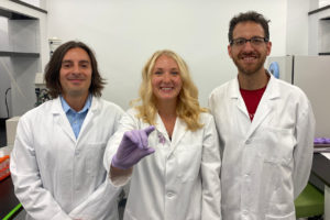
Michael Mitchell, Sarah Shepherd and David Issadore pose with their new device.

The COVID vaccines currently being deployed were developed with unprecedented speed, but the mRNA technology at work in some of them is an equally impressive success story. Because any desired mRNA sequence can be synthesized in massive quantities, one of the biggest hurdles in a variety of mRNA therapies is the ability to package those sequences into the lipid nanoparticles that deliver them into cells.

Now, thanks to manufacturing technology developed by bioengineers and medical researchers at the University of Pennsylvania, a hundred-fold increase in current microfluidic production rates may soon be possible.

The researchers’ advance stems from their design of a proof-of-concept microfluidic device containing 128 mixing channels working in parallel. The channels mix a precise amount of lipid and mRNA, essentially crafting individual lipid nanoparticles on a miniaturized assembly line.

This increased speed may not be the only benefit; more precisely controlling the nanoparticles’ size could make treatments more effective. The researchers tested the lipid nanoparticles produced by their device in a mouse study, showing they could deliver therapeutic RNA sequences with four-to-five times greater activity than those made by conventional methods.

The study was led by Michael Mitchell, Skirkanich Assistant Professor of Innovation in Penn Engineering’s Department of Bioengineering, and David Issadore, Associate Professor in Penn Engineering’s Department of Bioengineering, along with Sarah Shepherd, a doctoral student in both of their labs. Rakan El-Mayta, a research engineer in Mitchell’s lab, and Sagar Yadavali, a postdoctoral researcher in Issadore’s lab, also contributed to the study.

They collaborated with several researchers at Penn’s Perelman School of Medicine: postdoctoral researcher Mohamad-Gabriel Alameh, Lili Wang, Research Associate Professor of Medicine, James M. Wilson, Rose H. Weiss Orphan Disease Center Director’s Professor in the Department of Medicine, Claude Warzecha, a senior research investigator in Wilson’s lab, and Drew Weissman, Professor of Medicine and one of the original developers of the technology behind mRNA vaccines.

It was published in the journal Nano Letters.

“We believe that this microfluidic technology has the potential to not only play a key role in the formulation of current COVID vaccines,” says Mitchell, “but also to potentially address the immense need ahead of us as mRNA technology expands into additional classes of therapeutics.”

Read the full story in Penn Engineering Today.

“’Electronic Nose’ Accurately Sniffs Out Hard-to-Detect Cancers”

A.T. Charlie Johnson, Ph.D.

A.T. Charlie Johnson, Rebecca W. Bushnell Professor of Physics and Astronomy at the Penn School of Arts & Sciences, and member of the Penn Bioengineering Graduate Group has been working with a team of researchers on a new “electronic nose” that could help track the spread of COVID-19 based on the disease’s unique odor profile. Now, similar research shows that vapors emanating from blood samples can be tested to distinguish between benign and cancerous pancreatic and ovarian cells. Johnson presented the results at the annual American Society of Clinical Oncology meeting on June 4 (Abstract # 5544):

“It’s an early study but the results are very promising,” Johnson said. “The data shows we can identify these tumors at both advanced and the earliest stages, which is exciting. If developed appropriately for the clinical setting, this could potentially be a test that’s done on a standard blood draw that may be part of your annual physical.”

Read the full story in Penn Medicine News.

How HIV Infection Shrinks the Brain’s White Matter

by Katherine Unger Baillie

Researchers from Penn and CHOP detail the mechanism by which HIV infection blocks the maturation process of brain cells that produce myelin, a fatty substance that insulates neurons.

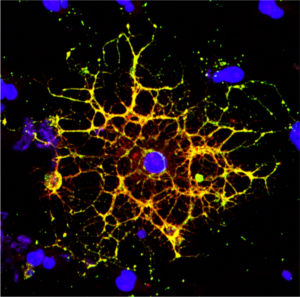
A confocal microscope image shows an oligodendrocyte in cell culture, labeled to show the cell nucleus in blue and myelin proteins in red, green, and yellow. Researchers from Penn and CHOP have shown that HIV infection prevents oligodendrocytes from maturing, leading to a reduction in white matter in the brain. (Image: Raj Putatunda)

It’s long been known that people living with HIV experience a loss of white matter in their brains. As opposed to gray matter, which is composed of the cell bodies of neurons, white matter is made up of a fatty substance called myelin that coats neurons, offering protection and helping them transmit signals quickly and efficiently. A reduction in white matter is associated with motor and cognitive impairment.

Earlier work by a team from the University of Pennsylvania and Children’s Hospital of Philadelphia (CHOP) found that antiretroviral therapy (ART)—the lifesaving suite of drugs that many people with HIV use daily—can reduce white matter, but it wasn’t clear how the virus itself contributed to this loss.

In a new study using both human and rodent cells, the team has hammered out a detailed mechanism, revealing how HIV prevents the myelin-making brain cells called oligodendrocytes from maturing, thus putting a wrench in white matter production. When the researchers applied a compound blocking this process, the cells were once again able to mature.

The work is published in the journal Glia.

“Even when people with HIV have their disease well-controlled by antiretrovirals, they still have the virus present in their bodies, so this study came out of our interest in understanding how HIV infection itself affects white matter,” says Kelly Jordan-Sciutto, a professor in Penn’s School of Dental Medicine and co-senior author on the study. “By understanding those mechanisms, we can take the next step to protect people with HIV infection from these impacts.”

“When people think about the brain, they think of neurons, but they often don’t think about white matter, as important as it is,” says Judith Grinspan, a research scientist at CHOP and the study’s other co-senior author. “But it’s clear that myelination is playing key roles in various stages of life: in infancy, in adolescence, and likely during learning in adulthood too. The more we find out about this biology, the more we can do to prevent white matter loss and the harms that can cause.”

Jordan-Sciutto and Grinspan have been collaborating for several years to elucidate how ART and HIV affect the brain, and specifically oligodendrocytes, a focus of Grinspan’s research. Their previous work on antiretrovirals had shown that commonly used drugs disrupted the function of oligodendrocytes, reducing myelin formation.

In the current study, they aimed to isolate the effect of HIV on this process. Led by Lindsay Roth, who recently earned her doctoral degree within the Biomedical Graduate Studies group at Penn and completed a postdoctoral fellowship working with Jordan-Sciutto and Grinspan, the investigation began by looking at human macrophages, one of the major cell types that HIV infects.

Read the full story in Penn Today.

Kelly Jordan-Sciutto is vice chair and professor in the University of Pennsylvania School of Dental Medicine’s Department of Basic & Translational Sciences and is director of Biomedical Graduate Studies. She is a member of the Penn Bioengineering Graduate Group.