Modified Nanoparticles Can Stop Osteoarthritis Development

Zhiliang Cheng

As we age, the cushioning cartilage between our joints begins to wear down, making it harder and more painful to move. Known as osteoathritis, this extremely common condition has no known cure; if the symptoms can’t be managed, the affected joints must be surgically replaced.

Now, researchers are exploring whether their specially designed nanoparticles can deliver a new inflammation inhibitor to joints, targeting a previously overlooked enzyme called sPLA2.

Zhiliang Cheng, a research associate professor in the Department of Bioengineering, recently collaborated with members of Penn Medicine’s McKay Orthopaedic Research Laboratory, on a study of this approach, published in the journal Science Advances.

The normal function of sPLA2 is to provide lipids (fats) that promote a variety of inflammation processes. The enzyme is always present in cartilage tissue, but typically in low levels. However, when the researchers examined mouse and human cartilage taken from those with osteoarthritis, disproportionately high levels of the enzyme were discovered within the tissue’s structure and cells.

“This marked increase strongly suggests that sPLA2 plays a role in the development of osteoarthritis,” said the study’s corresponding author, Zhiliang Cheng, PhD, a research associate professor of Bioengineering. “Being able to demonstrate this showed that we were on the right track for what could be a potent target for the disease.”

The next step was for the study team – which included lead author Yulong Wei, MD, a researcher in Penn Medicine’s McKay Orthopaedic Research Laboratory – to put together a nanoparticle loaded with an sPLA2 inhibitor. This would block the activity of sPLA2 enzyme and, they believed, inflammation. These nanoparticles were mixed with animal knee cartilage in a lab, then observed as they diffused deeply into the dense cartilage tissue. As time progressed, the team saw that the nanoparticles stayed there and did not degrade significantly or disappear. This was important for the type of treatment the team envisioned.

Continue reading at Penn Medicine News.

Originally posted in Penn Engineering Today.

“New Biosealant Can Stabilize Cartilage, Promote Healing After Injury”

New research from Robert Mauck, Mary Black Ralston Professor in Orthopaedic Surgery and Bioengineering and Director of Penn Medicine’s McKay Orthopaedic Research Laboratory, announces a “new biosealant therapy may help to stabilize injuries that cause cartilage to break down, paving the way for a future fix or – even better – begin working right away with new cells to enhance healing.” Their research was published in Advanced Healthcare Materials. The study’s lead author was Jay Patel, a former postdoctoral fellow in the McKay Lab and now Assistant Professor at Emory University and was contributed to by Claudia Loebel, a postdoctoral research in the Burdick lab and who will begin an appointment as Assistant Professor at the University of Michigan in Fall 2021. In addition, the technology detailed in this publication is at the heart of a new company (Forsagen LLC) spun out of Penn with support from the Penn Center for Innovation (PCI) Ventures Program, which will attempt to spearhead the system’s entry into the clinic. It is co-founded by both Mauck and Patel, along with study co-author Jason Burdick, Professor in Bioengineering, and Ana Peredo, a PhD student in Bioengineering.

Read the story in Penn Medicine News.

BE/MEAM Seminar: “Microbes in Biomechanics” (Christopher J. Hernandez)

Speaker: Christopher J. Hernandez, Ph.D.
Professor, Sibley School of Mechanical and Aerospace Engineering, Cornell University
Adjunct Scientist, Hospital for Special Surgery

Date: Thursday, February 4, 2021
Time: 3:00-4:00 PM EST
Zoom – check email for link or contact ksas@seas.upenn.edu

Title: “Microbes in Biomechanics”

This seminar is jointly hosted by the Department of Bioengineering and the Department of Mechanical Engineering and Applied Mechanics.

Abstract:

The idea that mechanical stresses influence the growth and form of organs and organisms originated in the 1800s and is the basis for the modern study of biomechanics and mechanobiology. Biomechanics and mechanobiology are well studied in eukaryotic systems, yet eukaryotes represent only a small portion of the diversity and abundance of life on Earth. Bacteria exhibit broad influences on human health (as both pathogens and as beneficial components of the gut microbiome) and processes used in biotechnology and synthetic biology. Over the past eight years my group has explored mechanobiology within individual bacteria and the effects of changes in the composition of commensal bacterial communities on the biomechanics in the musculoskeletal system.

The ability of the bacteria to not only resist mechanical loads (biomechanics) but also to respond to changes in the mechanical environment (mechanobiology) is necessary for survival. Here I describe a novel microfluidic platform used to explore the biomechanics and mechanobiology of individual, live bacteria. I discuss work from my group demonstrating that mechanical stress within the bacterial cell envelope can influence the assembly and function of multicomponent efflux pumps used by bacteria to resist toxins and antibiotics. Additionally, I share some of our more recent work showing that mechanical stress and strain within the bacterial cell envelope can stimulate a bacterial two-component system controlling gene expression. Our findings demonstrate that bacteria, like mammalian cells, have mechanosensitive systems that are key to survival.

In musculoskeletal disease, bacteria are commonly viewed as sources of infection. However, in the past decade the studies by my group and others have suggested that commensal bacteria – the microbiome – can modulate the pathogenesis of musculoskeletal disorders. My group is among the first to study the effects of the gut microbiome on orthopaedic disorders. Here I provide an introduction to the microbiome and current concepts of how modifications to the gut microbiome could influence the musculoskeletal system. Specifically, I discuss studies from my group which are the first to demonstrate that the gut microbiome influences bone biomechanics and the development of infection of orthopaedic implants.

Bio:

Dr. Hernandez is Professor in the Sibley School of Mechanical and Aerospace Engineering at Cornell University and is an Adjunct Scientist at the Hospital for Special Surgery. Dr. Hernandez is a Fellow of the American Institute for Medical and Biological Engineering (AIMBE), the American Society of Mechanical Engineers (ASME), and the American Society for Bone and Mineral Research (ASBMR). He is the 2018 recipient of the Fuller Albright Award for Scientific Excellence from the American Society for Bone and Mineral Research. He has served on the Board of Directors of the Orthopaedic Research Society and the American Society for Bone and Mineral Research. His laboratory’s research currently focuses on the effects of the microbiome on bone and joint disorders, periprosthetic joint infection and the biomechanics and mechanobiology of bacteria.

hernandezresearch.com

Bioengineering Faculty Contribute to New Treatment That “Halts Osteoarthritis-Like Knee Cartilage Degeneration”

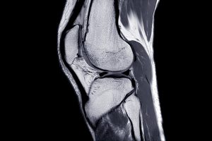
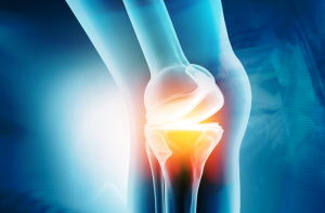
A recent study published in Science Translational Medicine announces a discovery which could halt cartilage degeneration caused by osteoarthritis: “These researchers showed that they could target a specific protein pathway in mice, put it into overdrive and halt cartilage degeneration over time. Building on that finding, they were able to show that treating mice with surgery-induced knee cartilage degeneration through the same pathway via the state of the art of nanomedicine could dramatically reduce the cartilage degeneration and knee pain.” This development could eventually lead to treating osteoarthritis with injection rather than more complicated surgery.

Among a team of Penn Engineering and Penn Medicine researchers, the study was co-written by Zhiliang Cheng, Research Associate Professor in Bioengineering, Andrew Tsourkas, Professor in Bioengineering, and Ling Qin, Associate Professor of Orthopaedic Surgery in the Perelman School of Medicine and member of the Bioengineering Graduate Group. The lead author was Yulong Wei of the Department of Orthopaedic Surgery and the McKay Orthopaedic Research Laboratory.

Read the press release in Penn Medicine News.

Student Spotlight: Sonia Bansal

Sonia Bansal, Ph.D.

Next up in the Penn Bioengineering student spotlight series is Sonia Bansal. Sonia got her B.S. in Biomedical Engineering at Columbia University in 2014. She then came to Penn, where she recently got her Ph.D. in September of 2020 in Bioengineering under the advisement of Robert Mauck, Mary Black Ralston Professor of Orthopaedic Surgery and Professor of Bioengineering. Her dissertation is entitled “Functional and Structural Remodeling of the Meniscus with Growth and Injury” and focuses on the ways the knee meniscus changes while being actively loaded (growth) and under aberrant loading (injurious) conditions. She has presented her work internationally and has first authored four papers, with two more in preparation. She is passionate about K-12 STEM outreach and teaching at the collegiate level. She has been on the teaching team for six classes in the department, and is the first recipient of the Graduate Fellowship for Teaching Excellence from the Bioengineering department.

What drew you to the field of Bioengineering?
I first got interested in Bioengineering when I realized that it would let me merge my interests in biology and the human body with my desire to solve big questions by building and creating solutions. I applied to college knowing it was what I wanted to study.

What kind of research do you conduct, and what is the focus of your thesis?
My research is focused on the knee meniscus, specifically the impacts of its complex extracellular matrix and how that matrix changes during growth and after meniscal injury. My interests are largely translational, and in the future, I’d like to think about how we can use preclinical animal models to create effective therapeutics and drive clinical decision making in the orthopedic space.

What did you study for your undergraduate degree? How does it pair with the work you’re doing now, and what advice would you give to your undergraduate self?
I studied Biomedical Engineering during my undergraduate education and worked in cartilage tissue engineering. These experiences helped guide me to my Ph.D. work here at Penn. The two pieces of advice I’d give my undergraduate self is to ask for help and that it’s important to get more than five hours of sleep a night.

What’s your favorite thing to do on Penn’s campus or in Philly?
My favorite thing to do on campus was to read papers/write lectures/work on grants at a local coffee shop. I used to go to HubBub when it still existed, Saxby’s, and United By Blue.

Have you done or learned anything new or interesting during quarantine?
I have embarked on a journey in culinary fermentation (variety of pickles and sourdough, of course), and recently started homebrewing!

Magnetic Field and Hydrogels Could Be Used to Grow New Cartilage

by Frank Otto

MRI Knee joint or Magnetic resonance imaging sagittal view for detect tear or sprain of the anterior cruciate ligament (ACL).

Using a magnetic field and hydrogels, a team of researchers in the Perelman School of Medicine have demonstrated a new possible way to rebuild complex body tissues, which could result in more lasting fixes to common injuries, such as cartilage degeneration. This research was published in Advanced Materials.

“We found that we were able to arrange objects, such as cells, in ways that could generate new, complex tissues without having to alter the cells themselves,” says the study’s first author, Hannah Zlotnick, a graduate student in bioengineering who works in the McKay Orthopaedic Research Laboratory at Penn Medicine. “Others have had to add magnetic particles to the cells so that they respond to a magnetic field, but that approach can have unwanted long-term effects on cell health. Instead, we manipulated the magnetic character of the environment surrounding the cells, allowing us to arrange the objects with magnets.”

In humans, tissues like cartilage can often break down, causing joint instability or pain. Often, the breakdown isn’t in total, but covers an area, forming a hole. Current fixes are to fill those holes in with synthetic or biologic materials, which can work but often wear away because they are not the same exact material as what was there before. It’s similar to fixing a pothole in a road by filling it with gravel and making a tar patch: The hole will be smoothed out but eventually wear away with use because it’s not the same material and can’t bond the same way.

What complicates fixing cartilage or other similar tissues is that their makeup is complex.

“There is a natural gradient from the top of cartilage to the bottom, where it contacts the bone,” Zlotnick explains. “Superficially, or at the surface, cartilage has a high cellularity, meaning there is a higher number of cells. But where cartilage attaches to the bone, deeper inside, its cellularity is low.”

So the researchers, which included senior author Robert Mauck, PhD, director of the McKay Lab and a professor of Orthopaedic Surgery and Bioengineering, sought to find a way to fix the potholes by repaving them instead of filling them in. With that in mind, the research team found that if they added a magnetic liquid to a three-dimensional hydrogel solution, cells, and other non-magnetic objects including drug delivery microcapsules, could be arranged into specific patterns that mimicked natural tissue through the use of an external magnetic field.

Read more at Penn Medicine News.

Penn Bioengineering Postdoc Brittany Taylor Appointed Assistant Professor at University of Florida


Brittany Taylor, PhD

The Department of Bioengineering is proud to congratulate Postdoctoral Researcher Brittany Taylor, PhD on her appointment as a tenure-track Assistant Professor in the J. Crayton Pruitt Family Department of Biomedical Engineering of the Herbert Wertheim College of Engineering at the University of Florida. Taylor’s appointment will begin in January 2021 after four years as a postdoc in Penn Medicine’s McKay Orthopaedic Research Laboratory where she worked under the supervision of Louis Soslowsky, Fairhill Professor in Orthopaedic Surgery and Professor in Bioengineering.

Taylor got her BS in Biomedical Engineering from the University of Virginia where she conducted research under Drs. Cato Laurencin and Edward Botchwey (the latter got his PhD in Penn Bioengineering in 2002). She went on to complete her PhD in Biomedical Engineering in 2016, studying with Dr. Joseph Freeman, in the Musculoskeletal Tissue Regeneration Laboratory at Rutgers University. During her time at Penn, she served as the Co-President of the Biomedical Postdoctoral Council, worked with the Perelman School of Medicine’s PennVIEW program on postdoctoral diversity recruitment, and spearheaded the mentoring circles program, which brings together postdoctoral researchers, graduate students, and undergraduates in informal groups that allow mentorship and learning to flow freely.

The foundation for Taylor’s research interests is a combination of her training in bone tissue engineering, bioactive biomaterials, and tendon injury and repair. Her graduate research focused on a three-dimensional biomimetic pre-vascularized scaffold that simultaneously promoted osteogenic and angiogenic differentiation of human mesenchymal stem cells in vitro and cellular infiltration and neovascularization in vivo without the addition of growth factors of cells. As a postdoctoral fellow, in addition to investigating the role of collagen type V on tendon inflammation and remodeling in a mouse patellar tendon injury model, she also elucidated the biological and mechanical implications of an implantable bilayer delivery system (BiLDS) for controlled and localized release of non-steroidal anti-inflammatory drugs (NSAIDs) to modulate tendon inflammation in a rat rotator cuff injury and repair model. This collection of work exploits the ability of these transformative technologies to provide physical and chemical regenerative cues without the use of exogenous cells; hence avoiding possible complications associated with autologous and allogeneic cell sources and simplifying the regulatory pathway towards clinical application. Taylor’s future research program at the University of Florida will focus on tailored cell-free combinatorial strategies, such as decellularized matrices, tunable delivery systems, and modified extracellular vesicles, to complement and improve the native musculoskeletal tissue regenerative and reparative process.

“Brittany has been an amazing postdoctoral fellow,” says her mentor Louis Soslowsky. “She has learned a lot and contributed to various projects in an exemplary manner. She has been a leader in many arenas here at Penn and I am so proud of what she has done so far. I look forward to following her continued accomplishments at the University of Florida! I know she’ll do great!”

In the course of her pre-faculty career, Taylor achieved an impressive list of accomplishments. She received a Postdoctoral Fellowship for Academic Diversity from the Office of the Vice Provost for Research; a Postdoctoral Enrichment Program (PDEP) award from the Burroughs Wellcome Fund; and a UNCF Bristol-Myers Squibb E.E. Just Postgraduate Fellowship. Additionally, she was named a Rising Star in Cell Mentor’s list of “100 inspiring Black scientists in America” in February 2020 and was given a Rising Star in Biomedical Science Award from MIT in 2019.

“I am grateful for the opportunity to complete my postdoctoral training at Penn,” Taylor says:

“[P]articularly in a lab that is affiliated with the Penn Bioengineering program and the Department of Orthopaedic Surgery, where I had the unique experience of addressing basic science questions using translational animal models, while utilizing my engineering background and having a direct interaction with clinicians. Additionally, I connected with some amazing people here at Penn who had a significant impact on my time at Penn, and will be lifelong friends, colleagues, and mentors.”

Congratulations Dr. Taylor from everyone at Penn Bioengineering!

Students in Penn’s Biomechatronics Course Create Robotic Hands for Their Final Project

by Sophie Burkholder

Andrew Chan (left, M.S.E. in Robotics ‘19) and Omar Abdoun (right, BE M.D./Ph.D. student) present “Cryogripper”

Almost every engineering school in the country offers a course in mechatronics — the overlap of mechanical, electrical, and computer engineering in electromechanical system design — but how many offer a course in biomechatronics? Taught by LeAnn Dourte, Ph.D., a Practice Associate Professor in Bioengineering, Penn Engineering’s Biomechatronics course (BE 570) gives students the chance to think about how the principles of mechatronic design can be used in biological settings involving orthopaedics, cardiovascular systems, and respiration, to name a few.

Throughout the course, students engage in different projects related to circuitry, signal processing, mechanics, motors, and analog controls, eventually applying all of these to biological examples before working on a final culminating project in design teams of two. In a simulation meant to mimic the sort of thinking and design processes that go behind innovations in robotic surgery, students create an electromechanical device that acts as a robotic hand. The catch? The “hand” has to have enough dexterity to pick up a water bead with a slipperiness similar to that of human tissue.

In addition to successfully performing this mechanical task using skills that the students learned throughout the semester, design teams also have to incorporate biological interfaces into the final project, such as using EMG signals to move part of the robotic hand, to give one example. Furthermore, each team needs to have a unique element to their design, whether in the use of a second biological interface, the application of Bluetooth to the system, or even a physical extension of the robotic hand to include the electromechanical equivalents of a shoulder, elbow, or wrist joint.

Carolyn Godone and Mike Furr (both M.S.E. in Bioengineering ‘19) model their design

Students Carolyn Godone and Mike Furr (both M.S.E. in Bioengineering ‘19) created a design inspired by the mechanical iris of a camera lens, using gears to push 3-D printed slices together in a symmetrical pattern to close around an object for pickup. They controlled their unique gripper with a thermal sensing camera that could employ a heat map of the device’s user to rotate, raise, and lower the gripper. Another pair of students, Omar Abdoun (BE M.D./Ph.D. student) and Andrew Chan (M.S.E. in Robotics ‘19), made what they called a “cryogripper”: a tissue moistened with water that freezes on demand when it contacts its target hydrogel. The ice allows the target to be lifted without falling, and the tissue can later be thawed with pumps of warm water to release hydrogel.

After weeks of working on their projects in the George H. Stephenson Foundation Educational Laboratory and Bio-MakerSpace, the class presented their final robotic hands during an open demonstration day (or Demo Day) in the lab. To see all the devices live and in action, watch the Facebook video below!

Week in BioE (March 15, 2019)

by Sophie Burkholder

Synthetic Spinal Discs from a Penn Research Team Might Be the Solution to Chronic Back Pain

Spinal discs, the concentric circles of collagen fiber found between each vertebra of the spine, can be the source of immense back pain when ruptured. Especially for truck and bus drivers, veterans, and cigarette smokers, there is an increased risk in spinal disc rupture due to overuse or deterioration over time. But these patients aren’t alone. In fact, spinal discs erode over time for almost everyone, and are one of the sources of back pain in older patients, especially when the discs erode so much that they allow direct bone-to-bone contact between two or more vertebrae.

Robert Mauck, Ph.D.

Robert Mauck, Ph.D., who is the director of the McKay Orthopaedic Research Laboratory here at Penn and a member of the Bioengineering Graduate Group Faculty, led a research team in creating artificial spinal discs, with an outer layer made from biodegradable polymer and an inner layer made with a sugar-like gel. Their findings appear in Science Translational Medicine. These synthetic discs are also seeded with stem cells that produce collagen over time, meant to replace the polymer as it degrades in vivo over time. Though Mauck and his time are still far from human clinical trials for the discs, they’ve shown some success in goat models so far. If successful, these biodegradable discs could lead to a solution for back pain that integrates itself into the human body over time, potentially eliminating the need of multiple invasive procedures that current solutions require. Mauck’s work was recently featured in Philly.com.

An Untethered, Light-Activated Electrode for Innovations in Neurostimulation

Neurostimulation, a process by which nervous system activity can be purposefully modulated, is a common treatment for patients with some form of paralysis or neurological disorders like Parkinson’s disease. This procedure is typically invasive, and because of the brain’s extreme sensitivity, even the slightest involuntary movement of the cables, electrodes, and other components involved can lead to further brain damage through inflammation and scarring. In an effort to solve this common problem, researchers from the B.I.O.N.I.C. Lab run by Takashi D.Y. Kozai, Ph. D., at the University of Pittsburgh replaced long cables with long wavelength light and a formerly tethered electrode with a smaller, untethered one.

The research team, which includes Pitt senior bioengineering and computer engineering student Kaylene Stocking, centered the device on the principle of the photoelectric effect – a concept first described in a publication by Einstein as the local change in electric potential for an object when hit with a photon. Their design, which includes a 7-8 micron diameter carbon fiber implant, is now patent pending, and Kozai hopes that it will lead to safer and more precise advancements in neurostimulation for patients in the future.

A New Microfluidic Chip Can Detect Cancer in a Drop of Blood

Many forms of cancer cannot be detected until the disease has progressed past the point of optimum treatment time, increasing the risk for patients who receive late diagnoses of these kinds of cancer. But what if the diagnostic process could be simplified and made more efficient so that even a single drop of blood could be enough input to detect the presence of cancer in a patient? Yong Zeng, Ph.D., and his team of researchers at the University of Kansas in Lawrence sought to answer that question.

They designed a self-assembled 3D-nanopatterned microfluidic chip to increase typical microfluidic chip sensitivity so that it can now detect lower levels of tumor-associated exosomes in patient blood plasma. This is in large part due to the nanopatterns in the structure of the chip, which promote mass transfer and increase surface area, which in turn promotes surface-particle interactions in the device. The team applied the device to their studies of ovarian cancer, one of the notoriously more difficult kinds of cancer to detect early on in patients.

A Wearable Respiration Monitor Made from Shrinky Dinks

Michelle Khine, Ph. D., a professor of biomedical engineering at the University of California, Irvine incorporates Shrinky Dinks into her research. After using them once before in a medical device involving microfluidics, her lab recently worked to incorporate them into a wearable respiration monitor – a device that would be useful for patients with asthma, cystic fibrosis, and other chronic pulmonary diseases. The device has the capability to track the rate and volume of its user’s respiration based on measurements of the strain at the locations where the device makes contact with the user’s abdomen.

Paired with Bluetooth technology, this sensor can feed live readings to a smartphone app, giving constant updates to users and doctors, as opposed to the typical pulmonary function test, which only provides information from the time at which the test takes a reading. Though Khine and her team have only tested the device on healthy patients so far, they look forward to testing with patients who have pulmonary disorders, in hopes that the device will provide more comprehensive and accessible data on their respiration.

People and Places

Ashley Kimbel, a high school senior from Grissom High School in Huntsville, Alabama, designed a lightweight prosthetic leg for local Marine, Kendall Bane, after an attack in Afghanistan led him to amputate one of his legs below the knee. Bane, who likes to keep as active as possible, said the new lighter design is more ideal for activities like hiking and mountain biking, especially as any added weight makes balance during these activities more difficult. Kimbel used a CAD-modeling software produced by Siemens called Solid Edge, which the company hopes to continue improving in accessibility so that more students can start projects like Kimbel’s.

This week, we would like to congratulate Angela Belcher, Ph.D., on being named the new head of the Department of Biological Engineering at the Massachusetts Institute of Technology (MIT). With her appointment to this role, now half of the MIT engineering department heads are women. Belcher’s research is in the overlap of materials science and biological engineering, with a particular focus on creating nanostructures based on the evolution of ancient organisms for applications in medical diagnostics, batteries, solar cells, and more.

We would also like to congratulate Eva Dyer, Ph.D., and Chethan Pandarinath, Ph.D., both of whom are faculty members at the Walter H. Coulter Department of Biomedical Engineering at Georgia Tech and Emory University, on receiving research fellowships from the Alfred P. Sloan Foundation. Dr. Dyer, who formerly worked with Penn bioengineering faculty member Dr. Konrad Kording while he was at Northwestern University, leads research in the field of using data analysis methods to quantify neuroanatomy. Dr. Pandarinth leads the Emory and Georgia Tech Systems Neural Engineering Lab, where he works with a team of researchers to use properties of artificial intelligence and machine learning to better understand large neural networks in the brain.