Congratulations to recent Penn Bioengineering graduate Gabriel DeSantis on being awarded a Fulbright grant for the 2021-22 academic year:
“The Fulbright Program is the United States government’s flagship international educational exchange program, awarding grants to fund as long as 12 months of international experience.
‘As an avenue for building cross-cultural understanding, the U.S. Student Fulbright Program is an unparalleled opportunity for American students to represent our country and our University across the world,’ says Jane Morris, executive director of Penn’s Center for Undergraduate Research and Fellowships, which supports applicants. ‘We are so proud of all our Penn Fulbright students who will be contributing to this important mission through their study, research, and English teaching as Fulbrighters.’
Gabriel DeSantis, from Wellesley, Massachusetts, received his bachelor’s degree from Penn Bioengineering in 2020 and will graduate in May with a master’s degree in bioengineering from the School of Engineering and Applied Science. He was awarded a Fulbright to conduct research in Portugal at the International Iberian Nanotechnology Laboratory. There he will be creating a 3D bio-printed model to optimize the texture and nutritional profiles of cultivated meat. At Penn his academic interests included biology, food science, and sustainability, which he hopes to use to develop new systems of food production. On campus, DeSantis was a Penn Abroad Leader and board member of the Graduate Association of Bioengineers. He is a past chair of the Mask and Wig Club. He currently works as a research assistant for Allevi, a Philadelphia-based bioprinting company at Pennovation Works.”
Read the full list of Fulbright awardees in Penn Today.
Speaker: Weihua Guan, Ph.D.
Assistant Professor
Department of Electrical Engineering & Department of Biomedical Engineering (courtesy)
Pennsylvania State University, University Park
Date: Thursday, April 8, 2021
Time: 3:00-4:00 PM EDT
Zoom – check email for link or contact ksas@seas.upenn.edu
Abstract:
Due to their conceptual simplicity, the nanopore sensors have attracted intense research interest in electronic single molecule detection. While considerable success has been achieved, the solid-state nanopores still face three significant challenges, including repeatable nanopore size control, introduction sensing specificity, and prolonged sensor response time at low concentrations. In this talk, I will discuss a calibration-free solid-state nanopore counting method and two representative applications in nucleic acid testing. One is an isothermal amplification-coupled nanopore counting for malaria analysis. The other is the CRISPR-cas12a-coupled nanopore counting for HIV analysis. Finally, I will also discuss how we can develop a fully integrated ‘sample-to-result’ nucleic acid testing device using the solid-state counting strategy. I believe the reaction-coupled solid-state nanopore digital counting could open a new avenue towards compact, robust, low-cost electronic nucleic acid testing at the point of care.
Weihua Guan Bio:
Weihua Guan received his Ph.D. degree in Electrical Engineering from Yale University in 2013 and did his postdoctoral training at Johns Hopkins University from 2013 to 2014. He joined the Department of Electrical Engineering at Pennsylvania State University in Jan 2015. He also held a courtesy appointment in the Department of Biomedical Engineering at Penn State. Dr. Guan’s research interests are in the multidisciplinary areas of micro- and nanotechnology, micro/nanofluidics, bioMEMS, lab-on-a-chip devices, and point-of-care devices. Dr. Guan’s research group at Penn State focuses on developing micro and nanoscale devices as well as novel sensing principles towards advanced medical diagnostics and testing. Dr. Guan is a member of IEEE, Engineering in Medicine and Biology Society, Biophysical Society, and American Physics Society. Among other honors, Dr. Guan is a recipient of the HHMI International Research Fellowship and NSF CAREER award.
A new approach uses “mechanoceuticals” to treat pain.
Drugs are commonly injected directly into an injury site to speed healing. For chronic pain, clinicians can inject drugs to reduce inflammation in painful joints, or can inject nerve blockers to block the nerve signals that cause pain. In a recent study, a group from UCLA developed a technique to deform a material surrounding nerve fibers to trigger a response in the fibers that would relieve pain. The combination of mechanics and treatment – i.e., ‘mechanoceuticals’ – is a clever way to trick fibers and reverse painful symptoms. Done without any injections and simply controlling magnetic fields outside the body, this approach can be reused as necessary.
The design of this mechanoceutical was completed by Dino Di Carlo, PhD, Professor of Bioengineering, and his team at UCLA’s Sameuli School of Engineering. By encasing tiny, magnetic nanoparticles within a biocompatible hydrogel, the group used magnetic force to stimulate nerve fibers and cause a corresponding decrease in pain signals. This promising development opens up a new approach to pain management, one which can be created with different biomaterials to suit different conditions, and delivered “on demand” without worrying about injections or, for that matter, any prescription drugs.
Understanding the Adolescent Brain
It’s no surprise that adults and adolescents often struggle to understand one another, but the work of neurologists and other researchers provides a possible physical reason for why that might be. Magnetic resonance elastrography (MRE) is a tool used in biomedical imaging to estimate the mechanical properties, or stiffness, of tissue throughout the body. Unexpectedly, a recent study suggests that brain stiffness correlates with cognitive ability, suggesting MRE may provide insight into patients’ behavior, psychology, and psychiatric state.
A new paper in Developmental Cognitive Neuroscience published the results of a study using MRE to track the relative “stiffness” vs. “softness” of adult and adolescent brains. The University of Delaware team, led by Biomedical Engineering Assistant Professor Curtis Johnson, PhD, and his doctoral student Grace McIlvain, sampled 40 living subjects (aged 12-14) and compared the properties to healthy adult brains.
The study found that children and adolescent brains are softer than those of adults, correlating to the overall malleability of childhood development. The team hopes to continue their studies with younger and older children, looking to demonstrate exactly when and how the change from softness to stiffness takes place, and how these properties correspond to individual qualities such as risk-taking or the onset of puberty. Eventually, establishing a larger database of measurements in the pediatric brain will help further studies into neurological and cognitive disorders in children, helping to understand conditions such as multiple sclerosis, autism, and cerebral palsy.
Can Nanoparticles Replace Stents?
Researchers and clinicians have made amazing advances in heart surgery. Stents, in particular, have become quite sophisticated: they are used to both prop open clogged arteries as well as deliver blood-thinning medication slowly over days to weeks in the area of the stent. However, the risk of blood clotting increases with stents and the blood vessels can constrict over time after the stent is placed in the vessel.
A recent NIH grant will support the design of a stent-free solution to unclog blood vessels. Led by Shaoqin Gong, PhD, Vilas Distinguished Professor of Biomedical Engineering at UW-Madison, the team used nanoparticles (or nanoclusters) to directly target the affected blood vessels and prevent regrowth of the cells post-surgery, eliminating the need for a stent to keep the pathways open. These nanoclusters are injected through an intravenous line, further reducing the risks introduced by the presence of the stent. As heart disease affects millions of people worldwide, this new material has far-reaching consequences. Their study is published in the September edition of Biomaterials.
NIST Grant Supports
The National Institute of Standards and Technology (NIST) awarded a $30 million grant to Johns Hopkins University, Binghamton University, and Morgan State University as part of their Professional Research Experience Program (PREP). Over five years, this award will support the collaboration of academics from all levels (faculty, postdoc, graduate, and undergraduate) across the three universities, enabling them to conduct research and attend NIST conferences.
The principal investigator for Binghamton U. is Professor and Chair of the Biomedical Engineering Department, Kaiming Ye, PhD. Dr. Ye is also the Director of the Center of Biomanufacturing for Regenerative Medicine (CBRM), which will participate in this collaborative new enterprise. Dr. Ye hopes that this grant will create opportunities for academics and researchers to network with each other as well as to more precisely define the standards for the fields of regenerative medicine and biomaterial manufacturing.
The gift honors the late A. James Clark, former CEO of Clark Enterprises and Clark Construction Group LLC, one of the country’s largest privately-held general building contractors. It is designed to prepare future engineering and business leaders, with an emphasis on low income families and first-generation college students. Clark never forgot that his business successes began with an engineering scholarship. This has guided the Clark family’s longstanding investments in engineering education and reflects its commitment to ensure college remains accessible and affordable to high-potential students with financial need.
We are proud to say that three incoming Clark Scholars from the Freshman Class of 2022 will be part of the Bioengineering Department here at Penn.
And finally, our congratulations to the new Dean of the School of Engineering at the University of Mississippi: David A. Puleo, PhD. Dr. Puleo earned his bachelor’s degree and doctorate in Biomedical Engineering from Rensselaer Polytechnic Institute. Most recently he served as Professor of Biomedical Engineering and Associate Dean for Research and Graduate Studies at the University of Kentucky’s College of Engineering. Building on his research in regenerative biomaterials, he also founded Regenera Materials, LLC in 2014. Over the course of his career so far, Dr. Puleo received multiple teaching awards and oversaw much departmental growth within his previous institution, and looks poised to do the same for “Ole Miss.”
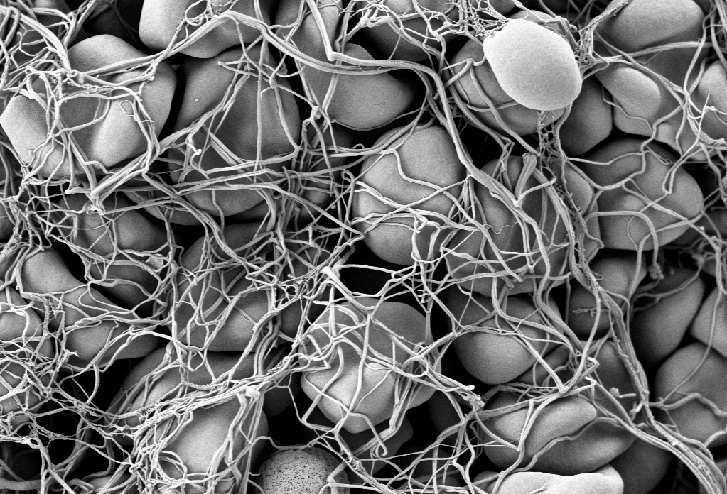
Electronic scanning microscopic image of red blood cells forming a clot
Hemostasis is the process by which blood stops flowing from damaged blood vessels. It is a complex process involving multiple molecules and forces, and our current understanding is limited by our inability to test these factors simultaneously in the laboratory. Some tests, for instance, can tell us much about clotting — a part of hemostasis — but little about the other elements at play. In particular, the role in hemostasis of the endothelium, which is the cell layer that lines the blood vessels, has generally been omitted from previous studies.
However, a new article in Nature Communications details the use of microfluidics technology, which is often used to model organ systems outside the body, to engineer a more complete model of hemostasis. Led by Wilbur A. Lam, M.D., Ph.D., Assistant Professor in the Wallace H. Coulter Department of Biomedical Engineering at Georgia Tech, the study authors fabricated microfluidics devices and then seeded vascular channels in the devices with human aortic and umbilical vein cells to simulate the endothelium.
Using the device, the authors were able to visualize hemostatic plug formation with whole blood and with blood from subjects with hemophilia. Although the authors concede that their model best represents capillary bleeding, rather than bleeding from larger vessels, they are confident that their model reliably represents the interaction of the endothelium with multiple varieties of blood cells.
Shedding Light on Cancer Response and Resistance
Penn’s founder, Benjamin Franklin, has a famous axiom: “an ounce of prevention is worth a pound of cure.” If Franklin were alive today, he would likely agree with two common axioms in cancer treatment: 1) if you can’t see it, you can’t treat it; and 2) if you treat it, treat all of it. Recent publications from investigators at Columbia and the University of Maryland reveal how imaging technologies can help predict to outcomes and how nanotechnology is offering a new therapeutic tools for fighting cancer.
Using diffuse optical tomography (DOT), which employs near-infrared spectroscopy to obtain three-dimensional images, scientists at Columbia have shown in an article in Radiology that treatment response could be predicted as early as two weeks after the start of therapy. The authors, led by Andreas H. Hielscher, PhD, Professor of Biomedical Engineering, Electrical Engineering, and Radiology at Columbia, applied DOT in 40 women with breast cancer undergoing chemotherapy. They found that DOT imaging features were associated, some very strongly, with treatment outcomes at 5 months. Given their positive findings, the authors intend to continue testing DOT in a larger cohort prospective study.
Another major issue in cancer chemotherapy is multidrug resistance (MDR), a highly frustrating complication resulting from lengthy treatment. In MDR, cancer types can find ways to overcome the effects of chemotherapy, resulting in relapse, often with deadly consequences. Therefore, among the challenges that oncologists face is the need to predict MDR, preferably before treatment even begins.
Based on the knowledge that adenosine triphosphate (ATP), a common organic molecule in energy generation, is involved in MDR, scientists at the University of Maryland engineered nanoparticles that could target cancer cells and, when exposed to near-infrared laser irradiation, reduce the amount of ATP in the cells . The scientists, led by Xiaoming Shawn He, Ph.D., Professor of Bioengineering at Maryland, published their findings in Nature Communications.
Dr. He’s team tested their nanoparticles in vitro and subsequently in mice and found decreased tumor sizes in mice treated with the particles, as well as more deaths of cancer cells. In addition, two of seven mice treated with the nanoparticles plus light experienced complete tumor eradication. The findings offer hope that MDR could be overcome with direct delivery of targeted treatment to resistant tumors.
Preserving the Tooth
A frustrating problem often encountered by dentists is the growth of new cavities around existing fillings. Microbes are often critical catalysts for these new cavities. Using antimicrobial agents at cavity-repair sites could make a real difference. However, mesoporous silica has proved suboptimal for this purpose.
However, help might be on the way. A study in a recent issue of Scientific Reports, written by a trio of authors led by Benjamin D. Hatton, Ph.D., Assistant Professor at the Institute of Biomaterials & Biomedical Engineering of the University of Toronto, reports the successful synthesis of 500-nm nanocomposite spheres combining silica with octenidine dihydrochloride, a common antiseptic. The newly synthesized nanospheres outperformed earlier attempts with mesoporous silica. The authors will continue to develop these nanoparticles to deliver other drugs for longer periods of time.
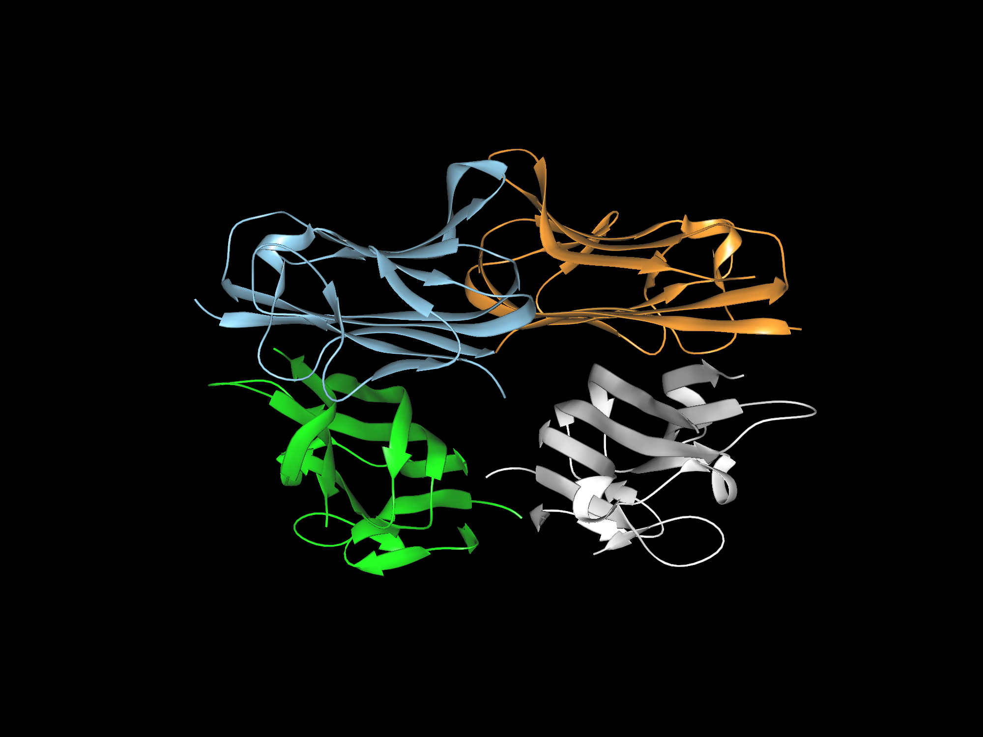
Cell signaling and the proteins involved in it participate in virtually every process in the body, whether normal or pathological. Much of this signaling involves proteins called cytokines, and of particular interest among them are tumor necrosis factors (TNFs), whose job it is to carry out apoptosis — the process by which cells die at predetermined time points as part of their normal life cycle. Among this family of cytokines, TNF-related apoptosis-inducing ligand (TRAIL) has been of particular interest to oncologists.
The process by which TRAIL combines with or binds to other molecules that modulate the life cycle of cancer cells can interfere with the ability of these molecules to facilitate the growth of cancer cells into tumors. However, attempts to deploy the cytokine to interfere in the process that produces cancer have been unsuccessful because of issues regarding inefficient delivery of TRAIL to the relevant sites, poor circulation of the cytokine in the blood, and the development of resistance to TRAIL. Bioengineers have been hard at work attempting to overcome these barriers.
In a new article published in ACS Nano coauthored by Michael J. Mitchell, Ph.D., Skirkanich Assistant Professor of Innovation at Penn Bioengineering, and Robert Langer, Ph.D., David H. Koch Institute Professor at MIT, these engineered solutions are reviewed and assessed. The review covers nanoparticle technologies with potential to solve the problems encountered thus far, including a range of materials (polymers, lipids, inorganic), cell-nanoparticle hybrids, and therapeutic cells genetically engineered using nanoparticles.
“The TRAIL protein is a essential component of our immune system,” Dr. Mitchell says, “and it kills tumor cells without harming normal ones. However, it remains challenging to deliver the protein into tumors, and tumors can also be resistant to the protein. We and others are now exploiting nanotechnology, genetic engineering, and immune cell-biomaterial hybrids to overcome these key biological barriers to cancer therapy.”