BE Seminar: “Emerging Technologies for Detection of Early Stage Bladder Cancer” (Audrey Bowden)

Audrey Bowden, PhD, Associate Professor of Biomedical Engineering. (Vanderbilt University / Steve Green)

Speaker: Audrey Bowden, Ph.D.
Dorothy J. Wingfield Phillips Chancellor’s Faculty Fellow and Associate Professor of Biomedical Engineering and Electrical Engineering & Computer Science
Vanderbilt University

Date: Thursday, November 19, 2020
Time: 3:00-4:00 PM EST
Zoom – check email for link or contact ksas@seas.upenn.edu

Title: “Emerging Technologies for Detection of Early Stage Bladder Cancer”

Abstract:

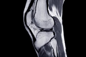
Bladder cancer (BC) —  the 4th most common cancer in men and the most expensive cancer to treat over a patient’s lifetime — is a lifelong burden to BC patients and a significant economic burden to the U.S. healthcare system. The high cost of BC stems largely from its high recurrence rate (>50%); hence, BC management involves frequent surveillance. Unfortunately, the current in-office standard-of-care tool for BC surveillance, white light cystoscopy (WLC), is limited by low sensitivity and specificity for carcinoma in situ (CIS), a high-grade carcinoma with high potential to metastasize. Early detection and complete eradication of CIS are critical to improve treatment outcomes and to minimize recurrence. The most promising macroscopic technique to improve sensitivity to CIS detection, blue light cystoscopy (BLC), is costly, time-intensive, has low availability and a high false-positive rate. Given the limitations of WLC, we aim to change the paradigm around how BC surveillance is performed by validating new tools with high sensitivity and specificity for CIS that are appropriate for in-office use. In this seminar, I discuss our innovative solutions to improve mapping the bladder for longitudinal tracking of suspicious lesions and to create miniature tools for optical detection based on optical coherence tomography (OCT). OCT and its functional variant, cross-polarized OCT, can detect early-stage BC with better sensitivity and specificity than WLC. We discuss the critical technical innovations necessary to make OCT and CP-OCT a practical tool for in-office use, and new results from recent explorations of human bladder samples that speak to the promise of this approach to change the management of patient care.

Bio:

Audrey K. Bowden is the Dorothy J. Wingfield Phillips Chancellor Faculty Fellow and Associate Professor of Biomedical Engineering (BME) and of Electrical Engineering and Computer Science (EECS) at Vanderbilt University. Prior to this, she served as Assistant and later Associate Professor of Electrical Engineering and Bioengineering at Stanford University. Dr. Bowden received her BSE in Electrical Engineering from Princeton University, her PhD in BME from Duke University and completed her postdoctoral training in Chemistry and Chemical Biology at Harvard University. During her career, Dr. Bowden served as an International Fellow at Ngee Ann Polytechnic in Singapore. From 2007-2008, she was the Arthur H. Guenther Congressional Fellow sponsored by the OSA and SPIE and served as a Legislative Assistant in the United States Senate through the AAAS Science and Technology Policy Fellows Program. Dr. Bowden is a Fellow of SPIE, a Fellow of AIMBE and is the recipient of numerous awards, including the Air Force Young Investigator Award, the NSF Career Award, the Hellman Faculty Scholars Award, the Phi Beta Kappa Teaching Award, Ford Foundation Postdoctoral Fellowship, and the NSBE Golden Torch Award. She is a former Associate Editor of IEEE Photonics Journal, former Lead Guest Editor of a Biomedical Optics Express Special Issue and is a member of numerous professional committees. Her research interests include biomedical optics – particularly optical coherence tomography and near infrared spectroscopy – microfluidics, and point of care diagnostics.

Magnetic Field and Hydrogels Could Be Used to Grow New Cartilage

by Frank Otto

MRI Knee joint or Magnetic resonance imaging sagittal view for detect tear or sprain of the anterior cruciate ligament (ACL).

Using a magnetic field and hydrogels, a team of researchers in the Perelman School of Medicine have demonstrated a new possible way to rebuild complex body tissues, which could result in more lasting fixes to common injuries, such as cartilage degeneration. This research was published in Advanced Materials.

“We found that we were able to arrange objects, such as cells, in ways that could generate new, complex tissues without having to alter the cells themselves,” says the study’s first author, Hannah Zlotnick, a graduate student in bioengineering who works in the McKay Orthopaedic Research Laboratory at Penn Medicine. “Others have had to add magnetic particles to the cells so that they respond to a magnetic field, but that approach can have unwanted long-term effects on cell health. Instead, we manipulated the magnetic character of the environment surrounding the cells, allowing us to arrange the objects with magnets.”

In humans, tissues like cartilage can often break down, causing joint instability or pain. Often, the breakdown isn’t in total, but covers an area, forming a hole. Current fixes are to fill those holes in with synthetic or biologic materials, which can work but often wear away because they are not the same exact material as what was there before. It’s similar to fixing a pothole in a road by filling it with gravel and making a tar patch: The hole will be smoothed out but eventually wear away with use because it’s not the same material and can’t bond the same way.

What complicates fixing cartilage or other similar tissues is that their makeup is complex.

“There is a natural gradient from the top of cartilage to the bottom, where it contacts the bone,” Zlotnick explains. “Superficially, or at the surface, cartilage has a high cellularity, meaning there is a higher number of cells. But where cartilage attaches to the bone, deeper inside, its cellularity is low.”

So the researchers, which included senior author Robert Mauck, PhD, director of the McKay Lab and a professor of Orthopaedic Surgery and Bioengineering, sought to find a way to fix the potholes by repaving them instead of filling them in. With that in mind, the research team found that if they added a magnetic liquid to a three-dimensional hydrogel solution, cells, and other non-magnetic objects including drug delivery microcapsules, could be arranged into specific patterns that mimicked natural tissue through the use of an external magnetic field.

Read more at Penn Medicine News.

An Ecosystem of Innovation Fosters Tech-based Solutions to COVID-19 Challenges

by Erica K. Brockmeier

GRASP lab researchers (from left) Bernd Pfrommer, Kenneth Chaney, and Caio Mucchiani assembling telemedicine cart prototypes in Levine hall earlier this spring. (Image courtesy of Kenneth Chaney and Bernd Pfrommer)

Since the start of the spring, members of the Penn community have been working to combat coronavirus and its many impacts. Some people are studying COVID-19 or developing vaccines, while others are 3D-printing face shields for health care workers and delivering fall courses online.

And while innovation in health care usually brings to mind new treatments and medicines, the efforts of clinicians, engineers, and IT specialists demonstrate the importance technological infrastructure for rapidly deployable, tech-based solutions so clinicians can provide the best care to patients amid social distancing and coronavirus restrictions.

The telemedicine revolution

In late March, telemedicine was key for allowing Penn Medicine clinicians to deliver care while avoiding potentially risky in-person interactions. Chief Medical Information Officer C. William Hanson III and his team helped set up the IT infrastructure for scaling up telemedicine capabilities and provided guidance to clinicians. Thanks to the quick pivot, Penn Medicine went from 300 telemedicine visits in February to more than 7,500 visits per day in a matter of weeks.

But far from seeing telemedicine as a temporary solution during the pandemic, Hanson has been a long-time advocate for this approach to health care. In his role as liaison between clinicians and the IT community in the past 10 years Hanson, helped establish remote ICU monitoring protocols and broadened opportunities for televisits with specialists. Now, with the pandemic removing many of the previous barriers to entry, be they technical, insurance-based, or simply a lack of familiarity, Hanson believes that telemedicine is here to stay.

“As the pandemic evolved, people were aware that telemedicine could help the health care system, as well as doctors and patients, during this crisis,” he says. “Now, there are definitely places where telemedicine makes good sense, and we will continue to use that as part of our way of handling a problem.” Other benefits include removing geographic barriers to entry for new patients, reduced appointment times, increased patient satisfaction, and reduced health care provider burnout.

Simple solutions for COVID-19 challenges

As the director of Penn’s Telestroke Program, neurologist Michael Mullen has experience diagnosing from a distance. This spring, telemedicine carts his group uses were repurposed in COVID ICUs. At the same time, Mullen and group wanted to expand their ability to assess stroke patients remotely, so he reached out to Brian Litt, faculty director of Penn Health-Tech, to see how he could collaborate to create an analogous telemedicine station using readily available, cost-effective components.

Rapid and simple solutions are at the heart of Penn’s ModLab, a subgroup of the GRASP lab focused on robots made of configurable individual components. As part of a COVID-19 rapid response initiative, engineers worked with Mullen to figure out a viable solution in record time. “The idea was to make it as simple and as fast as possible,” says graduate student Caio Mucchiani. “With robotics, usually you want to make things more sophisticated, however, given the situation, we needed to know how we could use off-the-shelf components to make something.”

Fellow graduate student Ken Chaney, postdoc Bernd Pfrommer, and Mucchiani came up with a plan that replicated the required specs of the existing telemedicine carts, including state-of-the-art cameras for detailed imaging as well as a reliable, easily rechargeable battery. The team then put together 10 telemedicine carts, assembling the prototypes with social distancing and masks at the GRASP lab in early April.

While changes to treatment approaches mean that these carts still require additional field testing, Mullen is still eager to expand the program, be it for diagnosing patients safely or educating medical students in an era of social distancing. “In the setting of COVID, when everything was getting crazy, it was remarkable to see the energy that GRASP brought to help,” adds Mullen. “Everyone was really busy, and it was amazing to see this group of people who wanted to use their expertise to help.”

Continue reading at Penn Today.

NB: Brian Litt is Professor in Neurology and Bioengineering.

Brian Litt Receives NIH Pioneer Award to Develop Implantable Neurodevices

Brian Litt, MD

Brian Litt, professor in Engineering’s Department of Bioengineering and the Perelman School of Medicine’s departments of Neurology and Neurosurgery, has received a five-year, $5.6 million Pioneer Award from the National Institutes of Health, which will support his research on implantable devices for monitoring, recording and responding to neural activity.

The Pioneer Award is part of the agency’s High-Risk, High-Reward Research Program honoring exceptionally creative scientists. It challenges investigators to pursue new research directions and develop groundbreaking, high-impact approaches to a broad area of biomedical or behavioral science. Litt’s neurodevice research represents a new frontier in addressing a wide variety of neurological conditions.

In epilepsy, for example, these devices would predict and prevent seizures; in Parkinson’s patients, implants will measure and communicate with patients to improve mobility, reduce tremor and enhance responsiveness. Other implants might improve hearing or psychiatric symptoms by querying patient perceptions, feelings, and altering stimulation patterns algorithmically to improve them

Continue reading about Litt’s Pioneer Award at Penn Medicine News.

A potential cause of CAR T side effects, and a path forward

Single cell sequencing aided researchers in identifying a previously undiscovered molecule in the brain.

Chimeric antigen receptor (CAR) T cell therapy has revolutionized treatment of leukemia, lymphoma, and multiple myeloma. But some people who have received this treatment experience neurotoxicity, or damage to the brain or nervous system.

New research from a team led by Avery Posey, an assistant professor of systems pharmacology and translational therapeutics in the Perelman School of Medicine, provides evidence that this side effect may owe to a molecule in the brain that scientists previously didn’t know was there.

The work, published in the journal Cell, revealed that the protein CD19 is present in brain cells that protect the blood-brain barrier. Prior to the finding, scientists believed CD19 was only expressed on B cells, and the protein served as a target for certain forms of CAR-T therapy. The discovery may chart a path forward for new strategies to effectively treat cancer while sparing the brain.

“The next question is,” says Posey, “can we identify a better target for eliminating B cell related malignancies other than CD19, or can we engineer around this brain cell expression of CD19 and build a CAR T cell that makes decisions based on the type of cell it encounters—for instance, CAR T cells that kill the B cells they encounter, but spare the CD19 positive brain cells?”

Read more at Penn Medicine News. Avery Posey is a member of the Department of Bioengineering Graduate Group.

BE Seminar: “Patients, Providers and Data: How the EMR and Data Science are Changing Clinical Care” (Kevin Johnson, Vanderbilt)

The Penn Bioengineering virtual seminar series continues on September 24th.

Kevin Johnson, MD, MS

Speaker: Kevin Johnson, M.D., M.S.
Cornelius Vanderbilt Professor and Chair
Department of Biomedical Informatics
Vanderbilt University Medical Center

Date: Thursday, September 24, 2020
Time: 3:00-4:00 pm
Zoom – check email for link or contact ksas@seas.upenn.edu

Title: “Patients, Providers and Data: How the EMR and Data Science are Changing Clinical Care”

Abstract:

The electronic health record (EHR) is a powerful application of Systems Engineering to healthcare. It is a byproduct of a host of pressures including cost, consolidation of providers into networks, uniform drivers of quality, and the need for timely care across disparate socioeconomic and geographic landscapes within health systems. The EHR is also a fulcrum for innovation and one of the most tangible examples of how data science affects our health and health care. In this talk I will showcase projects from my lab that demonstrate the multi-disciplinary nature of biomedical informatics/data science research and translation using the EHR, and our current understanding of its potential from my perspective as a pediatrician, a researcher in biomedical informatics, a Chief Information Officer, an educator, and an advisor to local and international policy. I will describe advances in applying human factors engineering to support medical documentation and generic prescribing, approaches to improve medication safety, and innovations to support precision medicine and interoperability. I will present our efforts to integrate EHR-enabled data science into the Vanderbilt health system and provide a vision for what this could mean for our future.

Bio:

Kevin B. Johnson, M.D., M.S. is Informatician-in-Chief, Cornelius Vanderbilt Professor and Chair of Biomedical Informatics, and Professor Pediatrics at Vanderbilt University Medical Center. He received his M.D. from Johns Hopkins Hospital in Baltimore and his M.S. in Medical Informatics from Stanford University. In 1992 he returned to Johns Hopkins where he served as a Pediatric Chief Resident. He was a member of the faculty in both Pediatrics and Biomedical Information Sciences at Johns Hopkins until 2002, when he was recruited to Vanderbilt University. He also is a Board-Certified Pediatrician.

Dr. Johnson is an internationally respected developer and evaluator of clinical information technology. His research interests have been related to developing and encouraging the adoption of clinical information systems to improve patient safety and compliance with practice guidelines; the uses of advanced computer technologies, including the Worldwide Web, personal digital assistants, and pen-based computers in medicine; and the development of computer-based documentation systems for the point of care. In the early phases of his career, he directed the development and evaluation of evidence-based pediatric care guidelines for the Johns Hopkins Hospital. He has been principal investigator on numerous grants and has been an invited speaker at most major medical informatics and pediatrics conferences. He also was the Chief Informatics Officer at Vanderbilt University Medical Center from 2015-2019.

See the full list of upcoming Penn Bioengineering fall seminars here.

MINS/BE Seminar: “Mapping emotions: discovering structure in mesoscale electrical brain recordings” (Kafui Dzirasa, Duke University)

The Mahoney Institute for Neurosciences (MINS) and Department of Bioengineering will co-host a seminar on September 16. Details and registration information are below. We hope you will join us to hear this upcoming talk.

Title: “Mapping emotions: discovering structure in mesoscale electrical brain recordings”

Kafui Dzirasa, MD, PhD

Speaker: Kafui Dzirasa,MD, PhD
K. Ranga Rama Krishnan Endowed Associate Professor
Department of Psychiatry and Behavioral Sciences
Duke University Medical Center


Date: Wednesday, September 16, 2020
Time: 4:00-5:30 PM Eastern Time
This event will be held virtually via Bluejeans (link here)

Hosted by Danielle Bassett and Joshua Gold


Avery Posey’s cancer research takes high risks for big rewards

by Melissa Moody

Avery Posey, PhD (Image: Penn Medicine Newsby Melissa Moody

Much of the world, including research at Penn Medicine, has focused its attention on how T cells–which play a central role in immune response—might shape the trajectory of COVID-19 infection, and how immunotherapy can shed light on treatment of the disease.

Already a leader in immunotherapy research and treatment, Penn Medicine pioneered the groundbreaking development of CAR T cell cancer therapy. Avery Posey, an assistant professor of systems pharmacology and translational therapeutics, trained as a postdoctoral fellow in the lab of Carl June, who pioneered CAR T cell immunotherapy to treat cancer. Now as a faculty member at Penn, Posey has maintained a focus on T cell therapeutics, mostly for the treatment of cancer.

“This research combines two of my biggest interests—the use of gene therapy to treat disease and the investigation of little known biology, such as the roles of glycans in cell behavior. The pursuit of new knowledge, the roads less traveled—those are my inspirations,” Posey says.

Read more at Penn Medicine News.

N.B.: Avery Posey and Carl June are members of the Department of Bioengineering Graduate Group. Learn more about BE’s Grad Group Faculty here.

Meet Bioengineering Sophomore and SNF Paideia Fellow Catherine Michelutti

Catherine Michelutti (BSE, BS ’23)

Rising Bioengineering Sophomore Catherine Michelluti (BSE 2023) has been featured on Penn’s SNF Paideia Program Instagram which discusses her diverse interests in machine learning in medicine, computer science, playing the violin and more. Catherine is a pre-med student who is pursuing an uncoordinated dual degree between the School of Engineering and Applied Science and the Wharton School of Business (BS in Economics 2023). She is also an incoming fellow in the SNF Paideia Program, which is supported by the Stavros Niarchos Foundation, is an interdisciplinary program which “encourage[s] the free exchange of ideas, civil and robust discussion of divergent views, and the integration of individual and community wellness, service, and citizenship through SNF Paideia designated courses, a fellows program, and campus events” (SNF Paideia website).

Read more about Catherine and other Fellows on the SNF Paideia Instagram.

César de la Fuente Wins Inaugural NEMO Prize, Will Develop Rapid COVID Virus Breath Tests

The paper-based tests could be integrated directly into facemasks and provide instant results at testing sites.

Cesar de la Fuente-Nunez, PhD

When Penn Health-Tech announced its Nemirovsky Engineering and Medicine Opportunity, or NEMO Prize, in February, the center’s researchers could only begin to imagine the impact the looming COVID-19 pandemic was about to unleash. But with the promise of $80,000 to support early-stage ideas at the intersection of engineering and medicine, the contest quickly sparked a winning innovation aimed at combating the crisis.

Judges from the University of Pennsylvania’s School of Engineering and Applied Sciences and Perelman School of Medicine awarded its first NEMO Prize to César de la Fuente, PhD, who proposed a paper-based COVID diagnostic system that could capture viral particles on a person’s breath, then give a result in a matter of seconds when taken to a testing site.

Similar tests for bacteria cost less than a dollar each to make. De la Fuente, a Presidential Assistant Professor in the departments of Psychiatry, Microbiology, and Bioengineering, is aiming to make COVID tests at a similar price point and with a smaller footprint so that they could be directly integrated into facemasks, providing further incentive for their regular use.

“Wearing a facemask is vital to containing the spread of COVID because, before you know you’re sick, they block your virus-carrying droplets so those droplets can’t infect others,” de la Fuente says. “What we’re proposing could eventually lead to a mask that can be infected by the virus and let you know that you’re infected, too.”

De la Fuente’s lab has conducted molecular dynamic simulations of the regions of the SARS-COV-2 spike protein (blue) that bind to the human ACE2 receptor (red and yellow).

De la Fuente’s expertise is in synthetic biology and molecular-scale simulations of disease-causing viruses and bacteria. Having such fine-grained computational models of these microbes’ binding sites allow de la Fuente to test them against massive libraries of proteins, seeing which bind best. Other machine learning techniques can then further narrow down the minimum molecular structures responsible for binding, resulting in functional protein fragments that are easier to synthesize and manipulate.

The spike-shaped proteins that give coronaviruses their crown-like appearance and name bind to a human receptor known as ACE2. De la Fuente and his colleagues are now aiming to characterize the molecular elements and environmental factors that would allow for the most precise, reliable detection of the virus.

Read the full story on the Penn Engineering blog.