Penn is fighting pancreatic cancer

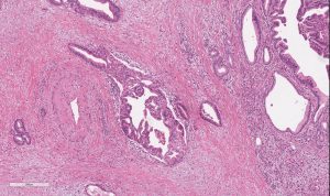
A microscopic view of pancreatic adenocarcinoma. (Image: Emma Furth)

Swept up in a pancreatic cancer diagnosis is inevitably a sense of fear and sadness.

But at Penn, researchers are bringing new hope to this disease. And with patients like Nick Pifani, it’s clear that they’re moving in the right direction.

Pifani, from Delran, New Jersey, first noticed some lingering stomach upset in February 2017. He called his family doctor, concerned—especially given that he was an otherwise healthy marathon runner who was only 42. He was sent to a gastrointestinal specialist. A few weeks later, some crippling stomach pain sent him back to the emergency room and he received an MRI that showed a mass on his pancreas—Stage Three, inoperable, he was told.

He was treated with chemotherapy, along with radiation and, eventually, and after receiving advice from doctors at Penn, his tumor was removed. Thereafter, he realized he had a PALB2 mutation—a cousin of the BRCA gene mutation. At that moment, his long-term needs changed and he found himself seeking specialized care at Penn, where he met Kim Reiss Binder, assistant professor of medicine at the Hospital of the University of Pennsylvania (HUP).

“I’m a planner; I want to understand what [my] potential options are,” Pifani says. “[Reiss Binder] asked why I was there to see her and I explained and quickly I could tell she was—outside of her being remarkably intelligent—a great listener and a compassionate doctor.”

“I have a feeling she worries about me more than I do,” he laughs.

Pifani has now been in remission for two years and four months; he sees Reiss-Binder every three months for checkups. His survival story is inspiring and a sign of momentum, even if a world without pancreatic cancer is still frustratingly out of reach.

Pancreatic cancer at Penn

Pancreatic cancer is the third-leading cause of cancer-related death in the United States, outmatched only by lung cancer (No. 1) and colorectal cancer (No. 2). A person diagnosed with pancreatic cancer is still unlikely to survive past five years—only 9% of survivors do, giving it the highest mortality rate among every major cancer.

In short, pancreatic cancer seldom paves the way for optimistic narratives. Some of the hope that has surfaced, though, is thanks to some talent, dedication to the cause, and hard work at Penn.

A key point of progress in the battle against the disease was made in 2002, when former Assistant Professor of Medicine David Tuveson established a standard model for examining human development of this disease in mice. This model has allowed for a reliable way to study the disease and has influenced progress made here at Penn and elsewhere since.

“There’s been a burst of activity in translational research, from bench to bedside,” explains Ben Stanger, the Hanna Wise Professor in cancer research and director of the Penn Pancreatic Cancer Research Center (PCRC) at the Abramson Cancer Center.

“And there’s a lot of momentum with community building, a dramatic increase in patient volumes, and a dramatic increase in what we know about the cancer,” he says of the status of pancreatic cancer today.

Reiss Binder, meanwhile, explains that one mark of progress at Penn and beyond has been learning about people like Pifani, who have the PALB2 gene, and why they respond differently to treatments than those without it. Platinum-based chemotherapies, for example, are especially effective for people with the PALB2 gene who are battling pancreatic cancer. An ongoing trial at Penn has tested and found some success with using PARP inhibitors—taken orally as an enzyme that fixes single-stranded breaks of DNA—as a maintenance therapy in that same PALB2 demographic after they’ve had chemotherapy. These are less toxic than chemotherapy for patients with the same mutations.

It’s all been slow progress toward better treatments, but there has been progress.

“This is the tip of the iceberg for a disease that we historically have treated with perpetual chemotherapy,” Reiss Binder says. “We owe it to patients to find better options to suppress the cancer but not ruin their quality of life.”

Catching cancer earlier

The consensus on why pancreatic cancer is so deadly? It just can’t be spotted fast enough.

Pancreatic cancer often presents well after it has developed and metastasized, and does so in a way that is not easy to recognize as cancer. Common symptoms include, for example, stomach upset and back pain. And by the time a harder-to-ignore symptom of the cancer surfaces, a sort of yellowing of the skin (a result of a bile duct blockage), it’s likely too late to stop the cancer in its tracks.

One approach to improved detection being tested at Penn, by Research Assistant Professor of Medicine Erica Carpenter, is a liquid biopsy—drawn from a standard blood test. Current means to test for pancreatic cancer—imaging through an endoscopic tube—are invasive and expensive, meaning a common liquid test could transform how many cases are detected early.

Carpenter explains that circulating tumor cells (CTCs) can shed from a tumor that’s adjacent to the wall of a blood vessel; what’s shed then shows up in a blood test. The cells, if detected, can explain more about the nature of the tumor, giving doctors an opportunity to examine characteristics of cancerous cells and decide how to effectively treat a tumor if it can’t be surgically removed. It also allows interpretations of disease burden and the effectiveness of medications—through genome sequencing—that imaging does not.

Ultimately, this gives doctors the potential to track the growth of a tumor before it’s fully developed, all through one tube of blood—detected through an innovative use of technology.
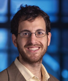
 

David Issadore, Ph.D.

David Issadore, associate professor of bioengineering and electrical and systems engineering in the School of Engineering and Applied Science, has worked since 2017 to develop a chip that detects cancer in the blood, using machine learning to sort through literally hundreds of billions of vesicles and cells, looking for these CTCs. The chip retrieves data and the machine learning developed interprets that data, attempting to make a diagnosis that not only finds pancreatic cancer but also provides information about its progression—and, importantly, whether a patient might benefit from surgery.

Read the full story at Penn Today. Media contact Brandon Baker.

Bioengineering Round-Up (December 2019)

by Sophie Burkholder

Positive results in first-in-U.S. trial of CRISPR-edited immune cells

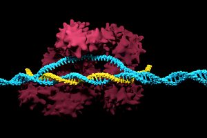
3D render of the CRISPR-Cas9 genome editing system

Genetically editing a cancer patient’s immune cells using CRISPR/Cas9 technology, then infusing those cells back into the patient appears safe and feasible based on early data from the first-ever clinical trial to test the approach in humans in the United States. Researchers from the Abramson Cancer Center have infused three participants in the trial thus far—two with multiple myeloma and one with sarcoma—and have observed the edited T cells expand and bind to their tumor target with no serious side effects related to the investigational approach. Penn is conducting the ongoing study in cooperation with the Parker Institute for Cancer Immunotherapy and Tmunity Therapeutics.

“This trial is primarily concerned with three questions: Can we edit T cells in this specific way? Are the resulting T cells functional? And are these cells safe to infuse into a patient? This early data suggests that the answer to all three questions may be yes,” says the study’s principal investigator Edward A. Stadtmauer, section chief of Hematologic Malignancies at Penn. Stadtmauer will present the findings next month at the 61st American Society of Hematology Annual Meeting and Exposition.

Read the rest of the story on Penn Today.

Tulane researchers join NIH HEAL initiative for research into opioid crisis

A Tulane University professor and researcher of biomedical engineering will join fellow researchers from over 40 other institutions in the National Institute of Health’s Help to End Addiction Long-Term (HEAL) Initiative. Of the $945 million that make up the project, Michael J. Moore, Ph.D. will receive a share of $1.2 million to advance research in modeling human pain through computer chips, with the help of fellow Tulane researchers Jeffrey Tasker, Ph.D., and James Zadina, Ph.D., each with backgrounds in neuroscience.

Because of the opioid epidemic sweeping the nation, Moore notes that there’s a rapid search going on to develop non-addictive painkiller options. However, he also sees a gap in adequate models to test those new drugs before human clinical trials are allowed to take place. Here is where he hopes to step in and bring some innovation to the field, by integrating living human cells into a computer chip for modeling pain mechanisms. Through his research, Moore wants to better understand not only how some drugs can induce pain, but also how patients can grow tolerant to some drugs over time. If successful, Moore’s work will lead to a more rapid and less expensive screening option for experimental drug advancements.

New machine learning-assisted microscope yields improved diagnostics

Researchers at Duke University recently developed a microscope that uses machine learning to adapt its lighting angles, colors, and patterns for diagnostic tests as needed. Most microscopes have lighting tailored to human vision, with an equal distribution of light that’s optimized for human eyes. But by prioritizing the computer’s vision in this new microscope, researchers enable it to see aspects of samples that humans simply can’t, allowing for a more accurate and efficient diagnostic approach.

Led by Roarke W. Horstmeyer, Ph.D., the computer-assisted microscope will diffuse light through a bowl-shaped source, allowing for a much wider range of illumination angles than traditional microscopes. With the help of convolutional neural networks — a special kind of machine learning algorithm — Horstmeyer and his team were able to tailor the microscope to accurately diagnose malaria in red blood cell samples. Where human physicians typically perform similar diagnostics with a rate of 75 percent accuracy, this new microscope can do the same work with 90 percent accuracy, making the diagnostic process for many diseases much more efficient.

Case Western Reserve University researchers create first-ever holographic map of brain

A Case Western Reserve University team of researchers recently spearheaded a project in creating an interactive holographic mapping system of the human brain. The design, which is believed to be the first of its kind, involves the use of the Microsoft HoloLens mixed reality platform. Lead researcher Cameron McIntyre, Ph.D., sees this mapping system as a better way of creating holographic navigational routes for deep brain stimulation. Recent beta tests with the map by clinicians give McIntyre hope that the holographic representation will help them better understand some of the uncertainties behind targeted brain surgeries.

More than merely providing a useful tool, McIntyre’s project also brings together decades’ worth of neurological data that has not yet been seriously studied together in one system. The three-dimensional atlas, called “HoloDBS” by his lab, provides a way of finally seeing the way all of existing neuro-anatomical data relates to each other, allowing clinicians who use the tool to better understand the brain on both an analytical and visual basis.

Implantable cancer traps reduce biopsy incidence and improve diagnostic

Biopsies are one of the most common procedures used for cancer diagnostics, involving a painful and invasive surgery. Researchers at the University of Michigan are trying to change that. Lonnie Shea, Ph.D., a professor of biomedical engineering at the university, worked with his lab to develop implants with the ability to attract any cancer cells within the body. The implant can be inserted through a scaffold placed under the patient’s skin, making it a more ideal option than biopsy for inaccessible organs like lungs.

The lab’s latest work on the project, published in Cancer Research, details its ability to capture metastatic breast cancer cells in vivo. Instead of needing to take biopsies from areas deeper within the body, the implant allows for a much simpler surgical procedure, as biopsies can be taken from the implant itself. Beyond its initial diagnostic advantages, the implant also has the ability to attract immune cells with tumor cells. By studying both types of cells, the implant can give information about the current state of cancer in a patient’s body and about how it might progress. Finally, by attracting tumor and immune cells, the implant has the ability to draw them away from the area of concern, acting in some ways as a treatment for cancer itself.

People and Places

Cesar de la Fuente-Nunez, PhD

The Philadelphia Inquirer recently published an article detailing the research of Penn’s Presidential Assistant Professor in Psychiatry, Microbiology, and Bioengineering, Cesar de la Fuente, Ph.D. In response to a growing level of worldwide deaths due to antibiotic-resistant bacteria, de la Fuente and his lab use synthetic biology, computation, and artificial intelligence to test hundreds of millions of variations in bacteria-killing proteins in the same experiment. Through his research, de la Fuente opens the door to new ways of finding and testing future antibiotics that might be the only viable options in a world with an increasing level of drug-resistant bacteria

Emily Eastburn, a Ph.D. candidate in Bioengineering at Penn and a member of the Boerckel lab of the McKay Orthopaedic Research Laboratory, recently won the Ashton fellowship. The Ashton fellowship is an award for postdoctoral students in any field of engineering that are under the age of 25, third-generation American citizens, and residents of either Pennsylvania or New Jersey. A new member of the Boerckel lab, having joined earlier this fall, Eastburn will have the opportunity to conduct research throughout her Ph.D. program in the developmental mechanobiology and regeneration that the Boerckel lab focuses on.

Week in BioE (July 12, 2019)

by Sophie Burkholder

DNA Microscopy Gives a Better Look at Cell and Tissue Organization

A new technique that researchers from the Broad Institute of MIT and Harvard University are calling DNA microscopy could help map cells for better understanding of genetic and molecular complexities. Joshua Weinstein, Ph.D., a postdoctoral associate at the Broad Institute, who is also an alumnus of Penn’s Physics and Biophysics department and former student in Penn Bioengineering Professor Ravi Radhakrishnan’s lab, is the first author of this paper on optics-free imaging published in Cell.
The primary goal of the study was to find a way of improving analysis of the spatial organization of cells and tissues in terms of their molecules like DNA and RNA. The DNA microscopy method that Weinstein and his team designed involves first tagging DNA, and allowing the DNA to replicate with those tags, which eventually creates a cloud of sorts that diffuses throughout the cell. The DNA tags subsequent interactions with molecules throughout the cell allowed Weinstein and his team to calculate the locations of those molecules within the cell using basic lab equipment. While the researchers on this project focused their application of DNA microscopy on tracking human cancer cells through RNA tags, this new method opens the door to future study of any condition in which the organization of cells is important.

Read more on Weinstein’s research in a recent New York Times profile piece.

Penn Engineers Demonstrate Superstrong, Reversible Adhesive that Works like Snail Slime

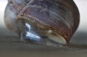
A snail’s epiphragm. (Photo: Beocheck)

If you’ve ever pressed a picture-hanging strip onto the wall only to realize it’s slightly off-center, you know the disappointment behind adhesion as we typically experience it: it may be strong, but it’s mostly irreversible. While you can un-stick the used strip from the wall, you can’t turn its stickiness back on to adjust its placement; you have to start over with a new strip or tolerate your mistake. Beyond its relevance to interior decorating, durable, reversible adhesion could allow for reusable envelopes, gravity-defying boots, and more heavy-duty industrial applications like car assembly.

Such adhesion has eluded scientists for years but is naturally found in snail slime. A snail’s epiphragm — a slimy layer of moisture that can harden to protect its body from dryness — allows the snail to cement itself in place for long periods of time, making it the ultimate model in adhesion that can be switched on and off as needed. In a new study, Penn Engineers demonstrate a strong, reversible adhesive that uses the same mechanisms that snails do.

This study is a collaboration between Penn Engineering, Lehigh University’s Department of Bioengineering, and the Korea Institute of Science and Technology.

Read the full story on Penn Engineering’s Medium blog. 

Low-Dose Radiation CT Scans Could Be Improved by Machine Learning

Machine learning is a type of artificial intelligence growing more and more popular for applications in bioengineering and therapeutics. Based on learning from patterns in a way similar to the way we do as humans, machine learning is the study of statistical models that can perform specific tasks without explicit instructions. Now, researchers at Rensselaer Polytechnic Institute (RPI) want to use these kinds of models in computerized tomography (CT) scanning by lowering radiation dosage and improving imaging techniques.

A recent paper published in Nature Machine Intelligence details the use of modularized neural networks in low-dose CT scans by RPI bioengineering faculty member Ge Wang, Ph.D., and his lab. Since decreasing the amount of radiation used in a scan will also decrease the quality of the final image, Wang and his team focused on a more optimized approach of image reconstruction with machine learning, so that as little data as possible would be altered or lost in the reconstruction. When tested on CT scans from Massachusetts General Hospital and compared to current image reconstruction methods for the scans, Wang and his team’s method performed just as well if not better than scans performed without the use of machine learning, giving promise to future improvements in low-dose CT scans.

A Mind-Controlled Robotic Arm That Requires No Implants

A new mind-controlled robotic arm designed by researchers at Carnegie Mellon University is the first successful noninvasive brain-computer interface (BCI) of its kind. While BCIs have been around for a while now, this new design from the lab of Bin He, Ph.D.,  a Trustee Professor and the Department Head of Biomedical Engineering at CMU, hopes to eliminate the brain implant that most interfaces currently use. The key to doing this isn’t in trying to replace the implants with noninvasive sensors, but in improving noisy EEG signals through machine learning, neural decoding, and neural imaging. Paired with increased user engagement and training for the new device, He and his team demonstrated that their design enhanced continuous tracking of a target on a computer screen by 500% when compared to typical noninvasive BCIs. He and his team hope that their innovation will help make BCIs more accessible to the patients that need them by reducing the cost and risk of a surgical implant while also improving interface performance.

People and Places

Daeyeon Lee, professor in the Department of Chemical and Biomolecular Engineering and member of the Bioengineering Graduate Group Faculty here at Penn, has been selected by the U.S. Chapter of the Korean Institute of Chemical Engineers (KIChE) as the recipient of the 2019 James M. Lee Memorial Award.

KIChE is an organization that aims “to promote constructive and mutually beneficial interactions among Korean Chemical Engineers in the U.S. and facilitate international collaboration between engineers in U.S. and Korea.”

Read the full story on Penn Engineering’s Medium blog.

We would also like to congratulate Natalia Trayanova, Ph.D., of the Department of Biomedical Engineering at Johns Hopkins University on being inducted into the Women in Tech International (WITI) Hall of Fame. Beginning in 1996, the Hall of Fame recognizes significant contributions to science and technology from women. Trayanova’s research specializes in computational cardiology with a focus on virtual heart models for the study of individualized heart irregularities in patients. Her research helps to improve treatment plans for patients with cardiac problems by creating virtual simulations that help reduce uncertainty in either diagnosis or courses of therapy.

Finally, we would like to congratulate Andre Churchwell, M.D., on being named Vanderbilt University’s Chief Diversity Officer and Interim Vice Chancellor for Equity, Diversity, and Inclusion. Churchwell is also a professor of medicine, biomedical engineering, and radiology and radiological sciences at Vanderbilt, with a long career focused in cardiology.

Week in BioE (February 2, 2018)

Broccoli + Yogurt = Cancer Prevention?

broccoliGeorge H.W. Bush refused to eat it, but maybe he should start. It turns out that broccoli, combined with bioengineered yogurt, could provide effect cancer prevention. We’ve known for some time that compounds in certain fresh vegetables can increase chemoprevention, but the levels are usually too low to be effective, or they can’t be assimilated optimally by the body.  However, scientists in Singapore found that engineered bacteria, when ingested by mice with colorectal cancer, had anticancer effects. The bacteria caused the secretion of an enzyme by the cancer cells that transformed glucosinolates — compounds found in vegetables — into molecules with anticancer efficacy. The scientists report their findings in Nature Biomedical Engineering.

The authors programmed an E. coli cell line to bind to heparan sulfate proteoglycan, a cell surface protein that occurs in colorectal cancer cells. Once the engineered bacteria bound to the cancer cells, the bacteria secreted myrosinase, an enzyme that commonly occurs in many plants to defend them against aphids. In the cell model employed by the authors, myrosinase caused the conversion of glucosinolates into sulforaphane, which in turn could inhibit cancer cell growth.

The scientists then applied their system in a mouse model of colorectal cancer, feeding the mice yogurt infused with the engineered bacteria. They found that the mice fed broccoli plus the yogurt developed fewer and smaller tumors than mice fed broccoli alone. Additional testing is necessary, of course, but the study authors believe that their engineered bacteria could be used both as a preventive tool in high-risk patients and as a supplement for cancer patients after surgery to remove their tumors.

The Gates of CRISPR

About two years ago, software giant Microsoft unveiled Azimuth, a gene-editing tool for CRISPR/Casa9 that it had developed in collaboration with scientists at the Broad Institute. Now, in response to concerns that CRIPR may edit more of the genome than a bioengineer wants, the team has introduced a tool called Elevation. A new article in Nature Biomedical Engineering discusses the new tool.

In the article, the team, co-led by John C. Doench, Ph.D., Institute Scientist at the Broad Institute, describes how it developed Azimuth and Elevation, both of which are machine learning models, and deployed the tools to compare their ability to predict off-target editing with the ability of other approaches. The Elevation model outperformed the other methods. In addition, the team has implemented a cloud-based service for end-to-end RNA design, which should alleviate some of the time and resource handicaps that scientists face in using CRISPR.

Reducing Infant Mortality With an App

Among the challenges still faced in the developing world with regard to health care is high infant mortality, with the most common cause being perinatal asphyxia, or lack of oxygen reaching the infant during delivery. In response, Nigerian graduate student Charles C. Onu, a Master’s student in the computer science lab of Doina Precup, Ph.D., at McGill University in Montreal, founded a company called Ubenwa, an Igbo word that means “baby’s cry.”

With Ubenwa and scientists from McGill, Onu developed a smartphone app and a wearable that apply machine learning to instantly diagnose birth asphyxia based on the sound of a baby’s cry. In initial testing, the device performed well, with sensitivity of more than 86% and specificity of more than 89%. You can read more about the development and testing of Ubenwa at Arxiv.

People and Places

Several universities have announced that they are introducing new centers for research in bioengineering. Purdue University secured $27 million in funding from Semiconductor Research Corp. for its Center for Brain-inspired Computing Enabling Autonomous Intelligence, or C-BRIC, which opened last month. The center will develop, among other technologies, robotics that can operate without human intervention.

In Atlanta, Emory University received a $400 million pledge from the Robert W. Woodruff Foundation for two new centers — the Winship Cancer Institute Tower and a new Health Sciences Research Building. The latter will host five research teams, including one specializing in biomedical engineering. Further north in Richmond, Virginia Commonwealth University announced that it will begin construction on a new $92 million Engineering Research Building in the fall.  The uppermost floors of the new building will include labs for the college’s Department of Biomedical Engineering.

Finally, North Carolina’s Elon College will introduce a bachelor’s degree program in engineering in the fall. The program will offer concentrations in biomedical engineering and computer engineering. Sirena Hargrove-Leak, Ph.D., is director of the program.

This Week in BioE (June 29, 2017)

Bioengineering Organ Systems

Two news stories this week detailed how bioengineering and biomedical engineering are transforming how human organ systems could be better manipulated for positive effects on health.

organ systemsOne of the critical organ transplant shortages  in medicine is the gap between patients needing a liver transplant (around 13,000 each year) and the those receiving a transplant (about 7,000). For many years, bioengineers tried to build liver tissue in sophisticated 2D and 3D structures. Yet we never really knew how nature ‘interpreted’ these structures. A research team at Cincinnati Children’s Hospital led by Takanori Takebe, MD, reported in Nature that mimicking the 3D shape of the liver was a critical part of making engineered organoids of liver show the same behavior as liver tissue in vivo. These findings show just how important form is for function in nature, bringing us a step closer to alleviating the pressure on organ transplants lists by providing engineering organs.

Not all organs need completely reconstructed replacements. Another critical target organ in the tissue engineering field is the pancreas, which is critical in regulating insulin release.  The nationwide increase in diabetes is only placing more emphasis on finding technologies to augment pancreatic function. Engineers at Duke report in Nature Biomedical Engineering that they could control glucose levels for over a week with a single injection of a new compound they synthesized in the lab.  Rather than many daily injections of insulin for controlling glucose levels in diabetics, this could lead to far less frequent injection.

Machine Diagnosis

We hear quite a bit about Big Data nowadays. This captures a very large field that includes methods to analyze bits of data reliably and quickly to establish patterns (i.e., machine learning) that can help us uncover very new and interesting relationships. Nearly all of this work focuses on narrow data streams, which means the data are largely linked to each other within a category. One example of a narrow data stream is the collection of different types of imaging scans (CT, MRI, PET) from the same patient, collated and compared to better establish how different areas of the brain function. Another example of a narrow data stream is the data contained in a patient’s electronic health record, where it includes facts from the patient’s visits with their physician and specialists.

One interesting thread that is emerging in Big Data is when one starts to cross narrow data streams and create ‘data fabric.’  This means that scientists and engineers are cross-correlating data that seem incompatible with each other, yet they are proving amazingly predictive.  One recent example is when we cross the analysis of speech — one of the earliest machine learning applications — with genetic screening data from patients. Remarkably, scientists at the University of Wisconsin-Madison developed an automated screening system that could analyze audio recordings and determine with 81% accuracy whether the speaker had Fragile X syndrome, a genetic disorder that can have a range of cognitive effects, indicated by genetic screening data. Creating these types of data fabrics could be very powerful in the future because it can use a relatively easy and accessible technology (speech recognition) as an early indicator for more through disease confirmation (genetic testing) and subsequent intervention.

Similarly, these data fabrics are allowing us to reduce our own variability in diagnosing diseases. Penn BE alum Anant Madabhushi developed an algorithm at Case Western Reserve University that was 100% accurate at identifying breast cancer by scanning mammograms, exceeding human performance. Technologies such as these that eliminate the possibility of human error could greatly decrease the rates of delayed or faulty diagnosis. Replacing physicians with computers ? I don’t think so. We all need the human touch, especially when it comes to finding out why we are sick. Capturing errors that humans make? I think so.

A Quick Note

Speaking of Penn alumni, Craig Simmons, Ph.D., who was a postdoctoral fellow in the lab of Penn BE secondary faculty member Peter F. Davies, has been named the interim director of the Institute of Biomaterials & Biomedical Engineering at the University of Toronto. His appointment begins next week. Congratulations to Dr. Simmons!