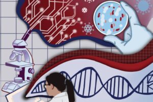
A Novel Method for Monitoring the ‘Engine’ of Pregnancy

Combining optical measurements with ultrasound, an interdisciplinary team from the School of Arts & Sciences, Perelman School of Medicine, and CHOP developed a device to better measure blood flow and oxygenation in the placenta. (Image: Lin Wang)

A study published in Nature Biomedical Engineering details a novel method for imaging the placenta in pregnant patients as well as the results of a pilot clinical study. By combining optical measurements with ultrasound, the findings show how oxygen levels can be monitored noninvasively and provides a new way to generate a better understanding of this complex, crucial organ. This research was the result of a collaboration of the groups of the University of Pennsylvania’s Arjun Yodh and Nadav Schwartz with colleagues from the Children’s Hospital of Philadelphia (CHOP) and was led by postdoc Lin Wang.

Schwartz describes the placenta as the “engine” of pregnancy, an organ that plays a crucial role in delivering nutrients and oxygen to the fetus. Placental dysfunction can lead to complications such as fetal growth restriction, preeclampsia, and stillbirth. To increase knowledge about this crucial organ, the National Institute of Child Health and Human Development launched the Human Placenta Project in 2014. One focus of the program is to develop tools to assess human placental structure and function in real time, including optical devices.

For three years, the researchers optimized the design of their instrument and tested it in preclinical settings. The process involved integrating optical fibers with ultrasound probes, exploring various ultrasound transducers, and improving the multimodal technology so that measurements were stable, accurate, and reproducible while collecting data at the bedside. The resulting instrumentation now enables researchers to study the anatomy of the placenta while also collecting detailed functional information about placenta blood flow and oxygenation, capabilities that existing commercially devices do not have, the researchers say.

Because the placenta is located far below the body’s surface, one of the key technical challenges addressed by Wang, a postdoc in Yodh’s lab, was reducing background noise in the opto-electronic system. Light is scattered and absorbed when it travels through thick tissues, Yodh says, and the key for success was to reduce background interference so that the small amount of light that penetrates deep into the placenta and then returns is still large enough for a high-quality measurement.

“We’re sending a light signal that goes through the same deep tissues as the ultrasound. The extremely small amount of light that returns to the surface probe is then used to accurately assess tissue properties, which is only possible with very stable lasers, optics, and detectors,” says Yodh. “Lin had to overcome many barriers to improve the signal-to-noise ratio to the point where we trusted our data.”

Read the full story in Penn Today.

The authors are Lin Wang, Jeffrey M. Cochran, Kenneth Abramson, Lian He, Venki Kavuri, Samuel Parry, Arjun G. Yodh, and Nadav Schwartz from Penn; Tiffany Ko, Wesley B. Baker, and Rebecca L. Linn from the Children’s Hospital of Philadelphia, and David R. Busch, previously a research associate at Penn and now at the University of Texas Southwestern Medical School.

Arjun Yodh is the James M. Skinner Professor of Science in the Department of Physics and Astronomy in the School of Arts & Sciences at the University of Pennsylvania. He is a member of the Penn Bioengineering Graduate Group.

Nadav Schwartz is an Associate Professor in the Department of Obstetrics and Gynecology in Penn’s Perelman School of Medicine.

Lin Wang is a postdoc in the Department of Physics and Astronomy in Penn’s School of Arts & Sciences.

This research was supported by National Institutes of Health grants F31HD085731, R01NS113945, R01NS060653, P41EB015893, P41EB015893, T32HL007915, and U01HD087180.

Shapeshifting Microrobots Can Brush and Floss Teeth

by Katherine Unger Baillie

In a proof-of-concept study, researchers from the School of Dental Medicine and School of Engineering and Applied Science shows that a hands-free system could effectively automate the treatment and removal of tooth-decay-causing bacteria and dental plaque. (Illustration: Melissa Pappas)

A shapeshifting robotic microswarm may one day act as a toothbrush, rinse, and dental floss in one.

The technology, developed by a multidisciplinary team at the University of Pennsylvania, is poised to offer a new and automated way to perform the mundane but critical daily tasks of brushing and flossing. It’s a system that could be particularly valuable for those who lack the manual dexterity to clean their teeth effectively themselves.

The building blocks of these microrobots are iron oxide nanoparticles that have both catalytic and magnetic activity. Using a magnetic field, researchers could direct their motion and configuration to form either bristlelike structures that sweep away dental plaque from the broad surfaces of teeth, or elongated strings that can slip between teeth like a length of floss. In both instances, a catalytic reaction drives the nanoparticles to produce antimicrobials that kill harmful oral bacteria on site.

Experiments using this system on mock and real human teeth showed that the robotic assemblies can conform to a variety of shapes to nearly eliminate the sticky biofilms that lead to cavities and gum disease. The Penn team shared their findings establishing a proof-of-concept for the robotic system in the journal ACS Nano.

“Routine oral care is cumbersome and can pose challenges for many people, especially those who have hard time cleaning their teeth” says Hyun (Michel) Koo, a professor in the Department of Orthodontics and divisions of Community Oral Health and Pediatric Dentistry in Penn’s School of Dental Medicine and co-corresponding author on the study. “You have to brush your teeth, then floss your teeth, then rinse your mouth; it’s a manual, multistep process. The big innovation here is that the robotics system can do all three in a single, hands-free, automated way.”

Read the full story in Penn Engineering Today.

Hyun (Michel) Koo is a professor in the Department of Orthodontics and divisions of Community Oral Health and Pediatric Dentistry in the School of Dental Medicine, co-director of the Center for Innovation & Precision Dentistry, and member of the Penn Bioengineering Graduate Group at the University of Pennsylvania.

Edward Steager is a senior research investigator in Penn’s School of Engineering and Applied Science.

Koo and Steager’s coauthors on the paper are Penn Dental Medicine’s Min Jun Oh, Alaa Babeer, Yuan Liu, and Zhi Ren and Penn Engineering’s Jingyu Wu, David A. Issadore, Kathleen J. Stebe, and Daeyeon Lee.

This work was supported in part by the National Institute for Dental and Craniofacial Research (grants DE025848 and DE029985), Procter & Gamble, and the Postdoctoral Research Program of Sungkyunkwan University.

Penn Startup Vittoria Biotherapeutics Raises $10M in Seed Funding

Marco Ruella, MD

A Philadelphia life sciences company spun out of Penn is emerging from stealth mode with nearly $10 million from a seed funding round. Vittoria Biotherapeutics’ mission is to overcome limitations of CAR T cell therapy by using unique cell engineering and gene editing technologies to create new therapies that address unmet clinical needs. The technology the company is attempting to commercialize was developed by Marco Ruella, M.D., Assistant Professor of Medicine in the Perelman School of Medicine and member of the Penn Bioengineering Graduate Group, who is the company’s scientific founder.

Read “Penn spinout Vittoria Biotherapeutics emerges from stealth mode with $10M seed round” in the Philadelphia Business Journal.

Penn Medicine and Children’s Hospital of Philadelphia Announce Partnership with Costa Rica for CAR T Cell Therapy

Carl June, MD

Carl June, MD, Professor in the Perelman School of Medicine and member of the Penn Bioengineering Graduate Group, was quoted in a recent press release  announcing a new international partnership between Penn Medicine (PSOM), the Children’s Hospital of Pennsylvania (CHOP), and Costa Rica’s CCSS, or the Caja Costarricense de Seguro Social (Social Security Program), to develop CAR T research in Costa Rica. June is a world renowned cancer cell therapy pioneer whose research led to the initial development and FDA approval of CAR T cell therapy:

“‘At least 15,000 patients across the world have received CAR T cells, and dozens more clinical trials using this approach are in progress, for almost every major tumor type, but people in many parts of the globe still do not have access to treatment with these transformative therapies,’ said Carl H. June, MD, the Richard W. Vague Professor in Immunotherapy and director of the Center for Cellular Immunotherapies in Penn’s Perelman School of Medicine. “We are honored to work with our colleagues in Costa Rica in hopes of building a path for patients in underserved areas to have the opportunity to benefit from clinical research programs offering this personalized therapy.’”

Read the the announcement in Penn Medicine News.


Yale Cohen Appointed Assistant Dean of Research Facilities and Resources at Penn Medicine

Yale E. Cohen, PhD

Yale E. Cohen, Professor of Otorhinolaryngology, with secondary appointments in Neuroscience and Bioengineering, was appointed Assistant Dean of Research Facilities and Resources at the Perelman School of Medicine at the University of Pennsylvania, effective April 1, 2022. Cohen is currently Chair of the Penn Bioengineering Graduate Group, and Director of the Hearing Sciences Center:

“Many of you are already quite familiar with Dr. Cohen, as his leadership roles in research training and education at PSOM and the University are far-reaching and impactful. Dr. Cohen is a Professor of Otorhinolaryngology with secondary appointments in the Department of Neuroscience and Engineering’s Department of Bioengineering. Recognized widely for his deep commitment to our teaching and training community, Dr. Cohen chairs the Bioengineering Graduate Group, and in 2020 received the prestigious Jane M. Glick Graduate Student Teaching Award, which honors clinicians and scientists who exemplify outstanding quality of patient care, mentoring, research, and teaching.”

Read the full announcement in the Penn Medicine archive.

How Bacteria Store Information to Kill Viruses (But Not Themselves)

by Luis Melecio-Zambrano

A group of bacteriophages, viruses that infect bacteria, imaged using transmission electron microscopy. New research sheds light on how bacteria fight off these invaders without triggering an autoimmune response. (Image: ZEISS Microscopy, CC BY-NC-ND 2.0)

During the last few years, CRISPR has grabbed headlines for helping treat patients with conditions as varied as blindness and sickle cell disease. However, long before humans co-opted CRISPR to fight genetic disorders, bacteria were using CRISPR as an immune system to fight off viruses.

In bacteria, CRISPR (Clustered Regularly Interspaced Short Palindromic Repeats) works by stealing small pieces of DNA from infecting viruses and storing those chunks in the genes of the bacteria. These chunks of DNA, called spacers, are then copied to form little tags, which attach to proteins that float around until they find a matching piece of DNA. When they find a match, they recognize it as a virus and cut it up.

Now, a paper published in Current Biology by researchers from the University of Pennsylvania Department of Physics and Astronomy shows that the risk of autoimmunity plays a key role in shaping how CRISPR stores viral information, guiding how many spacers bacteria keep in their genes, and how long those spacers are.

Ideally, spacers should only match DNA belonging to the virus, but there is a small statistical chance that the spacer matches another chunk of DNA in the bacteria itself. That could spell death from an autoimmune response.

“The adaptive immune system in vertebrates can produce autoimmune disorders. They’re very serious and dangerous, but people hadn’t really considered that carefully for bacteria,” says Vijay Balasubramanian, principal investigator for the paper and the Cathy and Marc Lasry Professor of Physics in the School of Arts & Sciences.

Balancing this risk can put the bacteria in something of an evolutionary bind. Having more spacers means they can store more information and fend off more types of viruses, but it also increases the likelihood that one of the spacers might match the DNA in the bacteria and trigger an autoimmune response.

Read the full story in Penn Today.

Vijay Balasubramanian is the Cathy and Marc Lasry Professor of Physics at the Department of Physics and Astronomy of the University of Pennsylvania, a visiting professor at Vrije Universiteit Brussel, and a member of the Penn Bioengineering Graduate Group.

FDA Approves Penn Pioneered CAR T Cell Therapy for Third Indication

The U.S. Food and Drug Administration has expanded its approval for Kymriah, a personalized cellular therapy developed at the Abramson Cancer Center, this time for the treatment of adults with relapsed/refractory follicular lymphoma who have received at least two lines of systemic therapy. “Patients with follicular lymphoma who relapse or don’t respond to treatment have a poor prognosis and may face a series of treatment options without a meaningful, lasting response,” said Stephen J. Schuster, the Robert and Margarita Louis-Dreyfus Professor in Chronic Lymphocytic Leukemia and Lymphoma in the Division of Hematology Oncology. It’s the third FDA approval for the “living drug,” which was the first of its kind to be approved, in 2017, and remains the only CAR T cell therapy approved for both adult and pediatric patients.

“In just over a decade, we have moved from treating the very first patients with CAR T cell therapy and seeing them live healthy lives beyond cancer to having three FDA-approved uses of these living drugs which have helped thousands of patients across the globe,” said Carl June, MD, the Richard W. Vague Professor in Immunotherapy in the department of Pathology and Laboratory Medicine in Penn’s Perelman School of Medicine and director of the Center for Cellular Immunotherapies in the Abramson Cancer Center and director of the Parker Institute for Cancer Immunotherapy at Penn. “Today’s news is new fuel for our work to define the future of cell therapy and set new standards in harnessing the immune system to treat cancer.”

Research from June, a member of the Penn Bioengineering Graduate Group, led to the initial FDA approval for the CAR T therapy (sold by Novartis as Kymriah) for treating acute lymphoblastic leukemia (ALL), one of the most common childhood cancers.

Read the full announcement in Penn Medicine News.

Exploring the History of CAR-T Cell Therapy

Carl June, MD

A new feature in Chemistry World explores the history of CAR (chimeric antigen receptor)-T cell therapy, a revolutionary type of therapeutic treatment for certain types of cancer. One of the pioneers of CAR-T cell therapy is Carl June, Richard W. Vague Professor in Immunotherapy in the Perelman School of Medicine and member of the Penn Bioengineering Graduate Group. His groundbreaking research opened the door for FDA approval of the CAR T therapy called Kymriah, which treats acute lymphoblastic leukemia (ALL), one of the most common childhood cancers.

Read “A decade of CAR-T cell therapy” in Chemistry World.

Vijay Balasubramanian Discusses Theoretical Physics in Quanta Magazine

Cathy and Marc Lasry Professor Vijay Balasubramanian at Penn’s BioPond.

In an interview with Quanta Magazine, Vijay Balasubramanian discusses his work as a theoretical physicist, noting his study of the foundations of physics and the fundamentals of space and time. He speaks of the importance of interdisciplinary study and about how literature and the humanities can contextualize scientific exploration in the study of physics, computer science, and neuroscience.

Balasubramanian is Cathy and Marc Lasry Professor in the Department of Physics and Astronomy in the Penn School of Arts and Sciences and a member of the Penn Bioengineering Graduate Group.

Read “Pondering the Bits That Build Space-Time and Brains” in Quanta Magazine.

Erin Berlew and Rhea Chitalia Receive Solomon R. Pollack Awards for Excellence in Graduate Bioengineering Research

The Solomon R. Pollack Award for Excellence in Graduate Bioengineering Research is given annually to the most deserving Bioengineering graduate students who have successfully completed research that is original and recognized as being at the forefront of their field. This year Penn Bioengineering recognizes the outstanding work of two graduate students in Bioengineering: Erin Berlew and Rhea Chitalia.

Erin Berlew, Ph.D. candidate in Bioengineering

Erin Berlew is a Ph.D. candidate in the lab of Brian Chow, Associate Professor in Bioengineering. She successfully defended her thesis, titled “Single-component optogenetic tools for cytoskeletal rearrangements,” in December 2021. In her research, she used the BcLOV4 optogenetic platform discovered/developed in the Chow lab to control RhoGTPase signaling. Erin earned a B.S. in Chemistry from Haverford College in 2015 and was an Americorps member with City Year Philadelphia from 2015-2016. “Erin is a world-class bioengineering with an uncommon record of productivity gained through her complementary expertise in molecular, cellular, and computational biology,” says Chow. “She embodies everything wonderful, both academically and culturally, about our graduate program and its distinguished history.” Erin’s hobbies outside the lab include spending time with family, reading mystery novels, enjoying Philadelphia, and crossword puzzles. In the future, she hopes to continue to teach for the BE department (she has already taught ENGR 105 and served as a TA for undergraduate and graduate courses) and to conduct further research at Penn.

Rhea Chitalia, Ph.D. candidate in Bioengineering

Rhea Chitalia is a Ph.D. candidate in Bioengineering and a member of the Computational Biomarker Imaging Group (CBIG), advised by Despina Kontos, Matthew J. Wilson Associate Professor of Research Radiology II in the Perelman School of Medicine. Rhea completed her B.S.E. in Biomedical Engineering at Duke University in 2015. Her doctoral research concerns leveraging machine learning, bioinformatics, and computer vision to develop computational imaging biomarkers for improved precision cancer care. In December 2021 she successfully defended her thesis titled “Computational imaging biomarkers for precision medicine: characterizing intratumor heterogeneity in breast cancer.” “It has been such a privilege to mentor Rhea on her dissertation research,” says Kontos. “Rhea has been a star graduate student. Her work has made fundamental contributions in developing computational methods that will allow us to gain important insight into tumor heterogeneity by utilizing a multi-modality imaging approach.” David Mankoff, Matthew J. Wilson Professor of Research Radiology in the Perelman School of Medicine, served as Rhea’s second thesis advisor. “It was a true pleasure for me to work with Rhea and to Chair her BE Thesis Committee,” Mankoff adds. “Rhea’s Ph.D. thesis and thesis presentation was one of the best I have had the chance to be involved with in my graduate mentoring career.” After graduation, Rhea hopes to further precision medicine initiatives through the use of real world, multi-omic data in translational industry settings. She will be joining Invicro as an Imaging Scientist. In her spare time, Rhea enjoys trying new restaurants, reading, and spending time with friends and family.