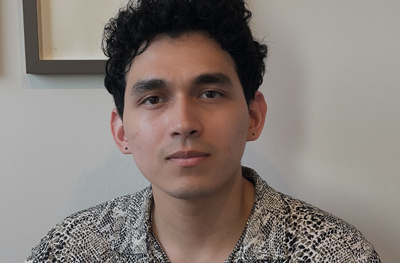
Sam Preza, a doctoral student in Bioengineering, was named one of two Penn graduate students and one of 50 graduate students nationwide to receive a 2024 Howard Hughes Medical Institute (HHMI) Gilliam Fellowship. The HHMI Gilliam Fellowship cohort is awarded annually to graduate students and their advisors for outstanding research and commitment to advancing equity and inclusion in science. The fellowship includes a one-year mentorship skills development course and support to promote healthy and inclusive graduate training environments at their home institution.
Preza is a member of lab of Juan Rene Alvarez Dominguez, Assistant Professor of Cell and Developmental Biology in the Perelman School of Medicine and member of the Bioengineering Graduate Group. He graduated from University of Maryland in 2019 with a degree in Chemical Engineering. After working for t three years at AstraZeneca in Bioprocess Development, he joined the J-RAD Lab where he researches technologies for unmet medical needs:
“[Preza’s] PhD program harnesses the power of stem cells and circadian rhythms to ultimately develop a cure for Type I diabetes, which he researches alongside his advisor, Juan Alvarez, PhD, an assistant professor in the Department of Cell and Developmental Biology. Their studies focus on beta cells, the type of cell found in the pancreas that helps regulate glucose. In the lab, they study how exposing cells to circadian rhythms could lead to functional beta cells that can be transplanted into diabetic patients to restore function. This work will be supported by their HHMI Fellowship grant.
The fellowship not only supports their scientific research but also helps foster an inclusive research environment, ensuring various backgrounds and ideologies contribute to their research. Preza is starting a DEI ‘potluck’, where bioengineering students can gather to discuss new research or career ideas. The meetups are catered by whichever student is hosting the meeting and can either showcase their nationality’s food or a cuisine they are passionate about, highlighting the celebration of diversity of ideas through food.
‘I believe STEM fields should look more like a mosaic of all our backgrounds rather than a melting pot, to add to the richness that is the art of science,’ Preza said.”
A multiracial Black and Asian self-described secular humanist, who was raised as one of Jehovah’s Witnesses and is now in an interracial, interfaith marriage, walked into a Passover seder.
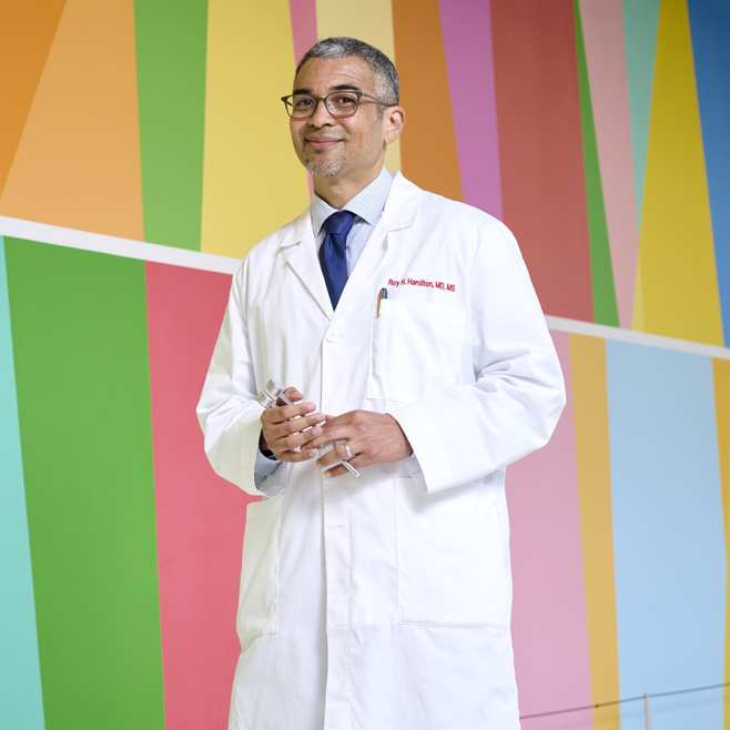
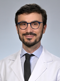
It’s not the setup for a groaner of a joke, or an epic fail of an evening. Rather, as Roy H. Hamilton, MD, tells it, this was his experience this spring as his Jewish in-laws—the family he has loved as his own for over two decades—came together to commemorate the universal human themes of freedom and deliverance from oppression reflected in the Passover narrative. Though he does this every year, this year he had some trepidation. In a time marked by tragic conflict and with tensions both abroad and at home, it seemed like having a frank discussion of these themes might invite acrimony. But what emerged instead was a profound opportunity to listen, to appreciate each other’s perspective, and to “exercise empathy for trauma that’s happening to everyone.”
It was a bit of a revelation for Hamilton, Penn Medicine’s new vice dean for Inclusion, Diversity, and Equity. “In the moment that you would have thought would be the worst to open up certain topics, we all ended up having a great dialogue across differences,” he said. Why? “Because we all felt connected enough to give each other respect, compassion, and grace, even when our thoughts and opinions differed. It made me think about how we can further cultivate a culture of empathy at Penn too.”
Today, as Hamilton begins his third decade on the faculty at Penn’s Perelman School of Medicine, he is devoted to making academia a safe, supportive space for students and colleagues alike. He serves as a professor of Neurology, with secondary appointments in Psychiatry and Physical Medicine and Rehabilitation. Hamilton is also director of both the Laboratory for Cognition and Neural Stimulation; and the Penn Brain Science, Translation and Modulation (BrainSTIM) Center. Previously, he was the Perelman School of Medicine’s assistant dean for Cultural Affairs and Diversity for almost a decade, and launched similar efforts in his field, serving as Penn Neurology’s vice chair for Diversity and Inclusion from 2017 until his recent elevation to the role for Penn Medicine as a whole.
Given his own diverse background and personal life, Hamilton wants everyone—trainees, faculty, patients—to feel valued and included. “I touch enough spaces in my personal life that when groups are being clearly systematically disadvantaged, it often feels like it’s touching on some piece of my own identity,” he said, in discussing his background and hope for his new leadership position. “I bring a lot of myself to this role.”
In a recent discussion, Hamilton shared perspectives on why supporting inclusion, diversity, and equity matters—particularly for an institution training future doctors—and what Penn Medicine is doing in this sphere.
The NSF AIRFoundry will accelerate RNA research using the power of AI and educate the next generation of RNA researchers. (DesignCells via Getty Images)
In a typical foundry, raw materials like steel and copper are melted down and poured into molds to assume new shapes and functions. The U.S. National Science Foundation Artificial Intelligence-driven RNA Foundry (NSF AIRFoundry), led by the University of Pennsylvania and the University of Puerto Rico and supported by an $18-million, six-year grant, will serve much the same purpose, only instead of smithing metal, the “BioFoundry” will create molecules and nanoparticles.
NSF AIRFoundry is one of five newly created BioFoundries, each of which will have a different focus. Bringing together researchers from Penn Engineering, Penn Medicine’s Institute for RNA Innovation, the University of Puerto Rico–Mayagüez (UPR-M), Drexel University, the Children’s Hospital of Philadelphia (CHOP) and InfiniFluidics, the facility, which will be physically located in West Philadelphia and at UPR-M, will focus on ribonucleic acid (RNA), the tiny molecule essential to genetic expression and protein synthesis that played a key role in the COVID-19 vaccines and saved tens of millions of lives.
The facility will use AI to design, optimize and synthesize RNA and delivery vehicles by augmenting human expertise, enabling rapid iterative experimentation, and providing predictive models and automated workflows to accelerate discovery and innovation.
“With NSF AIRFoundry, we are creating a hub for innovation in RNA technology that will empower scientists to tackle some of the world’s biggest challenges, from health care to environmental sustainability,” says Daeyeon Lee, Russell Pearce and Elizabeth Crimian Heuer Professor in Chemical and Biomolecular Engineering in Penn Engineering and NSF AIRFoundry’s director.
“Our goal is to make cutting-edge RNA research accessible to a broad scientific community beyond the health care sector, accelerating basic research and discoveries that can lead to new treatments, improved crops and more resilient ecosystems,” adds Nobel laureate Drew Weissman, Roberts Family Professor in Vaccine Research in Penn Medicine, Director of the Penn Institute for RNA Innovation and NSF AIRFoundry’s senior associate director.
The facility will catalyze new innovations in the field by leveraging artificial intelligence (AI). AI has already shown great promise in drug discovery, poring over vast amounts of data to find hidden patterns. “By integrating artificial intelligence and advanced manufacturing techniques, the NSF AIRFoundry will revolutionize how we design and produce RNA-based solutions,” says David Issadore, Professor in Bioengineering and in Electrical and Systems Engineering at Penn Engineering and the facility’s associate director of research coordination.
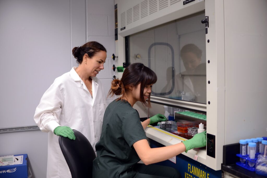
Rising second-year Sidney Wong, right, spent the summer working in the lab of Penn Vet professor Kyla Ortved, left, through the Penn Undergraduate Research Mentoring Program.
Roughly one in three Americans suffers from osteoarthritis, a progressive disease that causes joint cartilage to break down in a vicious cycle. The less cartilage, the more wear and tear on the joints, which further weakens the remaining connective tissue. In addition to joint pain, the condition can lead to loss of joint function, making it extremely hard to complete tasks of daily living.
At present, osteoarthritis has no cure. Zhiliang Cheng, Research Associate Professor in Bioengineering (BE), has studied the use of nanotechnology to treat the disease for years. In collaboration with Ling Qin, Professor in Orthopedic Surgery within the Perelman School of Medicine and member of the Penn Bioengineering Graduate Group, Cheng developed nanoparticles that activate the epidermal growth factor receptor (EGFR) pathway, increasing the expression of genes that promote healthy cartilage.
This summer, Sidney Wong, a rising second-year in the School of Arts and Sciences, built on Cheng and Qin’s research in the lab of Kyla Ortved, Jacques Jenny Endowed Term Chair of Orthopedic Surgery and Associate Professor in Large Animal Surgery at the School of Veterinary Medicine, studying the EGFR pathway in horses, whose joints resemble those of humans.
“What I’ve observed so far has been pretty promising,” says Wong, who found that equine cartilage treated with the nanoparticles appears healthier.
The effectiveness of CAR T cell therapy against a variety of cancers, including solid tumors, could be boosted greatly by using CRISPR-Cas9 technology to knock out the gene for CD5, a protein found on the surface of T cells, according to a preclinical study from investigators at the University of Pennsylvania’s Perelman School of Medicine and Abramson Cancer Center.
CAR T cells are T cells that have been engineered to attack specific targets found on cancer cells. They have had remarkable results in some patients with blood cancers. But they have not performed well against other cancers including solid-tumor cancers, such as pancreatic cancer, prostate cancer, and melanoma. Researchers have been searching for techniques to boost the effectiveness of CAR T cell therapy.
The study, published today in Science Immunology, suggests that knocking out CD5 could be a prime technique. Illuminating the protein’s previously murky role, the researchers found that it works as a powerful immune checkpoint, reining in T cell effectiveness. Removing it, they showed, dramatically enhanced CAR T cell anticancer activity in a variety of preclinical cancer models.
“We’ve discovered in preclinical models that CD5 deletion greatly enhances the function of CAR T cells against multiple cancers,” said senior author Marco Ruella, MD, an assistant professor of Hematology-Oncology, researcher with the Center for Cellular Immunotherapies and the scientific director of Penn Medicine’s Lymphoma Program. “The striking effects we observed across preclinical models suggest that CD5 knockout could be a general strategy for enhancing CAR T cell function.”
The study’s first author is Ruchi Patel, PhD, a recent graduate student from the Ruella Laboratory.
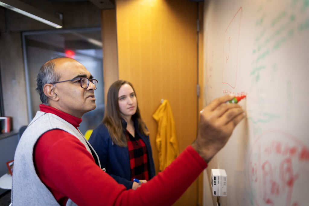
Younger scientists often ask him about exploring multiple fields, Balasubramanian says. The advice he offers is to “have a central line where you have credibility, where you’ve established that you’re really, really good at what you do, and you can be trusted.” (Image: Eric Sucar)
Academia is a long journey of specialization and behind any professor’s CV are long hours of research and study. While the path can be direct for some, for others there’s a pivot, a moment or experience that changes the course of that journey.
Penn Today spoke with four professors whose academic paths diverged, to learn about the trajectory of their interdisciplinary work. Vijay Balasubramanian traverses the boundaries of physics and neuroscience. Tukufu Zuberi is a demographer-turned-curator. Brittany Watson integrates education, research, and veterinary medicine. Amy Hillier began her career studying historical mortgage redlining and moved into supporting trans youth.
Vijay Balasubramanian The Cathy and Marc Lasry Professor of Physics in the School of Arts & Sciences
Wandering through Kolkata’s markets in India stimulates the mind. Hawkers’ cries pass through the inner ear as electrical signals; the pungent, earthy smell of turmeric enters the brain through olfactory sensory neurons. In 1976, a 7-year-old Vijay Balasubramanian had his own market revelation through a bookseller’s portico, where the cover of a slim volume showed a man peering through a microscope lens and a smattering of white paint scattered like stars across the firmament of man and machine.
“What is a scientist?” the book asked, running through a series of exciting adventure shots: archeologic discovery, venom extraction, missile control. In that moment, Balasubramanian knew he would be a scientist. It looked, he says, “amazingly cool.”
When he arrived at the Massachusetts Institute of Technology, Balasubramanian wanted to study the fundamental laws of nature. “So that’s physics,” he says. While earning his doctoral degree at Princeton University, a mentor suggested Balasubramanian read papers in the burgeoning field of neuroscience. It immediately resonated. “Oh my god, this stuff is so cool,” Balasubramanian thought. “But the final year of a Ph.D. is not the time to switch.”
He earned his degree and took a position as a junior fellow of the Harvard Society of Fellows. During the day, he worked on string theory and the information loss paradox for black holes. But in the evening, he would moonlight in a neuroscience lab.
As a young theoretical physicist at Penn, Balasubramanian met Peter Sterling. A former Freedom Rider and professor of neuroscience at the Perelman School of Medicine, Sterling was “a true intellectual,” Balasubramanian says. He knew everything, was interested in everything, and would talk with anybody.
The pair wrote a series of papers together regarding information processing and transmission. “He’s so quick and so much fun and so lively,” Sterling says of Balasubramanian. “He’s fearless; there’s nothing he won’t try.”
While in Cairo with wife Heather J. Sharkey, professor of modern Middle Eastern and North African history at Penn, Balasubramanian prepared a neuroscience grant and submitted it to the National Science Foundation, “sort of on a whim,” he says. “I put it in from an internet café on an island in the middle of the Nile.” He got the grant and started a research group.
After that, Balasubramanian says, “I was off and running.”
“I was certainly told,” Balasubramanian says of his work in neuroscience, “do not do this before tenure.” But, if he waited, “then I’d be too set my ways,” he says. “I just wouldn’t know enough; it would be too hard to learn; I wouldn’t have the time.”
Younger scientists often ask him about exploring multiple fields, Balasubramanian says. The advice he offers is to “have a central line where you have credibility, where you’ve established that you’re really, really good at what you do, and you can be trusted.” That gives you more latitude, he says.
After that, it’s just sheer discipline. “You’re going to have to wake up earlier than everybody else. You’re going to have to work longer days,” he says. “Otherwise, you know, everybody else is working hard too, and you’ll never be able to achieve the level of expertise and knowledge to be able to do things at that world-class level.”
Balasubramanian wants to see more interdisciplinary collaboration. “Each field trains its students with a certain body of techniques that has been found historically useful in that field,’ he says. “Very often, those techniques also have uses elsewhere, but they don’t know to apply it.”
Traversing borders can be helpful in producing new insights, Balasubramanian says. You can ask questions that people in the field won’t. You might experiment with new ideas or put two disjointed ideas together, he says. “If you’re coming from outside, you have the leeway to do all kinds of silly things. Sometimes, they’re not silly.”
Why not ask new questions and propose new answers? In the end, the data will tell you what’s true. “It gives me comfort to know how things tick.”
This post is adapted from a longer story in Penn Today. Read the full story here.
Balasubramanian is Cathy and Marc Lasry Professor in the Department of Physics and Astronomy in the Penn School of Arts and Sciences and is a member of the Penn Bioengineering Graduate Group. Read more stories featuring his research here.
Leaders and faculty from Penn Medicine, including Kevin Mahoney, Carl June, John Wherry, and Mike Mitchell (pictured left to right), speak on stage during the Penn London symposium.
Sharing the exciting work happening at Penn with alumni, parents, and friends throughout the world is a priority for Interim President J. Larry Jameson.
Shortly after challenging the graduating Class of 2024 to “keep reinventing, learning, and engaging” he brought that same spirit to the Penn community in London. He met with leadership volunteers from the region and welcomed approximately 200 attendees to an academic symposium titled “Frontiers of Knowledge and Discovery: Leading in a Changing World.”
Kevin Mahoney, CEO of the University of Pennsylvania Health System, moderated the first panel, on the genesis of breakthroughs. “When our faculty explain how landmark achievements like new fields of science or first-in-class cancer therapies come about, they never fail to emphasize how collaboration turns expertise into progress,” he said. “Hearing Mike Mitchell, John Wherry, and Carl June speak made plain how our brilliant, interconnected Penn faculty work together on one campus with results that are changing our world.”
Vijay Kumar, the Nemirovsky Family Dean of Penn Engineering, shared Mahoney’s perspective on collaboration—with a twist. “Non-engineers can be mystified, if not intimidated, by the complexities of the work we do,” he explained. “When a faculty member breaks down a project and talks it through, step by step, the engineering concepts become so much more understandable and relatable.” Kumar moderated a session with Dan Rader and Rene Vidal that focused on the increasing and powerful synergies among data science and AI, medical research, and clinical practice
Michael Mitchell is Associate Professor in Bioengineering. Read more stories featuring Mitchell in the BE Blog.
Carl June is Richard W. Vague Professor in Immunotherapy in the Perelman School of Medicine and is a member of the Penn Bioengineering Graduate Group. Read more stories featuring June in the BE Blog.
Congratulations to the fifteen Bioengineering students to receive 2024 National Science Foundation Graduate Research Fellowship Program (NSF GRFP) fellowships. The prestigious NSF GRFP program recognizes and supports outstanding graduate students in NSF-supported fields. The recipients were selected from a highly-competitive, nationwide pool. Further information about the program can be found on the NSF website.
The following Ph.D. students in Bioengineering received awards:
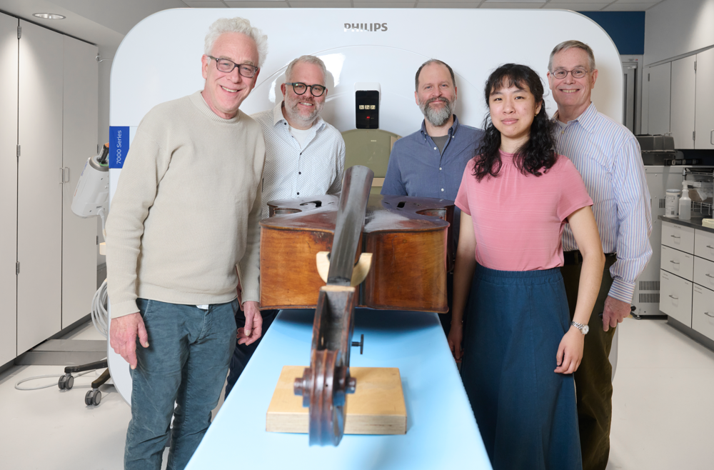
The instrument imaging team, from left: Philadelphia Orchestra bassist Duane Rosengard; Peter Noël, PhD, director of CT Research at the Perelman School of Medicine; luthier Zachary S. Martin; Leening Liu, a PhD student in Noël’s Laboratory of Advanced Computed Tomography Imaging; and Mark Kindig.
When you’re an expert in medical CT imaging, two things are bound to happen, says Peter Noël, PhD, associate professor of Radiology and director of CT Research at the Perelman School of Medicine. One: You develop an insatiable curiosity about the inner workings of all kinds of objects, including those unrelated to your research. And two: Both colleagues and complete strangers will ask for your help in imaging a wide variety of unexpected items.
Over the course of his career, in between managing his own research projects, Noël has imaged diverse objects ranging from animal skulls to tree samples from a German forest, all in the name of furthering scientific knowledge. But none has intrigued him as much as his current extracurricular project: the first known attempt to perform CT imaging of some of the world’s finest string basses.
The goal is to crack the code on what makes a world-class instrument. This knowledge could both increase the ability to better care for masterworks built between the 17th and 19th centuries, as well as providing insights into refining the building of new ones, including possibly shifting from older, scarcer European wood to the use of sustainably harvested U.S. wood.
That’s why Noël and Leening Liu, a PhD student in Noël’s Laboratory of Advanced Computed Tomography Imaging, have found themselves volunteering to run the basses through a Penn CT scanner occasionally, when they’re not developing next-generation CT technology.
“We always learn something out of projects like this … the more appealing part is that medical research can also be applied to non-medical things,” Noël said. “We have the opportunity to take what we learn in medicine and use it for something else—in this case, moving the arts forward.”
Leening Liu is a Ph.D. student in Bioengineering. She is a member of the Laboratory for Advanced Tomography Imaging (LACTI) with research interests including clinical applications of spectral CT and spectral CT thermometry.
Lasya Sreepada has always been fascinated by the brain and the underlying biology that shapes how people develop and age. “My curiosity traces back to observing differences between myself and my sister,” says Sreepada, a Ph.D. candidate in Bioengineering whose research unites efforts across Penn Medicine and Penn Engineering. “We grew up in the same environment but had remarkably different personalities, which led me to question what drove these differences and which brought me to the brain.”
Her academic journey began by applying medical imaging to understand how brain injuries sustained by professional athletes or military veterans impact their brain structure and chemistry over time. She became curious about how neurotrauma impacts aging and degeneration in the long term. Now, she leverages large, multimodal datasets to investigate neurodegenerative disease, with a particular focus on Alzheimer’s.