NASA’s James Webb Space Telescope has produced the deepest and sharpest infrared image of the distant universe to date. Known as Webb’s First Deep Field, this image of galaxy cluster SMACS 0723 is rich with detail. Thousands of galaxies—including the faintest objects ever observed in the infrared—have appeared in Webb’s view for the first time. The image shows the galaxy cluster SMACS 0723 as it appeared 4.6 billion years ago. The combined mass of this galaxy cluster acts as a gravitational lens, magnifying much more distant galaxies behind it. Webb’s Near-Infra Red Cam has brought those distant galaxies into sharp focus—they have tiny, faint structures that have never been seen before, including star clusters and diffuse features. (Image: NASA, ESA, CSA, and STScI)
Could the universe be twice as old as current estimates put forward? Rajendra Gupta of the University of Ottawa recently published a paper suggesting just that. Gupta claims the universe may be around 26.7 billion years rather than the commonly accepted 13.8 billion. The news has generated many headlines as well as criticism from astronomers and the larger scientific community.
Penn Today met with professors Vijay Balasubramanian and Mark Devlin to discuss Gupta’s findings and better understand the rationale of these claims and how they fit in the broader context of problems astronomers are attempting to solve.
How do we know how old the universe actually is?
Balasubramanian: The universe is often reported to be 13.8 billion years old, but, truth be told, this is an amalgamation of various measurements that factor in different kinds of data involving the apparent ages of ‘stuff’ in the universe.
This stuff includes observable or ordinary matter like you, me, galaxies far and near, stars, radiation, and the planets, then dark matter—the sort of matter that doesn’t interact with light and which makes up about 27% of the universe—and finally, dark energy, which makes up a massive chunk of the universe, around 68%, and is what we believe is causing the universe to expand.
And so, we take as much information as we can about the stuff and build what we call a consensus model of the universe, essentially a line of best fit. We call the model the Lambda Cold Dark Matter (ΛCDM).
Lambda represents the cosmological constant, which is linked to dark energy, namely how it drives the expansion of the universe according to Einstein’s theory of general relativity. In this framework, how matter and energy behave in the universe determines the geometry of spacetime, which in turn influences how matter and energy move throughout the cosmos. Including this cosmological constant, Lambda, allows for an explanation of a universe that expands at an accelerating rate, which is consistent with our observations.
Now, the Cold Dark Matter part represents a hypothetical form of dark matter. ‘Dark’ here means that it neither interacts with nor emits light, so it’s very hard to detect. ‘Cold’ refers to the fact that its particles move slowly because when things cool down their components move less, whereas when they heat up the components get excited and move around more relative to the speed of light.
So, when you consider the early formation of the universe, this ‘slowness’ influences the formation of structures in the universe like galaxies and clusters of galaxies, in that smaller structures like the galaxies form before the larger ones, the clusters.
Devlin: And then taking a step back, the way cosmology works and pieces how old things are is that we look at the way the universe looks today, how all the structures are arranged within it, and we compare it to how it used to be with a set of cosmological parameters like Cosmic Microwave Background (CMB) radiation, the afterglow of the Big Bang, and the oldest known source of electromagnetic radiation, or light. We also refer to it as the baby picture of the universe because it offers us a glimpse of what it looked like at 380,000 years old, long before stars and galaxies were formed.
And what we know about the physical nature of the universe from the CMB is that it was something really smooth, dense, and hot. And as it continued to expand and cool, the density started to vary, and these variations became the seeds for the formation of cosmic structures.
The denser regions of the universe began to collapse under their own gravity, forming the first stars, galaxies, and clusters of galaxies. So, this is why, when we look at the universe today, we see this massive cosmic web of galaxies and clusters separated by vast voids. This process of structure formation is still ongoing.
And, so, the ΛCDM model suggests that the primary driver of this structure formation was dark matter, which exerts gravity and which began to clump together soon after the Big Bang. These clumps of dark matter attracted the ordinary matter, forming the seeds of galaxies and larger cosmic structures.
So, with models like the ΛCDM and the knowledge of how fast light travels, we can add bits of information, or parameters, and we have from things like the CMB and other sources of light in our universe, like the ones we get from other distant galaxies, and we see this roadmap for the universe that gives us it’s likely age. Which we think is somewhere in the ballpark of 13.8 billion years.
Vijay Balasubramanian is the Cathy and Marc Lasry Professor in the Department of Physics and Astronomy in the School of Arts & Sciences at the University of Pennsylvania. He is a member of the Penn Bioengineering Graduate Group.
Mark Devlin is the Reese W. Flower Professor of Astronomy and Astrophysics in the Department of Physics and Astronomy in the School of Arts & Sciences at Penn.
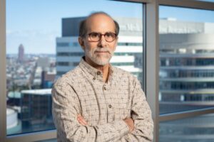
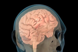
Traumatic brain injury (TBI) has disabled 1 to 2% of the population, and one of their most common disabilities is problems with short-term memory. Electrical stimulation has emerged as a viable tool to improve brain function in people with other neurological disorders.
Now, a new study in the journal Brain Stimulation shows that targeted electrical stimulation in patients with traumatic brain injury led to an average 19% boost in recalling words.
Led by University of Pennsylvania psychology professor Michael Jacob Kahana, a team of neuroscientists studied TBI patients with implanted electrodes, analyzed neural data as patients studied words, and used a machine learning algorithm to predict momentary memory lapses. Other lead authors included Wesleyan University psychology professor Youssef Ezzyat and Penn research scientist Paul Wanda.
“The last decade has seen tremendous advances in the use of brain stimulation as a therapy for several neurological and psychiatric disorders including epilepsy, Parkinson’s disease, and depression,” Kahana says. “Memory loss, however, represents a huge burden on society. We lack effective therapies for the 27 million Americans suffering.”
Michael Kahana is the Edmund J. and Louise W. Kahn Term Professor of Psychology at the University of Pennsylvania. He is a member of the Penn Bioengineering Graduate Group.
Kyle Vining, who is jointly appointed in the School of Dental Medicine and the School of Engineering and Applied Science, hopes that his research will help to push forward the state of clinical dentistry.
“During my training, I saw that there was overlap where I could do clinical work and science at the same time, and so that’s what I’ve been doing ever since,” Vining says. “As far back as middle school, I always wanted to be a biomedical engineer, and then the clinical side became interesting to me because I didn’t want to only do the theoretical or research side of things. I also wanted the hands-on, practical interaction of a skilled profession.”
The benefits of a dual career: Variety and opportunities to give back
Vining finds that wearing two hats offers the best of both worlds: opportunities to help both individual patients and to contribute to scientific and clinical progress.
“On the dentistry side, what I enjoy is getting to see patients, solving clinical problems, and trying to perform the best treatment I can; it has this rapid pace, which is kind of exciting and keeps you motivated,” Vining says. “And then research allows me to explore my interests and think about making an impact more broadly, not just in dentistry, but in medicine or in the world in general.”
Vining says dental school was demanding, yet a good time to explore his varied interests. He says he’d encourage others to pursue dentistry with an interdisciplinary approach. “Having exposure to different fields or different knowledge while you’re a student is really good for students and the profession in general,” he says.
The path towards a dual career
Vining first delved into research as a biomedical engineering undergraduate at Northwestern University. “I had the opportunity to work in a materials science lab studying the chemistry of surfaces. We would use molecules to modify the properties and surfaces that environments or cells could interact with,” he says.
Then, as a student at the University of Minnesota School of Dentistry, Vining realized that this same materials science research had many applications in dentistry. While in dental school, Vining conducted independent research in a materials science lab and also took the opportunity to do a yearlong fellowship in a cell and developmental biology lab at the National Institutes of Health.
Vining credits this fellowship with launching him towards a Ph.D., which he completed in bioengineering at Harvard in 2020. After earning his Ph.D., Vining conducted research at the Dana-Farber Cancer Institute prior to joining Penn.
Using biomaterials to understand how cells and tissues interact
Vining’s research at Penn aims to understand how the biophysical properties of materials impact cellular processes such as inflammation and fibrosis.
“Fibrosis is a physical change in tissues that produces a scar-like matrix that can inhibit healing, impair cancer treatment, and in general is not compatible with tissues regeneration,” Vining says. “There’s been a lot of effort on antifibrotic drugs, but we’re trying to look at fibrosis a little bit differently. Instead of directly inhibiting fibrosis, we’re trying to understand its consequences for the immune system because the immune system can be hijacked and become detrimental for your tissues.”
Through a better understanding the feedback loop between fibrotic tissue and the immune system, Vining hopes to design interventions to facilitate wound healing and tissue remodeling during restorative dental procedures and for treating diseases including head and neck cancer.
He’s also investigating how biomaterials like the resin used in tooth fillings interact with dental tissues. “Dental fillings rely on decades-old technologies that have been grandfathered in and contain toxic monomers that are not safe for biological systems,” Vining says. “We found a biocompatible resin chemistry that supports cells in vitro, and we’re trying to apply this to new types of dental fillings that promote repair or generation of dental tissues.”
Fostering interdisciplinary collaborations at Penn
“Dr. Vining is an ideal fit for the vision and mission of the CiPD,” says Penn Dental’s Hyun (Michel) Koo, co-founder and co-director of the CiPD. “With a secondary appointment in the School of Engineering, he will be instrumental in continuing to strengthen our engineering collaborations and teaching our students to work across disciplines to advance research, training, and entrepreneurship in this realm.”
Ultimately, Vining says it was Penn’s scientific community and the opportunities for interdisciplinary collaborations that drew him here.
“It was very apparent that there were a lot of potential research paths to pursue here and a lot of opportunities for collaborations,” Vining says. “One of the most exciting things for me so far has been meeting with faculty, whether it’s at Penn Medicine, the School of Engineering, Wharton, Penn Dental, or the Veterinary School. These meetings have already opened up new projects and collaborations.”
The collaboration sparked when Vining saw Mitchell present on a new technology that uses lipid nanoparticles to bind and target bone marrow cells at the 2022 CiPD first annual symposium. “It got me thinking because the dentin inside of teeth is a mineralized tissue very similar to bone, and the pulp inside the dentin is analogous to bone marrow tissue,” Vining says.
Rather than being the single disease class many people refer to, “cancer” is a blanket term that covers more than 100 distinct diseases, many of which have little in common aside from originating with rapidly dividing cells. Since different cancers demand different treatments, it follows that any given new therapy emerging from any institution would be likely to be a new cancer treatment.
But why so many in just this five-year period?
The volume of new cancer treatments makes sense, says Abramson Cancer Center (ACC) director Robert Vonderheide, attributing the flurry of new cancer drug approvals to a recent “explosion” in knowledge about cancer biology.
“Much of that knowledge is about the immune system’s ability to attack cancer, which people seriously doubted until about 20 years ago. As soon as we had a clinical validation for this Achilles heel in cancer, the dam burst for ideas about other ways to exploit that vulnerability to come forward,” he says. “The first drug that came out to activate the immune system inspired the rest of the field to find the next drug, and the one after that. We as a field have moved from serendipity and empiricism to science-driven drug design.”
The first CAR T cell therapy approval invigorated Penn faculty interested in finding new ways to harness the immune system to fight cancer.
“An approval like that makes what you’re working on more of a reality,” says Avery Posey, an assistant professor of systems pharmacology and translational therapeutics in the Perelman School of Medicine, whose lab team spends much of its time trying to identify more specific antigens for solid tumors and also studies ways to optimize engineered donor T cells. “It brings a new perspective, showing that your work is more than basic research and can actually become drugs that impact patients’ lives. That’s a real motivator to keep pushing forward.”
Honing new immunotherapies is a priority among Penn researchers, but not every recently approved new cancer treatment or detection tool developed at the institution engages the immune system. Faculty have explored and introduced widely varying approaches to improving the standard of care for cancer patients.
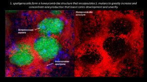
Collaborating researchers from the University of Pennsylvania School of Dental Medicine and the Adams School of Dentistry and Gillings School of Global Public Health at the University of North Carolina have discovered that a bacterial species called Selenomonas sputigena can have a major role in causing tooth decay.
Scientists have long considered another bacterial species, the plaque-forming, acid-making Streptococcus mutans, as the principal cause of tooth decay—also known as dental caries. However, in the study, published in Nature Communications, the Penn Dental Medicine and UNC researchers showed that S. sputigena, previously associated only with gum disease, can work as a key partner of S. mutans, greatly enhancing its cavity-making power.
“This was an unexpected finding that gives us new insights into the development of caries, highlights potential future targets for cavity prevention, and reveals novel mechanisms of bacterial biofilm formation that may be relevant in other clinical contexts,” says study co-senior author Hyun (Michel) Koo, a professor in the Department of Orthodontics and Divisions of Pediatrics and Community Oral Health and co-director of the Center for Innovation & Precision Dentistry at Penn Dental Medicine.
The other two co-senior authors of the study were Kimon Divaris, professor at UNC’s Adams School of Dentistry, and Di Wu, associate professor at the Adams School and at the UNC Gillings School of Global Public Health.
“This was a perfect example of collaborative science that couldn’t have been done without the complementary expertise of many groups and individual investigators and trainees,” Divaris says.
Michel Koo is a professor in the Department of Orthodontics and divisions of Community Oral Health and Pediatric Dentistry in Penn Dental Medicine and co-director of the Center for Innovation & Precision Dentistry. He is a member of the Penn Bioengineering Graduate Group.
Mustafa Mir, Assistant Professor of Cell and Developmental Biology in the Perelman School of Medicine and member of the Penn Bioengineering Graduate Group, was selected as one of Howard Hughes Medical Institute’s 31 new Freeman Hrabowski Scholars. The group consists of outstanding early career faculty in science who have potential to become leaders in their research fields and to create diverse and inclusive lab environments in which everyone can thrive. Mir and his lab develop and apply new microscopes to directly visualize the molecular scale events that underlie gene expression within live embryos.
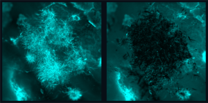
Candida albicans is a species of yeast that is a normal part of the human microbiota but can also cause severe infections that pose a significant global health risk due to their resistance to existing treatments, so much so that the World Health Organization has highlighted this as a priority issue. The picture above shows a before (left) and after (right) fluorescence image of fungal biofilms being precisely targeted by nanozyme microrobots without bonding to or disturbing the tissue sample. (Image: Min Jun Oh and Seokyoung Yoon)
Infections caused by fungi, such as Candida albicans, pose a significant global health risk due to their resistance to existing treatments, so much so that the World Health Organization has highlighted this as a priority issue.
Although nanomaterials show promise as antifungal agents, current iterations lack the potency and specificity needed for quick and targeted treatment, leading to prolonged treatment times and potential off-target effects and drug resistance.
“Candida forms tenacious biofilm infections that are particularly hard to treat,” Koo says. “Current antifungal therapies lack the potency and specificity required to quickly and effectively eliminate these pathogens, so this collaboration draws from our clinical knowledge and combines Ed’s team and their robotic expertise to offer a new approach.”
The team of researchers is a part of Penn Dental’s Center for Innovation & Precision Dentistry, an initiative that leverages engineering and computational approaches to uncover new knowledge for disease mitigation and advance oral and craniofacial health care innovation.
For this paper, published in Advanced Materials, the researchers capitalized on recent advancements in catalytic nanoparticles, known as nanozymes, and they built miniature robotic systems that could accurately target and quickly destroy fungal cells. They achieved this by using electromagnetic fields to control the shape and movements of these nanozyme microrobots with great precision.
“The methods we use to control the nanoparticles in this study are magnetic, which allows us to direct them to the exact infection location,” Steager says. “We use iron oxide nanoparticles, which have another important property, namely that they’re catalytic.”
Other authors include Min Jun Oh, Alaa Babeer, Yuan Liu, Zhi Ren, Zhenting Xiang, Yilan Miao, and Chider Chen of Penn Dental; and David P. Cormode and Seokyoung Yoon of the Perelman School of Medicine. Cormode also holds a secondary appointment in Bioengineering.
This research was supported in part by the National Institute for Dental and Craniofacial Research (R01 DE025848, R56 DE029985, R90DE031532 and; the Basic Science Research Program through the National Research Foundation of Korea of the Ministry of Education (NRF-2021R1A6A3A03044553).
Neil Sheppard, Adjunct Associate Professor of Pathology and Laboratory Medicine in the Perelman School of Medicine, and David Mai, a Bioengineering graduate student in the School of Engineering and Applied Science, explained the findings of their recent study, which offered a potential strategy to improve T cell therapy in solid tumors, to the European biotech news website Labiotech.
Brain technology offers all kinds of exciting possibilities — from treating conditions like epilepsy or depression, to simply maximizing brain health. But medical ethicists are concerned about potential dangers and privacy concerns. Roy Hamilton, Professor of Neurology in the Perelman School of Medicine, Director of the Penn Brain Science, Translation, Innovation, and Modulation (BrainSTIM) Center, and member of the Penn Bioengineering Graduate Group, spoke with WHYY about how brain stimulation is being used.
They join 120 members and 23 international members elected by their peers this year to NAS. Recognized for “distinguished and continuing achievements in original research,” this new class brings the total number of active members to 2,565 and of international members to 526.
David Brainard is the RRL Professor of Psychology, director of the Vision Research Center, and associate dean for the natural sciences in the School of Arts & Sciences. His research focuses on human vision, using both experiments and computer modeling of visual processing, to understand how the visual system deciphers information about objects from light entering the eye. Specifically, he and his lab are interested in color vision, conducting psychophysical experiments to investigate how the appearance of color is affected by an object’s surface properties and ambient light, and how color perception aids in identifying objects. Brainard is the recipient of many honors, including the Macbeth Award from the Inter-Society Color Council, Stein Innovation Award from Research to Prevent Blindness, and Edgard D. Tillyer Award from Optica. He is an elected member of the Society of Experimental Psychologists, a Silver Fellow of the Association for Research in Vision and Ophthalmology, and a Fellow of the Association for Psychological Science.
Kenneth Zaret
Kenneth S. Zaret is the Joseph Leidy Professor in the Department of Cell and Developmental Biology at the Perelman School of Medicine, director of the Institute for Regenerative Medicine, and a member of the Cell and Molecular Biology Graduate Program. His research focuses on gene regulation, cell differentiation, and chromatin structure, with a goal of elucidating these phenomena in the context of embryonic development and tissue regeneration. Pinpointing these aspects of development at the cellular level can serve as the basis for developing future therapeutics and experimental models that further scientists’ ability to understand and cure disease. Zaret has been the recipient of many honors, including a MERIT Award from the National Institutes of Health, the Stanley N. Cohen Biomedical Research Award, and election as a fellow of the American Association for the Advancement of Science.


 David Brainard is the RRL Professor of Psychology, director of the