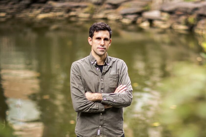
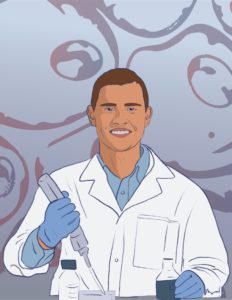
Cesar de la Fuente, Presidential Assistant Professor with appointments in the Perelman School of Medicine, School of Engineering and School of Arts & Sciences (Image: Eric Sucar)
In a significant advance against the growing threat of antibiotic-resistant bacteria, researchers have identified a novel class of antimicrobial agents known as encrypted peptides, which may expand the immune system’s arsenal of tools to fight infection. The findings, published in Trends in Biotechnology by Cell Press, reveal that many antimicrobial molecules originate from proteins not traditionally associated with immune responses.
Unlike conventional antibiotics that target specific bacterial processes, these newly discovered peptides disrupt the protective membranes surrounding bacterial cells. By inserting themselves into these membranes—much like breaching a fortress wall—the peptides destabilize and ultimately destroy the bacteria.
“Our findings suggest that these previously overlooked molecules could be key players in the immune system’s response to infection,” says César de la Fuente, presidential assistant professor in bioengineering and in chemical and biomolecular engineering in the School of Engineering and Applied Science, in psychiatry and microbiology in the Perelman School of Medicine, and in chemistry in the School of Arts & Sciences, who led the research team. “This may not only redefine how we understand immunity but also opens up new possibilities for treating drug-resistant infections.”
Lasya Sreepada has always been fascinated by the brain and the underlying biology that shapes how people develop and age. “My curiosity traces back to observing differences between myself and my sister,” says Sreepada, a Ph.D. candidate in Bioengineering whose research unites efforts across Penn Medicine and Penn Engineering. “We grew up in the same environment but had remarkably different personalities, which led me to question what drove these differences and which brought me to the brain.”
Her academic journey began by applying medical imaging to understand how brain injuries sustained by professional athletes or military veterans impact their brain structure and chemistry over time. She became curious about how neurotrauma impacts aging and degeneration in the long term. Now, she leverages large, multimodal datasets to investigate neurodegenerative disease, with a particular focus on Alzheimer’s.
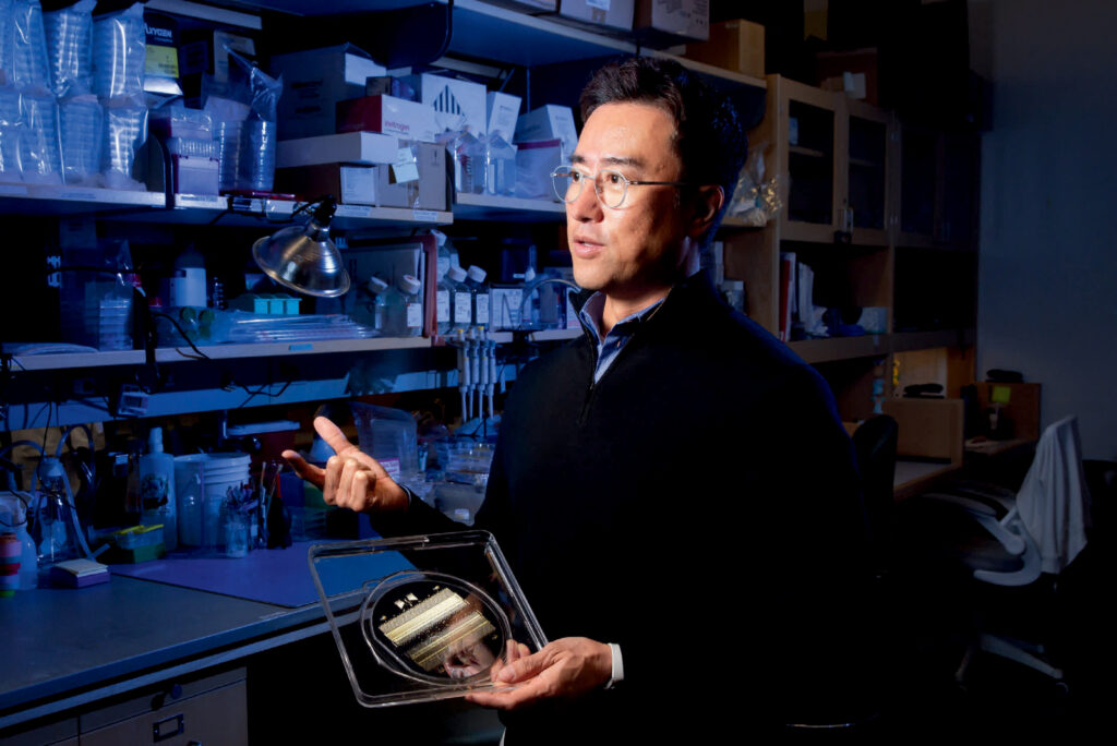
More than 34 million Americans suffer from pulmonary diseases like asthma, emphysema and chronic bronchitis. While medical treatments can keep these ailments in check, there are currently no cures. Part of the reason, notes Dan Huh, is that it’s incredibly hard to study how these diseases actually work. While researchers can grow cells taken from human lungs in a dish, they cannot expect them to act like they would in the body. In order to mimic the real deal, it’s necessary to recreate the complex, 3D environment of the lung — right down to its tiny air sacs and blood vessels — and to gently stretch and release the tissue to simulate breathing.
Huh, Associate Professor in Bioengineering, is the cofounder of Vivodyne, a Penn Engineering biotech spinoff that is creating tissues like these in the lab. Vivodyne uses a bioengineering technology that Huh has been developing for more than a decade. While a postdoctoral fellow at Harvard’s Wyss Institute, he played a central role in creating a novel device called an “organ on a chip,” which, as the name implies, assembles multiple cell types on a tiny piece of engineered plastic to create an approximation of an organ.
“While those chips represented a major innovation,” says Huh, “they still weren’t truly lifelike. They lacked many of the essential features of their counterparts in the human body, such as the network of blood vessels running between different kinds of tissue, which are essential for transporting oxygen, nutrients, waste products and various biochemical signals.”
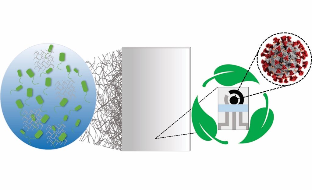
Fabrication steps of the biodegradable BC substrate and the electrochemical devices. (1) Incubation of the bacterium Gluconacetobacter hansenii. (2) BC substrate collected and treated, resulting in a clear sheet. (3) The biodegradable BC sheet is screen-printed, (4) resulting in a device with 3 electrodes, (4) which are cut out using a scissor, (5) resulting in a portable, biodegradable, and inexpensive electrochemical sensor.
The availability of rapid, accessible testing was integral to overcoming the worst surges of the COVID-19 pandemic, and will be necessary to keep up with emerging variants. However, these tests come with unfortunate costs.
Polymerase chain reaction (PCR) tests, the “gold standard” for diagnostic testing, are hampered by waste. They require significant time (results can take up to a day or more) as well as specialized equipment and labor, all of which increase costs. The sophistication of PCR tests makes them harder to tweak, and therefore slower to respond to new variants. They also carry environmental impacts. For example, most biosensor tests developed to date use printed circuit boards, or PCBs, the same materials used in computers. PCBs are difficult to recycle and slow to biodegrade, using large amounts of metal, plastic and non-eco-friendly materials.
In addition, most PCR tests end up in landfills, resulting in material waste and secondary contamination. An analysis by the World Health Organization (WHO) estimated that, as of February 2022, “over 140 million test kits, with a potential to generate 2,600 tonnes of non-infectious waste (mainly plastic) and 731,000 litres of chemical waste (equivalent to one-third of an Olympic-size swimming pool) have been shipped.”
In order to balance the need for fast, affordable and accurate testing while addressing these environmental concerns, César de la Fuente, Presidential Assistant Professor in Bioengineering and Chemical and Biomolecular Engineering in the School of Engineering and Applied Science, with additional primary appointments in Psychiatry and Microbiology within the Perelman School of Medicine, has turned his attention to the urgent need for “green” testing materials.
The de la Fuente lab has been working on creative ways to create faster and cheaper testing for COVID-19 since the outbreak of the pandemic. Utilizing his lab’s focus on machine biology and the treatment of infectious disease, they created RAPID, an aptly named test that generates results in minutes with a high degree of accuracy. An even more cost-effective version, called LEAD, was created using electrodes made from graphite. A third test, called COLOR, was a low-cost optodiagnostic test printed on cotton swabs.
The team’s latest innovation incorporates the speed and cost-effectiveness of previous tests with eco-friendly materials. In a paper published in Cell Reports Physical Science, the group introduces a new test made from Bacterial Cellulose (BC), an organic compound synthesized from several strains of bacteria, as a substitute for PCBs.
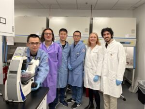
Members of the research team include (from left to right) Xuexiang Han, Michael J. Mitchell, Ningqiang Gong, Lulu Xue, Sarah J. Shepherd, and Rakan El-Mayta.
Since the success of the COVID-19 vaccine, RNA therapies have been the object of increasing interest in the biotech world. These therapies work with your body to target the genetic root of diseases and infections, a promising alternative treatment method to that of traditional pharmaceutical drugs.
Lipid nanoparticles (LNPs) have been successfully used in drug delivery for decades. FDA-approved therapies use them as vehicles for delivering messenger RNA (mRNA), which prompts the cell to make new proteins, and small interfering RNA (siRNA), which instruct the cell to silence or inhibit the expression of certain proteins.
The biggest challenge in developing a successful RNA therapy is its targeted delivery. Research is now confronting the current limitations of LNPs, which have left many diseases without an effective RNA therapy.
Liver fibrosis occurs when the liver is repeatedly damaged and the healing process results in the accumulation of scar tissue, impeding healthy liver function. It is a chronic disease characterized by the buildup of excessive collagen-rich extracellular matrix (ECM). Liver fibrosis has remained challenging to treat using RNA therapies due to a lack of delivery systems for targeting activated liver-resident fibroblasts. Both the solid fibroblast structure and the lack of specificity or affinity to target these fibroblasts has impeded current LNPs from entering activated liver-resident fibroblasts, and thus they are unable to deliver RNA therapeutics.
To tackle this issue and help provide a treatment for the millions of people who suffer from this chronic disease, Michael Mitchell, J. Peter and Geri Skirkanich Assistant Professor of Innovation in the Department of Bioengineering, and postdoctoral fellows Xuexiang Han and Ningqiang Gong, found a new way to synthesize ligand-tethered LNPs, increasing their selectivity and allowing them to target liver fibroblasts.
Lulu Xue, Margaret Billingsley, Rakan El-Mayta, Sarah J. Shepherd, Mohamad-Gabriel Alameh and Drew Weissman, Roberts Family Professor in Vaccine Research and Director of the Penn Institute for RNA Innovation at the Perelman School of Medicine, also contributed to this work.
Cynthia Reinhart-King, Cornelius Vanderbilt Professor of Engineering and Professor of Biomedical Engineering at Vanderbilt University, was one of a handful of experts invited to take part in the White House Summit in Biotechnology and Biomanufacturing on September 14, 2022 in Washington, D.C. Reinhart-King and her colleagues gathered to discuss “bio-based solutions to global challenges ranging from food security and climate change to health security and supply chain disruptions.”
Therapies that use engineered cells to treat diseases, infections and chronic illnesses are opening doors to solutions for longstanding medical challenges. Lukasz Bugaj, Assistant Professor in Bioengineering, has been awarded a National Science Foundation CAREER Award for research that may be key to opening some of those doors.
Such cellular therapies take advantage of the complex molecular mechanisms that cells naturally use to interact with one another, enabling them to be more precise and less toxic than traditional pharmaceutical drugs, which are based on simpler small molecules. Cellular therapies that use engineered immune system cells, for example, have recently been shown to be highly successful in treating certain cancers and protecting against viral infections.
However, there is still a need to further fine-tune the behavior of cells in these targeted therapies. Bugaj and colleagues are addressing that need by developing new ways to communicate with engineered cells once they are in the body, such as turning molecular events on and off at specific times.
The research team recently discovered that both temperature and light can act as triggers of a specific fungal protein, dynamically controlling its location within a mammalian cell. By using light or temperature to instruct that protein to migrate toward or away from the cell’s membrane, Bugaj and his colleagues showed how it could serve as a key component in controlling the behavior of human cells.
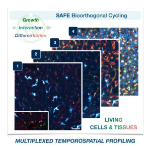
Cells in complex organisms undergo frequent changes, and researchers have struggled to monitor these changes and create a comprehensive profile for living cells and tissues. Historically researchers have been limited to only 3-5 markers due to spectral overlaps in fluorescence microscopy, an essential tool required for imaging cells. With only this small handful of markers, it is difficult to monitor protein expressions of live cells and a comprehensive profile of cellular dynamics cannot be created. However, a new study in Nature Biotechnology addresses these limitations by demonstrating a new method for comprehensive profiling of living cells.
Jina Ko, PhD
Jina Ko, Assistant Professor in Bioengineering in the School of Engineering and Applied Science and in Pathology and Laboratory Medicine in the Perelman School of Medicine, conducted postdoctoral research at Massachusetts General Hospital (MGH) and the Wyss Institute at Harvard University, and the work for this study was done under the supervision of Jonathan Carlson M.D., Ph.D. and Ralph Weissleder M.D., Ph.D. of MGH. Ko’s lab at Penn develops novel technologies using bioengineering, molecular biology, and chemistry to address diagnostic challenges for precision medicine.
To address these limitations in microscopy, the team developed a new chemistry tool which was highly gentle to cells. This “scission-accelerated fluorophore exchange (or SAFE)” method utilizes “click” chemistry, a type of chemistry that follows examples found in nature to create fast and simple reactions. This new SAFE method functions with non-toxic conditions to living cells and tissues, whereas previous methods have used harsh chemicals that would strip off fluorophores and consequently would not work with living cells and tissues.
With the development of SAFE, the authors demonstrated that researchers can now effectively perform multiple cycles of cell profiling and can monitor cellular changes over the course of their observations. Instead of the previous limitation of 3-5 markers total, SAFE allows for many more cycles and can keep track of almost as many markers as the researcher wants. One can now stain cells and quench/release fluorophores and repeat the cycle multiple times for multiplexing on living cells. Each cycle can profile 3 markers, and so someone interested in profiling 15 markers could easily perform 5 cycles to achieve this much more comprehensive cell profile. With this breakthrough in more detailed imaging of cells, SAFE demonstrates broad applicability for allowing researchers to better investigate the physiologic dynamics in living systems.
This study was supported by the Schmidt Science Fellows in Partnership with the Rhodes Trust and National Institutes of Health, National Cancer Institute (K99CA256353).
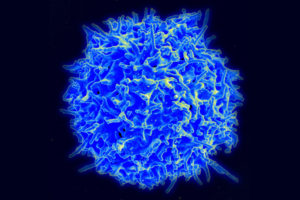
A scanning electron micrograph of a healthy human T cell. A better understanding the wide variety of antigen receptors that appear on the surfaces of these critical components of the immune system is necessary for improving a new class of therapies. (Credit: NIAID)
Our bodies are equipped with specialized white blood cells that protect us from foreign invaders, such as viruses and bacteria. These T cells identify threats using antigen receptors, proteins expressed on the surface of individual T cells that recognize specific amino acid sequences found in or on those invaders. Once a T cell’s antigen receptors bind to the corresponding antigen, it can directly kill infected cells or call for backup from the rest of the immune system.
We have hundreds of billions of T cells, each with unique receptors that recognize unique antigens, so profiling this T cell antigen specificity is essential in our understanding of the immune response. It is especially critical in developing targeted immunotherapies, which equip T cells with custom antigen receptors that recognize threats they would otherwise miss, such as the body’s own mutated cancer cells.
Jenny Jiang, Ph.D.
Jenny Jiang, Peter and Geri Skirkanich Associate Professor of Innovation in Bioengineering, along with lab members and colleagues at the University of Texas, Austin, recently published a study in Nature Immunology that describes their technology, which simultaneously provides information in four dimensions of T cell profiling. Ke-Yue Ma and Yu-Wan Guo, a former post doc and current graduate student in Jiang’s Penn Engineering lab, respectively, also contributed to this study.
This technology, called TetTCR-SeqHD, is the first to provide such detailed information about single T cells in a high-throughput manner, opening doors for personalized immune diagnostics and immunotherapy development.
There are many pieces of information needed to comprehensively understand the immune response of T cells, and gathering all of these measurements simultaneously has been a challenge in the field. Comprehensive profiling of T cells includes sequencing the antigen receptors, understanding how specific those receptors are in their recognition of invading antigens, and understanding T cell gene and protein expression. Current technologies only screen for one or two of these dimensions due to various constraints.
“Current technologies that measure T cell immune response all have limitations,” says Jiang. “Those that use cultured or engineered T cells cannot tell us about their original phenotype, because once you take a cell out of the body to culture, its gene and protein expression will change. The technologies that address T cell and antigen sequencing with mass spectrometry damage genetic information of the sample. And current technologies that do provide information on antigen specificity use a very expensive binding ligand that can cost more than a thousand dollars per antigen, so it is not feasible if we want to look at hundreds of antigens. There is clearly room for advancement here.”
The TetTCR-SeqHD technology combines Jiang’s previously developed T cell receptor sequencing tool, TetTCR-Seq, described in a Nature Biotechnology paper published in 2018, with the new ability of characterizing both gene and protein expression.
NextUp, a regular feature of Philadelphia Magazine that “highlights the local leaders, organizations and research shaping the Greater Philadelphia region’s life sciences ecosystem,” ran a profile of Philly-based agricultural startup Strella Biotechnology. Founded by Penn alumna Katherine Sizov (Bio 2019) and winner of a 2019 President’s Innovation Prize, Strella Biotech seeks to reduce food waste through innovative biosensors, and was initially developed in the George H. Stephenson Foundation Educational Laboratory, the biomakerspace and primary teaching lab of the Department of Bioengineering.
Sizov says the coronavirus pandemic has made the volatility of grocery stores’ offerings even more apparent. Last April, the Produce Marketing Association estimated that nearly $5 billion of fresh fruits and vegetables had gone to waste in the first month of the pandemic due to the complex supply chain’s inability to quickly redirect shipping and distribution. ‘In a way, I think COVID-19 has helped us realize how delicate and fragile supply chains are,’ she says. ‘We are working to create better, stronger supply chains that are economically and environmentally sustainable for everyone involved — researchers, growers, packagers, distributors, retailers, and consumers.'”


 NextUp, a regular feature of Philadelphia Magazine that “highlights the local leaders, organizations and research shaping the Greater Philadelphia region’s life sciences ecosystem,” ran a profile of Philly-based agricultural startup Strella Biotechnology. Founded by Penn alumna Katherine Sizov (Bio 2019) and winner of a 2019 President’s Innovation Prize, Strella Biotech seeks to reduce food waste through innovative biosensors, and was initially developed in the