Penn Health-Tech director Katie Reuther (center) with Glory Durham, director of operations, Penn Health-Tech (at left), and Courtney Houtsma, program manager, Penn Health-Tech (at right), at a recent symposium.
A new interview in Penn Medicine News examines Penn Health-Tech (PHT) five years after its founding. PHT began as an experimental collaborative effort between the Perelman School of Medicine, the School of Engineering and Applied Science, and the Office of the Vice Provost for Research to provide funding, advising, and resources to empower innovators to develop transformative devices and technologies in the Penn community. Specifically, PHT specializes in connecting innovators from across Penn’s campus and schools to connect and to develop technology and medical devices to answer some of the most pressing needs in healthcare. Katherine (Katie) Reuther, Practice Associate Professor in Bioengineering, was appointed Executive Director of PHT in 2021 and is leading this venture into the next phase of its growth. Reuther, an alumna of Penn Bioengineering, followed up her doctoral studies with a M.B.A. from Columbia University and subsequently stayed at Columbia as Senior Lecturer in Design, Innovation, and Entrepreneurship in the Department of Biomedical Engineering. As such, her experience and expertise in the fields of both biomedical engineering and entrepreneurship position her well to shepherd PHT into its fullest potential:
“What appealed to me most about the position was a strong foundation, deep resources, and the potential and room to do more, including the opportunity to elevate Penn and Philadelphia as a national hub for health-technology innovation.”
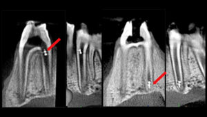
Controlled and actuated by magnetic fields, these mircrorobots are capable of precisely targeting the apical region — the opening where blood vessels and nerve enter the tooth — in a root canal.
With its irregularities and anatomical complexities, the root canal system is one of the most clinically challenging spaces in the oral cavity. As a result, biofilm not fully cleared from the nooks and crannies of the canals remains a leading cause of treatment failure and persistent endodontic infections, and there are limited means to diagnose or assess the efficacy of disinfection. One day, clinicians may have a new tool to overcome these challenges in the form of microrobots.
In a proof-of-concept study, researchers from Penn Dental Medicine and its Center for Innovation & Precision Dentistry (CiPD), have shown that microrobots can access the difficult to reach surfaces of the root canal with controlled precision, treating and disrupting biofilms and even retrieving samples for diagnostics, enabling a more personalized treatment plan. The Penn team shared their findings on the use of two different microrobotic platforms for endodontic therapy in the August issue of the Journal of Dental Research;the work was selected for the issue’s cover.
“The technology could enable multimodal functionalities to achieve controlled, precision targeting of biofilms in hard-to-reach spaces, obtain microbiological samples, and perform targeted drug delivery, ” says Dr. Alaa Babeer, lead author of the study and a Penn Dental Medicine Doctor of Science in Dentistry (DScD) and endodontics graduate, who is now within the lab of Dr. Michel Koo, co-director of the CiPD .
In both platforms, the building blocks for the microrobots are iron oxide nanoparticles (NPs) that have both catalytic and magnetic activity and have been FDA approved for other uses. In the first platform, a magnetic field is used to concentrate the NPs in aggregated microswarms and magnetically control them to the apical area of the tooth to disrupt and retrieve biofilms through a catalytic reaction. The second platform uses 3D printing to create miniaturized helix-shaped robots embedded with iron oxide NPs. These helicoids are guided by magnetic fields to move within the root canal, transporting bioactives or drugs that can be released on site.
“This technology offers the potential to advance clinical care on a variety of levels,” says Dr. Koo, co-corresponding author of the study with Dr. Edward Steager, a senior research investigator in Penn’s School of Engineering and Applied Science. “One important aspect is the ability to have diagnostic as well as therapeutic applications. In the microswarm platform, we can not only remove the biofilm, but also retrieve it, enabling us identify what microorganisms caused the infection. In addition, the ability to conform to the narrow and difficult-to-reach spaces within the root canal allows for a more effective disinfection in comparison to the files and instrumentation techniques presently used.”
Michel Koo is a professor in the Department of Orthodontics and divisions of Community Oral Health and Pediatric Dentistry in Penn Dental Medicine and co-director of the Center for Innovation & Precision Dentistry. He is a member of the Penn Bioengineering Graduate Group.
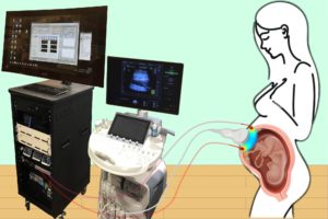
Combining optical measurements with ultrasound, an interdisciplinary team from the School of Arts & Sciences, Perelman School of Medicine, and CHOP developed a device to better measure blood flow and oxygenation in the placenta. (Image: Lin Wang)
A study published in Nature Biomedical Engineering details a novel method for imaging the placenta in pregnant patients as well as the results of a pilot clinical study. By combining optical measurements with ultrasound, the findings show how oxygen levels can be monitored noninvasively and provides a new way to generate a better understanding of this complex, crucial organ. This research was the result of a collaboration of the groups of the University of Pennsylvania’s Arjun Yodh and Nadav Schwartz with colleagues from the Children’s Hospital of Philadelphia (CHOP) and was led by postdoc Lin Wang.
Schwartz describes the placenta as the “engine” of pregnancy, an organ that plays a crucial role in delivering nutrients and oxygen to the fetus. Placental dysfunction can lead to complications such as fetal growth restriction, preeclampsia, and stillbirth. To increase knowledge about this crucial organ, the National Institute of Child Health and Human Development launched the Human Placenta Project in 2014. One focus of the program is to develop tools to assess human placental structure and function in real time, including optical devices.
For three years, the researchers optimized the design of their instrument and tested it in preclinical settings. The process involved integrating optical fibers with ultrasound probes, exploring various ultrasound transducers, and improving the multimodal technology so that measurements were stable, accurate, and reproducible while collecting data at the bedside. The resulting instrumentation now enables researchers to study the anatomy of the placenta while also collecting detailed functional information about placenta blood flow and oxygenation, capabilities that existing commercially devices do not have, the researchers say.
Because the placenta is located far below the body’s surface, one of the key technical challenges addressed by Wang, a postdoc in Yodh’s lab, was reducing background noise in the opto-electronic system. Light is scattered and absorbed when it travels through thick tissues, Yodh says, and the key for success was to reduce background interference so that the small amount of light that penetrates deep into the placenta and then returns is still large enough for a high-quality measurement.
“We’re sending a light signal that goes through the same deep tissues as the ultrasound. The extremely small amount of light that returns to the surface probe is then used to accurately assess tissue properties, which is only possible with very stable lasers, optics, and detectors,” says Yodh. “Lin had to overcome many barriers to improve the signal-to-noise ratio to the point where we trusted our data.”
The authors are Lin Wang, Jeffrey M. Cochran, Kenneth Abramson, Lian He, Venki Kavuri, Samuel Parry, Arjun G. Yodh, and Nadav Schwartz from Penn; Tiffany Ko, Wesley B. Baker, and Rebecca L. Linn from the Children’s Hospital of Philadelphia, and David R. Busch, previously a research associate at Penn and now at the University of Texas Southwestern Medical School.
This research was supported by National Institutes of Health grants F31HD085731, R01NS113945, R01NS060653, P41EB015893, P41EB015893, T32HL007915, and U01HD087180.
Though the technology for brain-computer interfaces (or BCI’s) has existed for decades, recent strides have been made to create BCI devices which are safer, smaller, and more effective. Konrad Kording, Nathan Francis Mossell University Professor in Bioengineering, Neuroscience, and Computer and Information Science, helps to elucidate the potential future of this technology in a recent feature in Wired. In the article, he discusses the “invasive” aspects of previous BCI technology, in contrast to recent innovations, such as a new device by Synchron, which do not require surgery and are consequently much less risky:
“The device, called a Stentrode, has a mesh-like design and is about the length of a AAA battery. It is implanted endovascularly, meaning it’s placed into a blood vessel in the brain, in the region known as the motor cortex, which controls movement. Insertion involves cutting into the jugular vein in the neck, snaking a catheter in, and feeding the device through it all the way up into the brain, where, when the catheter is removed, it opens up like a flower and nestles itself into the blood vessel’s wall. Most neurosurgeons are already up to speed on the basic approach required to put it in, which reduces a high-risk surgery to a procedure that could send the patient home the very same day. ‘And that is the big innovation,” Kording says.
Congratulations to Kevin B. Johnson, David L. Cohen University Professor, on his recent appointed as a Senior Fellow in the Leonard Davis Institute of Health Economics at the University of Pennsylvania (Penn LDI). Johnson, an expert in health care innovation and health information technology, holds appointments in Biostatistics, Epidemiology and Informatics in the Perelman School of Medicine and Computer and Information Science in the School of Engineering and Applied Science. He also holds secondary appointments in Bioengineering, Pediatrics, and in the Annenberg School of Communication and is Vice President for Applied Informatics in the University of Pennsylvania Health System.
Penn LDI is Penn’s hub for health care delivery, health policy, and population health, we connect and amplify experts and thought-leaders and train the next generation of researchers. Johnson joins over 500 Fellows from across all of Penn’s schools, the University of Pennsylvania Health System, and the Children’s Hospital of Philadelphia. Johnson brings expertise in Health Care Innovation, Health Information Technology, Medication Adherence, and Social Media to his new fellowship and has extensively studied healthcare informatics with the goal of improving patient care.
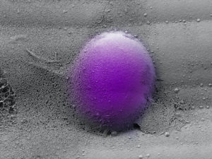
Acollaborative team developed an alginate-based hydrogel system that mimics the viscoelasticity of the natural extracellular matrix in bone marrow. By tweaking the balance between elastic and viscous properties in these artificial ECMs, they could recapitulate the viscoelasticity of healthy and scarred fibrotic bone marrow, and study the effects on human monocytes placed into these artificial ECMs. (Image: Adam Graham/Harvard CNS/Wyss Institute at Harvard University)
Fibrosis is the thickening of various tissues caused by the deposition of fibrillar extracellular matrix (ECM) in tissues and organs as part of the body’s wound healing response to various forms of damage. When accompanied by chronic inflammation, fibrosis can go into overdrive and produce excess scar tissue that can no longer be degraded. This process causes many diseases in multiple organs, including lung fibrosis induced by smoking or asbestos, liver fibrosis induced by alcohol abuse, and heart fibrosis often following heart attacks. Fibrosis can also occur in the bone marrow, the spongy tissue inside some bones that houses blood-producing hematopoietic stem cells (HSCs) and can lead to scarring and the disruption of normal functions.
Chronic blood cancers known as “myeloproliferative neoplasms” (MPNs) are one example, in which patients can develop fibrotic bone marrow, or myelofibrosis, that disrupts the normal production of blood cells. Monocytes, a type of white blood cell belonging to the group of myeloid cells, are overproduced from HSCs in neoplasms and contribute to the inflammation in the bone marrow environment, or niche. However, how the fibrotic bone marrow niche itself impacts the function of monocytes and inflammation in the bone marrow was unknown.
Now, a collaborative team from Penn, Harvard, the Dana-Farber Cancer Institute (DFCI), and Brigham and Women’s Hospital has created a programmable hydrogel-based in vitro model mimicking healthy and fibrotic human bone marrow. Combining this system with mouse in vivo models of myelofibrosis, the researchers demonstrated that monocytes decide whether to enter a pro-inflammatory state and go on to differentiate into inflammatory dendritic cells based on specific mechanical properties of the bone marrow niche with its densely packed ECM molecules. Importantly, the team found a drug that could tone down these pathological mechanical effects on monocytes, reducing their numbers as well as the numbers of inflammatory myeloid cells in mice with myelofibrosis. The findings are published in Nature Materials.
“We found that stiff and more elastic slow-relaxing artificial ECMs induced immature monocytes to differentiate into monocytes with a pro-inflammatory program strongly resembling that of monocytes in myelofibrosis patients, and the monocytes to differentiate further into inflammatory dendritic cells,” says co-first author Kyle Vining, who recently joined Penn’s School of Dental Medicine and School of Engineering and Applied Science as an assistant professor of preventive and restorative sciences. “More viscous fast-relaxing artificial ECMs suppressed this myelofibrosis-like effect on monocytes. This opened up the possibility of a mechanical checkpoint that could be disrupted in myelofibrotic bone marrow and also may be at play in other fibrotic diseases.”
Vining worked on the study as a postdoctoral fellow at Harvard in the lab of David Mooney. “Our study shows that the differentiation state of monocytes, which are key players in the immune system, is highly regulated by mechanical changes in the ECM they encounter,” says Mooney, who co-led the study with DFCI researcher Kai Wucherpfennig. “Specifically, the ECM’s viscoelasticity has been a historically under-appreciated aspect of its mechanical properties that we find correlates strongly between our in vitro and the in vivo models and human disease. It turns out that myelofibrosis is a mechano-related disease that could be treated by interfering with the mechanical signaling in bone marrow cells.”
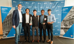
(From left to right) Startup Challenge sponsor Eric Aroesty with members of Toxisense: Aravind Krishnan, Udit Garg, Andrew Diep-Tran, and Aarush Sahni. (Image: The Wharton School)
Penn’s Venture LabStartup Challenge awarded its 2022 prize to a sustainable and cost-effective water-testing startup. The venture, ToxiSense, was awarded at a ceremony on April 29, at Tangen Hall, Penn’s hub for student entrepreneurship and innovation.
Co-founded by four first-year students—Aravind Krishnan, Udit Garg, Andrew Diep-Tran, and Aarush Sahni—ToxiSense aims to improve the endotoxin testing required for drinking water and biopharma products through genetically engineering plants with bioluminescent properties. Biopharmaceutical products and drinking water must be tested for endotoxins, the sickness-causing molecule from bacteria. The current method relies on expensive horseshoe crab blood and is environmentally damaging. ToxiSense genetically engineered the Arabidopsis plant to luminesce based on the endotoxin concentration applied to it, serving as a sustainable, cost-effective solution.
ToxiSense was selected from a field of eight finalist teams—including DeToXyFi, Groov, Impact Local, Miren, Nemu, Ossum Technologies, and Shinkei Systems Corp.—who advanced from 30 ventures during the semi-finals portion of the competition, which consisted of a day of virtual pitching and Q&A in front of alumni entrepreneur and investor panels. For the finals, teams pitched to a panel of alumni judges and in front of a live audience of nearly 200 attendees as they competed for over $150,000 in cash and prizes to launch their startups.
“The Startup Challenge is Venture Lab’s premier yearly event, showcasing Penn’s most promising teams of student entrepreneurs,” says Lori Rosenkopf, vice dean of entrepreneurship and Simon and Midge Palley Professor at the Wharton School. “This year’s finalists included undergraduate and graduate students from across the University, and their products offered solutions for environmental, financial, health, and social challenges. These motivated teams capture the spirit of Penn entrepreneurship—innovative, interdisciplinary, inclusive—and we offer our congratulations and our optimistic wishes for their futures.”
Brian Litt, MD, Professor in Neurology, Neurosurgery and Bioengineering and Director of the Penn Epilepsy Center, has received a 2022 Landis Award for Outstanding Mentorship from the National Institute of Neurological Disorders and Stroke (NINDS). This award honors Litt’s dedication to superior mentorship and training in neuroscience research. The award includes $100,000 in the form of a supplement to an existing NINDS grant to support his efforts to foster the career advancement of additional trainees.
Buffone got his Ph.D. in Chemical Engineering from SUNY Buffalo in Buffalo, NY in 2012, working with advisor Sriram Neelamegham, Professor of Chemical and Biological Engineering. Buffone completed previous postdoctoral studies at Roswell Park Comprehensive Cancer Center with Joseph T.Y. Lau, Distinguished Professor of Oncology in the department of Cellular and Molecular Biology. Upon coming to Penn in 2015, Buffone has worked in the Hammer Lab under advisor Daniel A. Hammer, Alfred G. and Meta A. Ennis Professor in Bioengineering and in Chemical and Biomolecular Engineering, first as a postdoc and later a research associate. Buffone also spent a year as a Visiting Scholar in the Center for Bioengineering and Tissue Regeneration, directed by Valerie M. Weaver, Professor at the University of California, San Francisco in 2019.
While at Penn, Buffone was a co-investigator on an R21 grant through the National Institutes of Health (NIH) which supported his time as a research associate. Buffone is excited to start his own laboratory where he plans to train a diverse set of trainees.
Buffone’s research area lies at the intersection of genetic engineering, immunology, and glycobiology and addresses how to specifically tailor the trafficking and response of immune cells to inflammation and various diseases. The work seeks to identify and subsequently modify critical cell surface and intracellular signaling molecules governing the recruitment of various blood cell types to distal sites. The ultimate goal of his research is to tailor and personalize the innate and adaptive immune response to specific diseases on demand.
“None of this would have been possible without the unwavering support of all of my mentors, past and present, and most especially Dan Hammer,” Buffone says. “His support in helping me transition into an independent scientist and his understanding of my outside responsibilities as a dad with two young children is truly the reason why I am standing here today. It’s a testament to Dan as both a person and a mentor.”
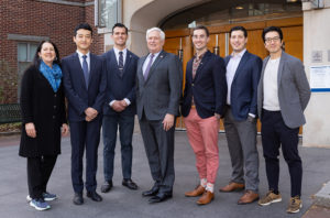
The inaugural class of the CiPD NIDCR T90/R90 Postdoctoral Training Program Fellows with Dean Mark Wolff (center); Dr. Michel Koo, Founding Director of CiPD (far right); and CiPD Co-Director Dr. Kathleen Stebe of Penn’s School of Engineering and Applied Science (far left).
With one of its key missions to develop a new generation of scientists at the interface of dental medicine and engineering, the Center for Innovation & Precision Dentistry (CiPD) has selected its inaugural class of fellows for its new postdoctoral training program.
The CiPD was awarded a $2.5 million T90/R90 grant from the National Institute of Dental and Craniofacial Research (NIDCR) last summer to establish the program, recently naming this first cohort of fellows that includes Justin Burrell, Marshall Padilla, Zhi Ren, and Dennis Sourvanos.
“We’re hoping this program will promote cross-pollination and create a culture between these two fields to help dentists develop innovative strategies with engineers,” says Penn Dental Medicine’s Michel Koo, Co-Director of CiPD, who launched the Center in 2021 with Co-Director Kathleen Stebe, Richer & Elizabeth Goodwin Professor in Penn Engineering’s Department of Chemical and Biomolecular Engineering. “Dentists can learn from engineering principles and tools, and engineers can understand more about the needs of the dental and craniofacial fields. We’re providing a platform for them to work together to address unmet clinical needs and develop careers in that interface.”
The NIDCR T90/R90 Postdoctoral Training Program aims to specifically focus on the oral microbiome, host immunity, and tissue regeneration, each of which ties into different aspects of oral health, from tooth decay and periodontal disease to the needs of head and neck cancer patients. To advance these areas, emerging approaches, from advanced materials, robotics, and artificial intelligence to tissue engineering, chloroplast- and nanoparticle-based technologies, will be leveraged.
As part of the two-year training, each postdoc will receive co-mentorship from faculty from each school in conjunction with a career development committee of clinicians, basic scientists, as well as engineers. These mentorships will be focused on research outcomes and readying participants to submit grants and compete for positions in academia or industry.
The inaugural class of fellows includes Justin Burrell, a postdoctoral student in the lab of D. Kacy Cullen, Associate Professor of Neurosurgery; Marshall Padilla, a postdoc in the lab of Michael J. Mitchell, Skirkanich Assistant Professor of Innovation in Bioengineering; and Zhi Ren, a postdoc in the lab of Michael Koo; and Dennis Sourvanos, an Advanced Graduate Dental Education resident at Penn Dental Medicine whose research has been co-directed by Timothy C. Zhu, Professor of Radiation Oncology in the Perelman School of Medicine. Cullen, Mitchell, Koo and Zhu are all members of the Penn Bioengineering Graduate Group.