Week in BioE (October 13, 2017)

Seeing and Repairing Damaged Heart Vessels

angiography
Immunofluorescent staining for F-actin filaments (green) and nuclei (blue) in neonatal cardiomyocyte

Two common diagnostic procedures in cardiology are intravascular ultrasound and cardiac angiography. These procedures are performed to quantify the amount of plaque affecting a patient’s blood vessels. This information is vital because it helps to determine how advanced heart degree is, as well as guiding treatment planning and even the course of bypass surgery. However, the current technologies used for these procedures have significant limitations. Although conventional angiography can help to quantify the plaque burden, it does not offer any information about how much of the diameter of a vessel is blocked. Intravascular ultrasound is very good at quantifying plaque burden, but it is poor at identifying smaller features of compromised blood vessels.

One solution suggested to these issues is the combination of these imaging technologies into a single multimodal technique. Scientists led by Laura Marcu, Ph.D., professor of biomedical engineering at the University of California, Davis, invented a method combining intravascular ultrasound with multispectral fluorescence lifetime imaging (FLIM). As published in Scientific Reports, the device resembles a typical cardiac catheter but contains an optical fiber within the catheter that emits fluorescent light to characterize the plaque components before treatment.

Dr. Marcu and her colleagues tested their new device in live pigs and in human coronary arteries obtained from cadavers. The fluorescence data acquired with the device were comparable to those acquired with traditional fluorescence angiography. Moreover, the device could acquire data without having to administer a contrast agent, which can be dangerous in some patients due to allergies or weakened kidneys. The authors are currently seeking FDA approval to test their combined catheter in humans.

In addition to treating vessels before a heart attack can occur, there is new work showing how to efficiently repair heart tissue after a heart attack. A team of scientists collaborating among Clemson University, the Medical University of South Carolina,  the University of South Carolina, and the University of Chicago has received a $1.5 million grant from the National Institutes of Health to examine a treatment that combines stem cells with nanowires. The principal investigator on the grant is Ying Mei, Ph.D., who is assistant professor of bioengineering at Clemson. Dr. Mei’s team mixes stem cells with nanowires so that they form spheroids that are larger than single cells and thus less likely to wash away. In addition, the investigators hope that the spheroids will mitigate the issue of the transplanted cells and the recipient’s heart beating at different rhythms.  If successful, the group’s treatment paradigm could be a major step forward in stem cell therapies and cardiology.

Look, Up in the Sky!

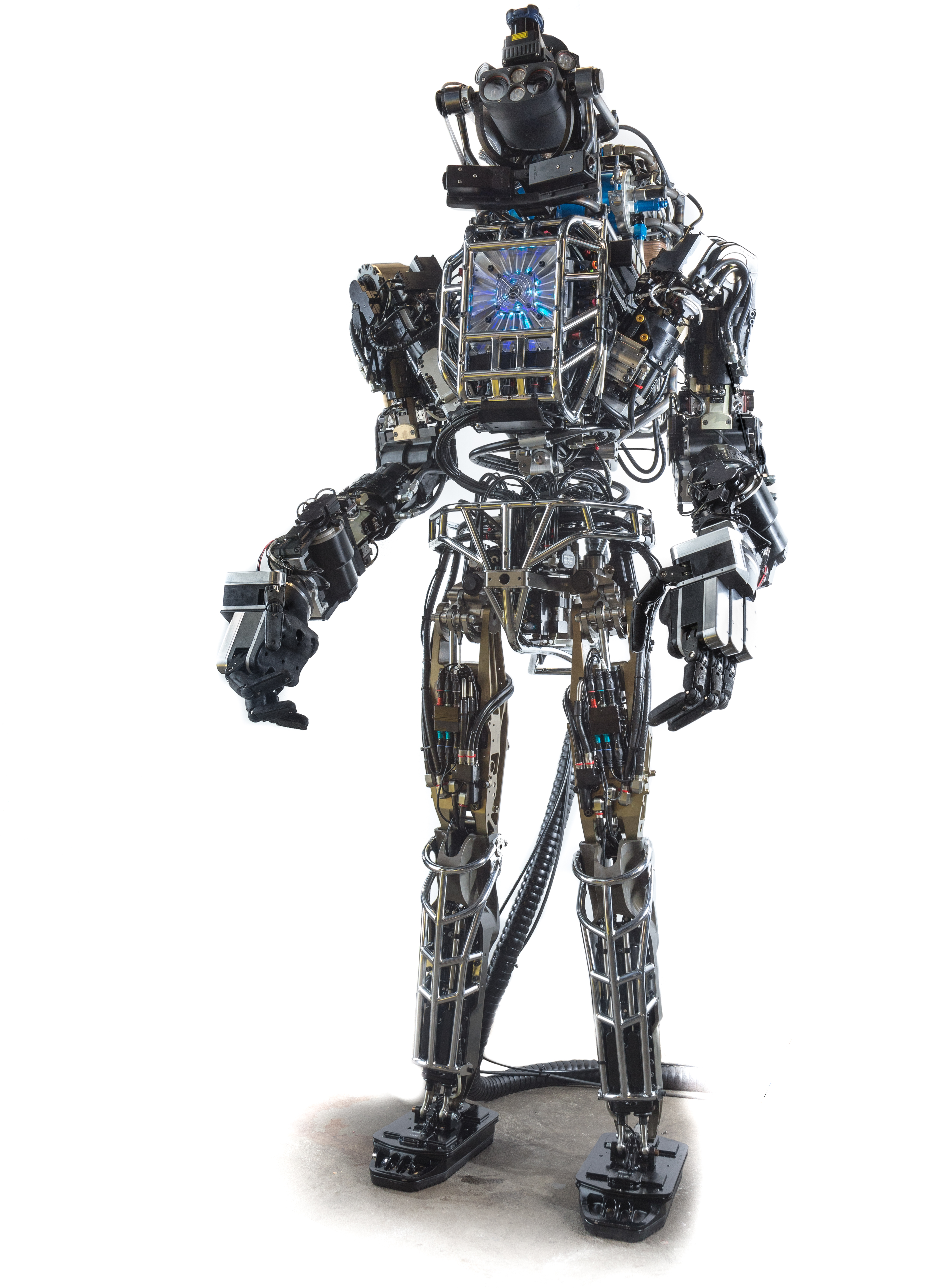
Drones became famous when deployed on battlefields for the first time a decade ago. Since then, they’ve been adopted as a technology for a variety of purposes. For example, Amazon introduced delivery drones almost a year ago, and it has plans to expand its drone fleet enormously in coming years. It was only a matter of time before engineers began to imagine medical applications for drones.

Engineers in Australia and Iraq recently investigated whether a drone could be used to monitor cardiorespiratory signals remotely. They reported their findings in BioMedical Engineering OnLine. The authors used imaging photoplethysmography (PPG), which employs a video camera to detect visual indications on the skin of heart activity. They also applied advanced digital processing technology due to the tendency of PPG to be affected by sound and movement in the area of detection. By testing the combined technologies in 15 healthy volunteers, the authors found that their data compared well with several traditional techniques for monitoring vital signs. Among the possible applications that the authors imagine for this technology is battlefield triage performed remotely using drones. In the meantime, they will seek to fine-tune the technology’s abilities.

Concussion Distressingly Common

A research letter published in a recent issue of JAMA reports that a study conducted in Canada found that one in five adolescents sustained a concussion on at least one occasion. Of the approximately 20% of the study respondents who had experienced concussions, one quarter had suffered more than one. The letter is particularly relevant to the United States because of the similar popularity in Canada of contact and semicontact sports such as ice hockey and football. In addition, the study included more than 13,000 teenagers, lending significantly reliability to the conclusions.

Ending the Time of Cholera

Although largely eradicated in the developed world, cholera remains a major public health issue in the Global South and other parts of the developing world. The disease is a bacterial infection that causes severe gastrointestinal distress. Because the disease is transmitted via water, effective public sanitation is a core requirement of an effective prevention campaign.

One technology being deployed in this fight is a smartphone microfluidics platform that can determine the presence of the pathogen that causes cholera in a sample and report the data almost immediately to public health authorities. This technology was produced by a company called PathVis, which was spun off at Purdue University based on science produced the laboratories of Tamara Kinzer-Ursem, Ph.D., and Jacqueline Linnes, Ph.D., both of whom are assistant professors in Purdue’s Weldon School of Biomedical Engineering. There are plans to test PathVis in Haiti and to expand it to detect other diseases in the future.

The Latest on CRISPR

CRISPR/Cas9 is the biggest bioengineering story to come along in some time — certainly the biggest in genetic engineering. But the mere fact that it’s here and already being used in animals and in human cell lines doesn’t mean that the story is over.  For instance, the Cas9 protein, which CRISPR deploys as part of its gene editing process, is currently developed most often using a viral vector. However, this system of delivery has certain drawbacks, not the least of which is a host immune system response when levels of the deployed viral vector reach the levels necessary for CRISPR to work.

A recent study published in Nature Biomedical Engineering reports on the successful use of gold nanoparticles to deliver Cas9. The new delivery system, called CRISPR-Gold, could obviate the need to use a viral vector as part of the CRISPR induction process. So far, the authors, led by University of California, Berkeley, bioengineers Irina Conboy, Ph.D., and Niren Murthy, Ph.D., have only used CRISPR-Gold in mice, but their successful results indicate that nonviral delivery with CRISPR is possible, so CRISPR could be used for more than previously thought.


Week in BioE (October 6, 2017)

We Bleed Green

field goalBiomechanics is a subdiscipline within bioengineering with many applications that include studying how tissue forms and grows during development (see our profile of incoming faculty member Alex Hughes to learn more) and determining how the ‘imprint’ of spine-based pain can be treated with anti-inflammatory medication (see story here on work from the lab of Dr. Beth Winkelstein). Understanding and analyzing human performance are other areas of biomechanics application. On September 24, Eagles kicker Jake Elliott set a team record when he kicked a 61 yard field goal at the end of regulation time to beat the Giants. Marking the achievement, the Philadelphia Inquirer featured an interview with Dr. Chase Pfeifer, a biomedical engineer from the University of Nebraska-Lincoln (UNL), to find out what factors contribute to a successful field goal from this distance. Pfeifer has both knowledge and experience with this question; he was a backup kicker for the Florida State Seminoles as an undergraduate. In the Inquirer, Dr. Pfeifer explained to readers the biophysics behind Elliott’s record kick.

Dr. Pfeifer assures readers that there’s more to kicking a 61-yard field goal than strong muscles. He explains, “The timing of muscle activation was actually more important than muscle strength in achieving that higher foot velocity.” Four muscle groups were activated by Elliott to set his record. In addition, Pfeifer’s colleague at UNL, Professor Tim Gay of the physics department, explained to the Inquirer how the climatic conditions favored Elliott’s success. Specifically, since it was a hot day, the air was less dense, allowing for longer kick distances.

Speaking of football, a new article in Annals of Biomedical Engineering offers some hope for NFL players and the league itself in the wake of growing awareness and concern about chronic traumatic encephalopathy (CTE). The increased focus on CTE has resulted in increased research on the condition and its prevention. Real-time measurement of impact energy in head injuries has, until now, relied on measurements of the motion of the head or helmet, rather than measuring the impact energy directly.

In the Annals article, scientists from Brigham Young University, including David T. Fullwood, Ph.D., professor of mechanical engineering, report on the testing of a new sensor used to measure the impact event. The authors implanted nano-composite foam (NCF) sensors into football helmets and then submitted the helmets to two dozen drop tests. The data collected from the foam sensors were compared to two of the most widely used indices to measure head injury risk, achieving excellent correlation (>90%) with injury risk indicators. The authors intend to test newer models of the sensors in the future, as well as in vivo testing.

Breast Cancer News

Despite significant advances in diagnosis and treatment, breast cancer remains the most common cancer in women. More than 300,000 women are newly diagnosed in the United States each year. Maximizing the effectiveness of treatment involves early detection of the tumor and its metastasis. Imaging plays a key role in this process. However, early tumors are very small, making their detection quite difficult.

In response to this challenge, Zheng-Rong Lu, Ph.D., the M. Frank Rudy and Margaret Domiter Rudy Professor of Biomedical Engineering at Case Western Reserve University, has coauthored a paper recently published in Nature Communications showing that a newly developed type of molecule could be used for highly sensitive magnetic resonance imaging (MRI) to detect and stratify early breast tumors.

Dr. Lu and his colleagues engineered molecules called fullerenes, which are hollow, spherical molecules of carbon. The team embedded gadolinium, a rare earth metal that is easily detected with MR imaging, into the fullerene to create metallofullerenes. They tested these metallofullerenes both in vitro and in a mouse model to determine how well they enhanced the ability of MRI to detect tumors. Metallofullerene particles could not only distinguish cancers more effectively on MRI, but they could also distinguish more aggressive tumor types from less aggressive ones. In addition, they found that metallofullerenes rapidly cleared from the body via the kidneys, ensuring that these agents would pose minimal toxicity risk. If this technology proves effective in humans, it could significantly improve the early detection of breast cancer and, in turn, survival rates.

One possible risk for breast cancer patients is the metastasis of the cancer to other body regions.  Scientists at Cornell identified a mineral process underlying breast cancer metastasis to bones, reporting their findings in PNAS. Led by Claudia Fischbach-Teschl, Ph.D., associate professor of biomedical engineering at Cornell, the authors investigated how hydroxylapatite — a naturally occurring mineral — participates in metastasis.

Dr. Fischbach-Teschl and her colleagues used X-ray scattering and Raman spectroscopy to examine the nanostructures of hydroxylapatite in the bones of mice with and without breast cancer. They found that bones were more likely to be metastasized by primary breast tumors if the hydroxylapatite crystals in them were less mature. Before the cancer spreads to the bone, the authors found that the cancer “communicates” with these immature crystals, preparing sites in the bone to which the cancer can spread.

With 80% of metastatic breast cancer cases spreading to the bones, the discovery of the Cornell team could contribute enormously to preventing metastasis — not only of breast cancer but also of any cancer with a greater likelihood of spreading to the bones.

A Review of Glucose Monitoring Technology

Our ability to treat diabetes has consistently improved over the years, but the need for patients with the disease to monitor their blood glucose requires either multiple needle pricks or invasive insulin pumps.  However, several technologies are in development around the world to make blood glucose monitoring less cumbersome. In a new article published in Bioengineering, engineers in the United Arab Emirates offer a review of the latest technologies, including analytic methods that could streamline decisions on doses of insulin to offset high glucose levels.

People and Places

The University of California, Davis, opened its Molecular Prototyping and BioInnovation Laboratory (MPBIL) in 2014. Since then, the “biomaker lab” has stood as an example of the future of biotechnology education. In collaboration with UCD’s First-Year Seminar Program, the MPBIL has launched a student-designed pilot course that has thus far yielded three seminars. You can read more about UCD’s innovative program at their Web site.

To promote diversity in engineering, the cosmetics company L’Oréal started the L’Oréal USA For Women in Science Program, which awards Changing the Face of STEM mentoring grants. Among this year’s recipients is John Hopkins University’s Sridevi Sarma, Ph.D. Dr. Sarma, who is an associate professor of biomedical engineering, will use her grant in collaboration with the Girls Scouts of Central Maryland to promote physics education for young women. Congratulations to her!

Week in BioE (September 29, 2017)

An Immune Cell Atlas

atlas
A human B cell

The human immune system deploys a variety of cells to counteract pathogens when they enter the body. B cells are a type of white blood cell specific to particular pathogens, and they form part of the adaptive immune system. As these cells develop, the cells with the strongest reactions to antigens are favored over others. This process is called clonal selection. Given the sheer number of pathogens out there, the number of different clonal lineages for B cells is estimated to be around 100 billion. A landscape like that can be difficult to navigate without a map.

Luckily, an atlas was recently published in Nature Biotechnology.  It is the work of scientists collaborating between Penn’s own Perelman School of Medicine and faculty from the School of Biomedical Engineering, Science and Health Systems at our next-door neighbor, Drexel University. Using tissue samples from an organ donor network, the authors, led by Nina Luning Prak, MD, PhD, of Penn and Uri Heshberg, Ph.D., of Drexel, submitted the samples to a process called deep immune repertoire profiling to identify unique clones and clonal lineages. In total, they identified nearly a million lineages and mapped them to two networks: one in the gastointestinal tract and one that connects the blood, bone marrow, spleen, and lungs. This discovery suggests that the networks might be less complicated than initially thought. Also, it confirms a key role for the immune system in the gut.

Not only does this B cell atlas provide valuable information to the scientific community, but it also could serve as the basis for immune-based therapies for diseases. If we can identify these lineages and how clonal selection occurs, we could identify the most effective immunological cells and perhaps engineer them in the lab. At the very least, the extent to which scientists understand how B cells are formed and develop has received an enormous push with this research.

Understanding Muscle Movement

Natural movements of limbs require the coordinated activation of several muscle groups. Although the molecular composition of muscle is known, there remains a poor understanding of how these molecules coordinate their actions to confer power, strength, and endurance to muscle tissue. New fields of synthetic biology require this new knowledge to efficiently produce naturally inspired muscle substitutes.

Responding to this challenge, scientists at Carnegie Mellon University, including Philip R. LeDuc, Ph.D.,  William J. Brown Professor of Mechanical Engineering and Professor of Biomedical Engineering, have developed a computational system to better understand how mixtures of specific myosins affect muscle properties. Their method, published in PNAS, uses a computer model to show that mixtures of myosins will unexpectedly produce properties that are not the average of myosin molecular properties. Instead, the myosin mixtures coordinate and complement each other at the molecular level to create emergent behaviors, which lead to a robustness in how the muscle functions across a broad range. Dr. LeDuc and his colleagues then confirmed their model in lab experiments using muscle tissue from chickens. In the future, this new computational method could be used for other types of tissue, and it could prove useful in developing treatments for a variety of disorders.

Determining Brain Connectivity

How the brain forms and keeps memories is one of the greatest challenges in neuroscience. The hippocampus is a brain region considered critical for remembering sequences and events. The connections made by the hippocampus to other brain regions is considered critical for the hippocampus to integrate and remember experiences. However, this broad connectivity of the hippocampus to other brain areas raises a critical question: What connections are essential for rewiring the brain for new memories?

To offer an explanation for this question, a team of scientists in Hong Kong published a paper in PNAS in which they report on a study conducted in rats using resting-state function MRI. The study team, led by Ed X. Wu, Ph.D., of the University of Hong Kong, found that stimulation of a region deep in the hippocampus would propagate more broadly out into many areas of the cortex. The stimulation frequency affected how far this signal propagated from the hippocampus and pointed out the ability for frequency-based information signals to selectively connect the hippocampus to the rest of the brain. Altering the frequency of stimulation could affect visual function, indicating that targeted stimulation of the brain could have widespread functional effects throughout the brain.

Although human and rodent brains are obviously different, these findings from rats offer insights into how brain connectivity emerges in general. Similar studies in humans will be needed to corroborate these findings.

Seeing Inside a Tumor

Years of research have yielded the knowledge that the most effective treatments for cancer are often individualized. Knowing the genetic mutation involved in oncogenesis, for instance, can provide important information about the right drug to treat the tumor. Another important factor to know is the tumor’s chemical makeup, but far less is known about this factor due to the limitations of imaging.

However, a new study published in Nature Communications is offering some hope in this regard. In the study, scientists led by Xueding Wang, Ph.D., associate professor of biomedical engineering and radiology at the University of Michigan, used pH-sensing nanoprobes and multiwavelength photoacoustic imaging to determine tumor types in phantoms and animals. This new technology is based on the principle that cancerous cells frequently lower the pH levels in tissue, and designing probes with properties that are pH sensitive provides a method to find tumors with imaging methods and also treat these tumors.

With this technology, Dr. Wang and his colleagues were able to obtain three-dimensional images of pH levels inside of tumors. Importantly, it allowed them to noninvasively view the changes in a dye injected inside the tumor. Although a clinical application is years away, the information obtained using the Michigan team’s techniques could add significantly to our knowledge about tumorigenesis and tumor growth.

The Role of Bacteria in MS

The growing awareness of how bacteria interact with humans to affect health has led to the emergence of new scientific areas (e.g., human microbiome). Research findings from scientists collaborating between Caltech and UCSF suggest bacteria can play a role in the onset of multiple sclerosis. These investigators include Sarkis K. Mazmanian, Ph.D., Luis B. and Nelly Soux Professor of Microbiology and a faculty member in the Division of Biology and Biological Engineering at Caltech. Reporting their research results in PNAS, the researchers found several bacteria elevated in the MS microbiome. Study results showed that these bacteria regulated adaptive immune responses and helped to create a proinflammatory milieu. The identification of the bacteria interacting with immunity in MS patients could result in better diagnosis and treatment of this disabling disease.

People and Places

Faculty members at the University of California, Irvine, including biomedical engineer Zoran Nenadic, Ph.D., have received an $8 million grant to develop a brain-computer interface. The research using this grant aims to restore function in people with spinal cord injuries. Also, at the University of Texas, Austin, the lab of Amy Brock, Ph.D., an assistant professor in the Department of Biomedical Engineering, has received a three-year $180,000 R21 grant from the National Cancer Institute to develop a barcoding platform to isolate cancer cell lineages and to identify genetic targets for treatment.

Undergraduates Converge at Penn for REU

REU
This year’s summer students

This past summer, 10 undergraduate from 10 colleges came to Penn for 10 weeks (May 30 to August 4) for the Summer Undergraduate Research Experience (SURE), also known as the Research Experience for Undergraduates (REU). During the program, the students were hosted in the laboratories of faculty in Penn’s Schools of Engineering and Applied Science (including Penn Bioengineering faculty Beth Winkelstein, Dan Huh, and Jason Burdick) and Arts and Sciences and the Perelman School of Medicine. These students were hosted under the aegis of the Center for Engineering MechanoBiology (CEMB), a National Science Foundation-funded collaboration among Penn, Washington University (WashU) in St. Louis, New Jersey Institute of Technology (NJIT), Alabama State University, Bryn Mawr College, Boston University, and the University of Texas at Austin.

The students all worked on individual research projects. At the end of the 10-week term, three abstracts from this research were chosen for presentation at the forthcoming annual meeting of the Biomedical Engineering Society (BMES), which will be held October 11-14 in Phoenix. The three students are Kimberly DeLuca (NJIT), John Durel (Univ. of Virginia), and Olivia Leavitt (Worcester Polytech).

The CEMB Web site at WashU has a nice page up featuring the program and this summer’s students.

https://cemb.wustl.edu/penns-2017-summer-undergraduate-research-experience-at-a-glance/

Bioengineers Get Support to Study Chronic Pain

chronic pain
Zhiliang Cheng, Ph.D.

Zhiliang Cheng, Ph.D., a research assistant professor in the Department of Bioengineering at the University of Pennsylvania, has received an R01 grant from the National Institute of Neurological Disorders and Stroke to study chronic pain. The grant, which provides nearly $1.7 million over the next five years, will support the work of Dr. Cheng, Bioengineering Professor Andrew Tsourkas, and Vice Provost for Education and Professor Beth Winkelstein, in developing a novel nanotechnology platform for greater effectiveness in radiculopathy treatment.

Based on the idea that phospholipase-A2 (PLA2) enzymes, which modulate inflammation, play an important role in pain due to nerve damage, the group’s research seeks to develop PLA2-responsive multifunctional nanoparticles (PRMNs) that could both deliver anti-inflammatory drugs and magnetic resonance contrast agents to sites of pain so that the molecular mechanisms at work in producing chronic pain can be imaged, as well as allowing for the closer monitoring of treatment.

This research builds on previous findings by Drs. Cheng, Tsourkas, and Winkelstein. In a 2011 paper, Drs. Tsourkas and Winkelstein used superparamagnetic iron oxide nanoparticles to enhance magnetic resonance imaging of neurological injury in a rat model. Based on the theory of reactive oxygen species playing a role in pain following neural trauma, a subsequent paper published in July with Sonia Kartha as first author and Dr. Cheng as a coauthor found that a type of nanoparticle called polymersomes could be used to deploy superoxide dismutase, an antioxidant, to sites of neuropathic pain. The current grant-supported study combines the technologies developed in the previous studies.

“To the best of our knowledge, no studies have sought to combine and/or leverage this aspect of the inflammatory and PLA2 response for developing effective pain treatment. We hypothesize that this theranostic agent, which integrates both diagnostic and therapeutic functions into a single system, offers a unique opportunity and tremendous potential for monitoring and treating patients with direct, clinically translational impact,” Dr. Cheng said.

Noordergraaf Fellows Conduct Summer Research

Each year, the Penn Department of Bioengineering chooses undergraduate students to receive fellowships for summer research. These fellowships, which provide a $3,500 stipend for use over 10 weeks, were endowed by the Abraham Noordergraaf Student Summer Bioengineering Research Fund. Dr. Noordergraaf, who died in 2014, was a founding member and first chair of the Penn BE Department. In keeping with Dr. Noordergraaf’s research focus on the cardiovascular system, fellows with a focus on this system are favored but not exclusively awarded.

Noordergraaf
Brianna Karpowicz

The fellows for the summer of 2017 were Brianna Karpowicz, Jacqueline Valeri, and Alejandro Villasmil. Brianna is a junior bioengineering major working in the lab of Professor Yale Cohen. In her research, Brianna worked with Dr. Cohen in the Auditory Research Laboratory, examining the modeling of multisensory perceptual decision making and specifically seeking to better understand the mechanisms underlying the relationship between sensory information and perception.

Noordergraaf
Alejandro Villasmil

Alejandro Villasmil, who is a senior bioengineering major working in Professor Beth Winkelstein’s lab, used his Noordergraaf’s grant to study chronic pain in neck injury. To better understand this problem, Alejandro helped to model injury to the facet capsular ligament — one of the structures in the neck — by examining how painful and nonpainful stimuli affected the axonal structure. He found using fluorescence technology that uniaxial tension resulted in axonal changes resulting in pain.

Noordergraaf
Jacqueline Valeri

Finally, Jacqueline Valeri is a senior bioengineering major doing research in the lab of Professor Jennifer Phillips-Cremins. In Professor Cremins’s lab, Jackie undertook research on stem cells, specifically examining the question of whether light could be used to control and modulate the fate of these cells — a field called optogenetics. She helped to design two light boxes to stimulate the interaction between two proteins as a first step toward ultimately attempting to control pluripotent stem cells using light, specifically determining what cell lines these stem cells ultimately produce.

We congratulate our Noordergraaf award winners!

Week in BioE (September 8, 2017)

A Breath of Fresh Air

lung grafts
A macrophage in the alveolus of a lung.

At Columbia, a new way of treating lung disease is under development. As reported recently in Science Advances, a Columbia research group, headed by Gordana Vunjak-Novakovic, Ph.D., from the Department of Biomedical Engineering, developed a way to prepare grafted lung tissue for transplantation that could make the process easier. The challenge has been removing the epithelial cells, which ultimately make up the surface of the organ, from potential grafts without damaging the blood vessels. Applying a detergent solution to lung tissue from rats, Dr. Vunjak-Novakovic’s team was able to obtain grafts that could subsequently be used as scaffolds for human pulmonary cells and stem cell-derived lung epithelial cells.  Although this approach remains in a very early state, the results here indicate promise for this technology for end-stage lung diseases such as emphysema.

Eliminating Obesity and Diabetes With Injections

You’ve probably heard that there’s an epidemic of obesity in the United States. Obesity carries an enormous health cost because it is linked to a variety of major health complications, including diabetes and heart disease. At a cell level, white fat cells require more energy to work off than brown fat cells. Approaches to fight obesity now include efforts to increase the number of brown fat cells. Scientists at Purdue University might have found a significant shortcut to creating more brown fat cells. By inhibiting the Notch signaling pathway, Meng Deng, Ph.D., of the Weldon School of Biomedical Engineering and his colleagues were able to cause white fat cells to convert into brown cells. Reporting their results in Molecular Therapy, the team used nanoparticles loaded with dibenazapine, a chemical used widely in pharmacology, to treat obese mice with targeted injections of the drug-laden nanoparticles. Results showed that the reduction of white fat in the mice was correlated with improved glucose metabolism and reduced body weight. While it’s not yet time to cancel the gym membership, an easier way to combat obesity could be on the horizon.

Diabetes is a chronic health condition with treatments that include diet management and/or insulin injections. In a new twist on diabetes treatments, scientists at the University of Toronto have shown, in a recent PNAS study, that pancreatic islets cells, which produce insulin, could be injected subcutaneously to reverse diabetes in mice. While the idea of transplanting islets into the pancreas has been investigated for some time, this is the first time that transplants were placed under the skin, far away from the pancreas. Impressively, the modules could be retrieved and reused. If future investigations are successful, these modules could form the basis of a treatment for type 1 (so-called juvenile) diabetes, which is caused by autoimmune destruction of the pancreatic islets.

News from New England

Feng Zhang, Ph.D., associate professor in the Departments of Brain and Cognitive Sciences and of Biological Engineering at MIT, is one of five scientists to receive the Albany Medical Prize in Medicine and Biomedical Research for his work on CRISPR-Cas9 gene editing technology. We offer Dr. Zhang our heartfelt congratulations.

Across the river from Cambridge in Medford, Tufts University has announced that its newly completed Science and Engineering Complex (SEC) will open this semester and will combine classrooms and laboratories — specifically what the developers are calling “lab neighborhoods,” or spaces for collaboration among laboratories working on related research questions. Bruce Panilaitis, Ph.D., a research assistant professor in the Department of Biomedical Engineering, is the director of the SEC, and his department will also have offices there.

Week in BioE (September 1, 2017)

Overcoming CP With Robotics

robotic exoskeletonCerebral palsy (CP) remains one of the most common congenital birth defects, affecting 500,000 American newborns per year. Gait disorders from CP are common, and crouch gait — characterized by misdirection and improper bending of the feet, causing excessive knee bending and the appearance of crouching — is among the most difficult to correct.

Researchers at Northern Arizona University recently developed a new exoskeleton to treat crouch gait. In an article published in Science Translational Medicine, Zach Lerner, Ph.D., assistant professor of mechanical engineering and a faculty member with NAU’s Center for Bioengineering Innovation, tested a robotic, motorized exoskeleton in seven patients with crouch gait. Six of the seven participants using the exoskeleton show improvements on par with surgical procedures to correct crouch gait. Although commercial availability of the exoskeleton will require testing in much larger patient groups, the device is an encouraging development in the treatment of a difficult disorder.

Brain Science News

A couple of weeks ago, we discussed here how the Department of Defense supports research using electrical stimulation of the scalp to direct brain activity. At the University of Texas at Arlington (UTA), bioengineering professor Hanli Liu, Ph.D., received a NIH grant to test how infrared light, rather than electrical stimulation, can achieve similar effects on the brain. In collaboration with two other UTA professors, Professor Liu uses Transcranial Infrafred Brain Stimulation (TIBS) to project infrared light onto the forehead to enhance blood flow and oxygen supply to the underlying area of the brain. With the grant, she and her colleagues intend to develop imaging tools that will provide greater insight into how both TIBS and the brain itself work.

Even as we learn more about the brain, the devastating effects of neurodegenerative diseases show us how much we still don’t know. Certain drugs can slow the inevitable advance of the disease, but beginning treatment early is important to maintaining a sense of normalcy. At Case Western Reserve University, Anant Madabhushi, Ph.D., professor of biomedical engineering, is developing computer technology to distinguish Alzheimer’s from other disorders and to predict onset earlier and more accurately.  Reporting their outcomes in Scientific Reports from testing in nearly 150 patients, Dr. Madabhushi and his colleagues used a variety of clinical measures (blood biomarkers, imaging data, neuropsychological testing) instead of a single test and developed a much more accurate test for detecting Alzheimer’s disease. Their approach, called cascaded multiview canonical correlation (CaMCCo), used the ordered analysis of different tests to stratify different patient groups at each stage, rather than developing a single combined measure all at once. More work will be needed to determine how this approach can lead to earlier detection of Alzheimer’s, but its accuracy is very encouraging for future studies.

Causes for Congratulations

Rose-Hulman Institute of Technology has announced that Kay C. Dee, Ph.D., is among the recipients of this year’s Inspiring Leaders in STEM Award from Insight Into Diversity magazine.  Professor Dee, Associate Dean of Learning and Technology and Professor of Biology and Biomedical Engineering at Rose-Hulman, is the former head of her department. As a dean, she has focused on several issues, including easier access for students with disabilities. Congratulations to Dr. Dee!

Also, several bioengineering and biomedical engineering departments across the country are celebrating birthdays. The departments at both the University of Virginia (biomedical engineering) and the University of Michigan (bioengineering) are 50 years old, with Michigan also celebrating the 20th birthday of their biomedical engineering department. The comparative baby of the group, the Department of Biomedical Engineering at Tulane University, turns 40. Happy birthday all! 

Penn Engineers Develop “WorMotel”

The roundworm C. elegans is one of the most important model organisms in biological research. With a transparent, millimeter-long body containing only about a thousand cells and a lifespan of a few weeks, there is no better way of deciphering the role of a given gene on a living creature’s anatomy or behavior. In addition, many of the genes discovered in the worm have been shown to have similar roles in other animals and humans.

In the era of big data, however, a single worm isn’t enough. Thousands upon thousands of individual organisms are necessary to compare many different genes and ensure the reliability of experimental results.

Engineers at the University of Pennsylvania have taken strides to make this type of high-throughput experiment feasible by developing a system they have dubbed “the WorMotel.” To demonstrate its effectiveness, the researchers have studied the role of a set of mutations and stress-inducing drugs on the aging of 1,935 of these organisms, specifically, what percentage of their lifespans they remain healthy and active.

The WorMotel system features index-card-sized plates made out of a transparent polymer. Each plate features 240 individual wells, in which a single worm lives its entire life. Automated systems keep them fed and stimulated while machine vision algorithms track and record their behavior.

The WorMotel system is also designed to be highly scalable. Robotic carousels can automatically swap hundreds of WorMotel plates in and out of analysis chambers, studying up to 57,600 worms in a single experiment. 

WorMotel
Christopher Fang-Yen, Ph.D.

The study, published in the journal eLife, was led by Christopher Fang-Yen, Wilf Family Term Assistant Professor in Bioengineering in Penn’s School of Engineering and Applied Science, and Matthew Churgin, a former graduate student (now a postdoctoral fellow) in his lab. They collaborated with David Raizen, an Associate Professor of Neurology in Penn’s Perelman School of Medicine. Former Fang-Yen lab members Sang-Kyu Jung, Chih-Chieh (Jay) Yu, and Xiangmei Chen also contributed to the research.


New Faculty: Interview With Joel Boerckel

Boerckel
Joel Boerckel, Ph.D.

Continuing with our series of interviews with new faculty members, we feature this interview with Dr. Joel Boerckel, who has a dual appointment in the Department of Bioengineering at Penn and the Perelman School of Medicine’s Department of Orthopaedic Surgery.  Dr. Boerckel’s research concerns the mechanobiology of development and regeneration. Here, he speaks with Andrew Mathis about his career to this point and where he sees the fields of tissue engineering and regenerative medicine heading over the future. Enjoy!