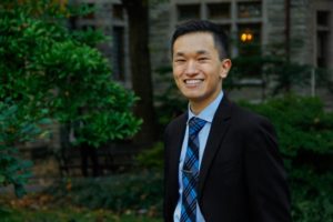
Sevile Mannickarottu, Director of Educational Labs, Penn Bioengineering
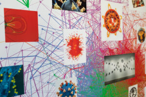
Sevile Mannickarottu, Director of Educational Laboratories in the Department of Bioengineering (BE), was interviewed in a recent episode of Shifting Schools, a weekly podcast that hosts educators and thought-leaders in conversations about the latest trends in education and EdTech. Mannickarottu, a Penn Engineering alumnus, runs the George H. Stephenson Foundation Educational Laboratory & Bio-MakerSpace, also known as the Penn BE Labs. In addition to being the primary teaching lab for Penn Bioengineering, the Penn BE Labs has grown into “the world’s only interdisciplinary Bio-MakerSpace.”
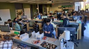
MakerSpaces–collaborative, educational work environments–have recently grown in popularity. Penn BE Labs distinguishes itself as a Bio-MakerSpace, embracing the interdisciplinary character of bioengineering by offering itself freely as a space for both academic and personal projects. It is stocked with tools ranging from 3D printers, laser cutters, and electrical equipment, including supplies to support work in molecular biology, physiology, chemistry, and microfluidics.
In the episode, hosts Tricia Friedman and Jeff Utecht talk with Mannickarottu about the organic process by which the Penn BE Labs evolved from a standard teaching space for undergraduate engineering laboratory courses into a student-driven hub of creativity and entrepreneurial spirit that is open to the entire Penn community regardless of discipline or major.
Mannickarottu and his team have found that “creativity needs to let go of control – that’s when fun things happen.” As the lab staff and faculty started to allow more creative freedom in the undergraduate bioengineers’ education, the requests for more supplies started pouring in and the lab’s activities and resources grew. “Honestly, we’re driven almost entirely by student requests and student demands,” says Mannickarottu. So when a student requested a sewing machine for a project? They went out and bought one, adding to their ever-growing stockpile of tools. Over time, more and more diverse projects have emerged from the BE Labs, many of them going on to win awards and grow beyond Penn’s campus as independent startups.
In case this sounds out of reach for smaller institutions, Mannickarottu shares words of encouragement. “The biggest thing,” he says, “is to allow for creativity on the part of the students.” A lab or program can start their own MakerSpace surprisingly inexpensively and build their inventory over time. His number one recommendation for those looking to replicate the success of Penn BE Labs is to allow students freedom to innovate, and administrators will be drawn to invest in the MakerSpace to allow for even more opportunities for them to create and thrive.
To help others get started, the Penn BE Labs staff have put a wide range of resources online, including extensive video and photo archives, FAQ’s, tutorials, information about student projects and startups, and equipment inventories. A 2019 post written for the BE Blog by BE alumna Sophie Burkholder (BSE ‘20 & MSE ‘21) gives the reader tips on “how to build your own MakerSpace for under $1500.”
Though it may currently be “the world’s only interdisciplinary Bio-MakerSpace,” the greatest legacy of the Penn BE Labs would be to be known as the first of many.
Listen to “The legacy of your lab” in Shifting Schools to learn more about the Penn BE Labs and for tips on starting your own MakerSpace.
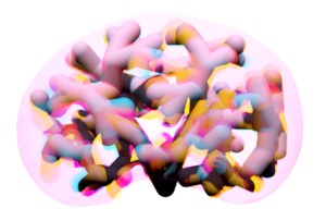
Artist-in-residence and visiting scholar Rebecca Kamen has blended AI and art to produce animated illustrations representing how a dyslexic brain interprets information.
A work that Penn artist-in-residence Rebecca Kamen produced for the show, “Dyslexic Dictionary” at Arion Press in San Francisco. Here, she reinterprets Ph.D. candidate Dale Zhou’s network visualization. (Image: Cat Fennell)
Communicating thoughts with words is considered a uniquely human evolutionary adaptation known as language processing. Fundamentally, it is an information exchange, a lot like data transfer between devices, but one riddled with discrete layers of complexity, as the ways in which our brains interpret and express ideas differ from person to person.
Learning challenges such as dyslexia are underpinned by these differences in language processing and can be characterized by difficulty learning and decoding information from written text.
Artist-in-residence in Penn’s Department of Physics and Astronomy Rebecca Kamen has explored her personal relationship with dyslexia and information exchange to produce works that reflect elements of both her creative process and understanding of language. Kamen unveiled her latest exhibit at Arion Press Gallery in San Francisco, where nine artists with dyslexia were invited to produce imaginative interpretations of learning and experiencing language.
The artists were presented with several prompts in varying formats, including books, words, poems, quotes, articles, and even a single letter, and tasked with creating a dyslexic dictionary: an exploration of the ways in which their dyslexia empowered them to engage in information exchange in unique ways.
Undiagnosed dyslexia
“[For the exhibit], each artist selected a word representing the way they learn, and mine was ‘lens,’” explains Kamen. “It’s a word that captures how being dyslexic provides me with a unique perspective for viewing and interacting with the world.”
From an early age, Kamen enjoyed learning about the natural sciences and was excited about the process of discovery. She struggled, however, with reading at school, which initially presented an obstacle to achieving her dreams of becoming a teacher. “I had a difficult time getting into college,” says Kamen. “When I graduated high school, the word ‘dyslexia’ didn’t really exist, so I assumed everyone struggled with reading.”
Kamen was diagnosed with dyslexia well into her tenure as a professor. “Most dyslexic people face challenges that may go unnoticed by others,” she says, “but they usually find creative ways to overcome them.”
This perspective on seeing and experiencing the world through the lens of dyslexia not only informed Kamen’s latest work for the exhibition “Dyslexic Dictionary,” but also showcased her background in merging art and science. For decades, Kamen’s work has investigated the intersection of the two, creating distinct ways of exploring new relationships and similarities.
“Artists and scientists are curious creatures always looking for patterns,” explains Kamen. “And that’s because patterns communicate larger insights about the world around us.”
The researchers studied different information-seeking approaches by monitoring how participants explore Wikipedia pages and categorically related these to two ideas rooted in philosophical understandings of learning: a “busybody,” who typically jumps between diverse ideas and collects loosely connected information; and a more purpose-driven “hunter,” who systematically ties in closely related concepts to fill their knowledge gaps.
They used these classifications to inform their computational model, the knowledge network. This uses text and context to determine the degree of relatedness between the Wikipedia pages and their content—represented by dots connected with lines of varying thickness to illustrate the strength of association.
In an adaption of the knowledge network, Kamen was classified as a dancer, an archetype elaborated on in an accompanying review paper by Dale Zhou, a Ph.D. candidate in Bassett’s Complex Systems Lab, who had also collaborated with Kamen on “Reveal.”
“The dancer can be described as an individual that breaks away from the traditional pathways of investigation,” says Zhou. “Someone who takes leaps of creative imagination and in the process, produces new concepts and radically remodels knowledge networks.”
Dani Smith Bassett is J. Peter Skirkanich Professor in Bioengineering with secondary appointments in the Departments of Physics & Astronomy, Electrical & Systems Engineering, Neurology, and Psychiatry.
David Lydon-Staley is an Assistant Professor in the Annenberg School for Communications and Bioengineering and is an alumnus of the Bassett Lab.
The model of tubule packing developed by the Hughes Lab shows the tubules repelling each other and shifting around.
A recent study by Penn Bioengineering researchers sheds new light on the role of physics in kidney development. The kidney uses structures called nephrons and tubules to filter blood and pass urine to the bladder. Nephron number is set at birth and can vary over an order of magnitude (anywhere from 100,000 to over a million nephrons in an individual kidney). While the reasons for this variability remain unclear, low numbers of nephrons predispose patients to hypertension and chronic kidney disease.
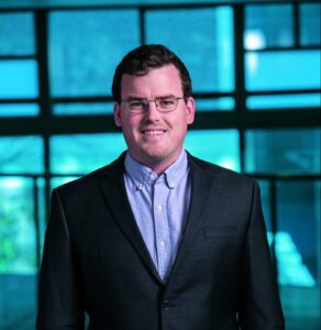
Now, research published in Developmental Cell led by Alex J. Hughes, Assistant Professor in the Department of Bioengineering, demonstrates a new physics-driven approach to better visualize and understand how a healthy kidney develops to avoid organizational defects that would impair its function. While previous efforts have typically approached this problem using molecular genetics and mouse models, the Hughes Lab’s physics-based approach could link particular types of defects to this genetic information and possibly highlight new treatments to prevent or fix congenital defects.
Alex J. Hughes, Assistant Professor in Bioengineering
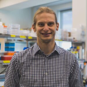
Louis Prahl, NIH F32 Postodctoral Fellow
During embryonic development, kidney tubules grow and the tips divide to make a branched tree with clusters of nephron stem cells surrounding each branch tip. In order to build more nephrons, the tree needs to grow more branches. To keep the branches from overlapping, the kidney’s surface grows more crowded as the number of branches increase. “At this point, it’s like adding more people to a crowded elevator,” says Louis Prahl, first author of the paper and Postdoctoral Fellow in the Hughes Lab. “The branches need to keep rearranging to accommodate more until organ growth stops.”
To understand this process, Hughes, Prahl and their team investigated branch organization in mouse kidneys as well as using computer models and a 3D printed model of tubules. Their results show that tubules have to actively restructure – essentially divide at narrower angles – to accommodate more tubules. Computer simulations also identified ‘defective’ packing, in which the simulation parameters caused tubules to either overlap or be forced beneath the kidney surface. The team’s experimentation and analysis of published studies of genetic mouse models of kidney disease confirmed that these defects do occur.
This study represents a unique synthesis of different fields to understand congenital kidney disease. Mathematicians have studied geometric packing problems for decades in other contexts, but the structural features of the kidney present new applications for these models. Previous models of kidney branching have approached these problems from the perspective of individual branches or using purely geometric models that don’t account for tissue mechanics. By contrast, The Hughes Lab’s computer model demonstrates the physics of how tubule families interact with each other, allowing them to identify ‘phases’ of kidney organization that either relate to normal kidney development or organizational defects. Their 3D printed model of tubules shows that these effects can occur even when one sets the biology aside.
Hughes has been widely recognized for his research in the understanding of kidney development. This new publication is the first fruit of his 2021 CAREER Award from the National Science Foundation (NSF) and he was recently named a 2023 Rising Star by the Cellular and Molecular Bioengineering (CMBE) Special Interest Group. In 2020 he became the first Penn Engineering faculty member to receive the Maximizing Investigators’ Research Award (MIRA) from the National Institutes of Health (NIH) for his forward-thinking work in the creation of new tools for tissue engineering.
Pediatric nephrologists have long worked to understand the cause of these childhood kidney defects. These efforts are often confounded by a lack of evidence for a single causative mutation. The Hughes Lab’s approach presents a new and different application of the packing problem and could help answer some of these unsolved questions and open doors to prevention of these diseases. Following this study, Hughes and his lab members will continue to explore the physics of kidney tubule packing, looking for interesting connections between packing organization, mechanical stresses between neighboring tubule tips, and nephron formation while attempting to copy these principles to build stem cell derived tissues to replace damaged or diseased kidney tissue. Mechanical forces play an important role in developmental biology and there is much scope for Hughes, Prahl and their colleagues to learn about these properties in relation to the kidney.
Other authors include Bioengineering Ph.D. students and Hughes Lab members John Viola and Jiageng Liu.
This work was supported by NSF CAREER 2047271, NIH MIRA R35GM133380, Predoctoral Training Program in Developmental Biology T32HD083185, and NIH F32 fellowship DK126385.
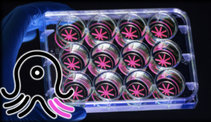
With OCTOPUS, Dan Huh’s team has significantly advanced the frontiers of organoid research, providing a platform superior to conventional gel droplets. OCTOPUS splits the soft hydrogel culture material into a tentacled geometry. The thin, radial culture chambers sit on a circular disk the size of a U.S. quarter, allowing organoids to advance to an unprecedented degree of maturity.
When it comes to human bodies, there is no such thing as typical. Variation is the rule. In recent years, the biological sciences have increased their focus on exploring the poignant lack of norms between individuals, and medical and pharmaceutical researchers are asking questions about translating insights concerning biological variation into more precise and compassionate care.
What if therapies could be tailored to each patient? What would happen if we could predict an individual body’s response to a drug before trial-and-error treatment? Is it possible to understand the way a person’s disease begins and develops so we can know exactly how to cure it?
Dan Huh, Associate Professor in the Department of Bioengineering at the University of Pennsylvania’s School of Engineering and Applied Science, seeks answers to these questions by replicating biological systems outside of the body. These external copies of internal systems promise to boost drug efficacy while providing new levels of knowledge about patient health.
An innovator of organ-on-a-chip technology, or miniature copies of bodily systems stored in plastic devices no larger than a thumb drive, Huh has broadened his attention to engineering mini-organs in a dish using a patient’s own cells.
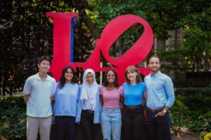
The 2022 iGEM team from left to right: June Ahn, Shreya Villimanalan, Adiva Daniar, Wangari Mbuthia, Cristina Perez and Moses Zeidan.
Congratulations to the 2022 University of Pennsylvania iGEM Team who took home a gold medal in the iGEM Grand Jamboree. This international competition of multidisciplinary teams of graduate and undergraduate students presenting original projects in synthetic biology culminated in the in-person Jamboree event held in Paris, France in October 2022. Over 370 judges awarded prizes and medals to the 350+ teams representing over 40 countries.
The 2022 Penn team was awarded a Gold Medal for their project “Photocreate,” a toolbox to control intercellular communication using optogenetics. Their plasmid constructs are designed to control protein secretion, display and shedding using a photocleavable protein, Phocl. The full abstract reads:
Intercellular communication is primarily studied using synthetic protein-level circuits. These circuits currently lack the spatial and temporal control necessary for targeted and time-sensitive applications. To address this gap, we developed Photocrete, a toolbox of protein constructs for light-inducible control of protein display, secretion, and shedding. We expanded upon RELEASE (Vlahos et al.), a modular and generalizable protein circuit which utilizes an ER retention motif and an exogenous protease to control protein secretion. We optogenetically modified RELEASE by replacing different components with the photocleavable protein PhoCl, allowing us to control the mammalian secretion pathway at distinct nodes with finely-tuned light administration regimens. Preliminary results indicate integration of Photocrete into the secretion pathway, but more research is necessary to determine optimal light administration settings. The potential for high spatial and temporal control of Photocrete could allow researchers to perform various signaling studies and develop therapeutics at a new level of precision.
The 2022 iGEM team includes undergraduates June Ahn (B.S. in Biochemistry, Physics and Nutrition), Adiva Daniar (B.S.E. in Bioengineering, minor in Engineering Entrepreneurship), Wangari Mbuthia (B.S.E. in Bioengineering), Cristina Perez (B.S.E. in Bioengineering, minor in Physics), Shreya Vallimanalan (B.S.E. in Bioengineering, minor in Computational Neuroscience), an d Moses Zeidan (B.S.E. in Bioengineering, minor in Chemistry and Spanish). They were mentored by graduate students David Gonzalez-Martinez, Gabrielle Ho, Zikang Huang, and Will Benman. Their faculty advisor is Lukasz Bugaj, Assistant Professor in Bioengineering.
Read the full results of the 2022 iGEM Competition here.
Penn Bioengineering alumnus Jiaqi Liu has been named to the eighth class of Schwarzman Scholars and will enroll at Tsinghua University in Beijing in August.
The program’s core curriculum focuses on leadership, China, and global affairs, according to the Schwarzman program. The academic program is updated each year to align with current and future geopolitical priorities. The coursework, cultural immersion, and personal and professional development opportunities are designed to equip students with an understanding of China’s changing role in the world.
This year, approximately 151 Schwarzman Scholars were selected from a pool of 3,000 applicants from 36 countries and 121 universities.
Jiaqi Liu earned his master’s degree in bioengineering in the School of Engineering and Applied Science in 2021. After graduation, he returned to China and works in global early-stage Venture Capital. According to the Schwarzman Scholars program, Liu is passionate about promoting medical equality and affordable health care solutions and has experience in medtech startup, global pharmaceutical company, health care consulting, and health care venture capital.
This story is by Amanda Mott. Read more about the Schwarzman Scholars at Penn Today.
Bella Mirro, a fourth year student in Bioengineering who also minors in Chemistry, spoke with 34th Street Magazine about her many roles at Penn, including being Co–President of Shelter Health Outreach Program (SHOP), a Research Assistant in lab of Michal A. Elovitz, the Hilarie L. Morgan and Mitchell L. Morgan President’s Distinguished Professor in Women’s Health at Penn Medicine, and a Penn Engineering Council Marketing Team Member. In this Q&A, she discusses her research in women’s health and her passions for accessible healthcare, serving Philadelphia’s homeless community, and good food.
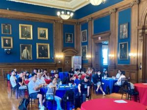
Retreat participants in Mitchell Hall at the College of Physicians
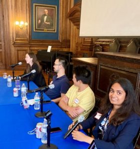
This year, the lineup of new student orientation activities included a new event: the first bioengineering retreat for incoming Ph.D. graduate students. Sitting in the historic Mitchell Hall at the College of Physicians, the 2022 Ph.D. cohort participated in a fun and educational half-day program that included a series of bonding activities, small-group conversations, and panel discussions. Current members of the Graduate Association of Bioengineers (GABE) planned the program to strengthen personal connections among students and to lend some advice to the newcomers as they embarked on their scholastic journey.
Prior to the retreat, participants read The Immortal Life of Henrietta Lacks by Rebecca Skloot, a work that delves into the human story of Henrietta Lacks, a Black woman from Virginia whose cancer cells were obtained for scientific study in the early 1950s without her knowledge. Today, “HeLa” cells have become one of the most significant tools in cell biology, enabling the development of polio vaccines, research into radiation effects, and even research on COVID-19. Together at the retreat, we discussed the intersection of ethics and scientific discovery, and reflected on our responsibility as scientists to consider the impact of our work beyond the immediate scientific question.
“Surviving the PhD 101” Panel Discussion. From left to right: Aoifa O’Farrell, Mosha Deng, David Mai, Lasya Sreepada
Current Ph.D. students volunteered their afternoons to share in two additional activities. Aoife O’Farrell, David Mai, Lasya Sreepada, and Mosha Deng imparted sage advice about using on-campus resources, handling advisor-advisee conflicts, and finding the best bites in Philly in the “Surviving the Ph.D. 101” panel discussion. Seven other students presented a series of flash talks about their research areas and musings on the best hypothetical mascot to represent their lab. The afternoon finished with an after-hours visit to the Mütter Museum, which holds an extensive and unique collection of anatomical specimens and antique medical equipment previously used for medical education.
If the WhatsApp group formed by the new cohort during the event is any indication, the retreat was an overall success! GABE looks forward to continuing the event in the future.
Brittany H. Scheid is a Ph.D. candidate studying Bioengineering in the lab of Brian Litt, Professor in Bioengineering and Neurology, and she is Co-President of GABE at Penn.
Yi-An Hsieh, a fourth year Bioengineering student from Anaheim, California, worked remotely this summer on a team that spanned three labs, including the Kamoun Lab at the Hospital of the University of Pennsylvania. Hsieh credits her research on kidney graft failure with enriching her scientific skill set, exposing her to machine learning and real-time interaction with genetic datasets. In a guest post for the Career Services Blog, Hseih writes about her remote summer internship experience. “It showed me that this type of research energy that could not be dampened despite the distance,” she writes.
Each year, the the Department of Bioengineering seeks exceptional candidates to conduct summer research in bioengineering with the support of two scholarships: the Abraham Noordergraaf Student Summer Bioengineering Research Fund and the Blair Undergraduate Research Fund in the Department of Bioengineering. These scholarships provide a living stipend for students to conduct research on campus in a Penn research lab under the mentorship of a faculty member. The Abraham Noordergraaf Student Summer Bioengineering Research Fund provides financial support for undergraduate or graduate summer research opportunities in bioengineering with a preference for study in the area of cardiovascular systems. Dr. Noordergraaf, who died in 2014, was a founding member and first chair of Penn Bioengineering. The Blair Undergraduate Research Fund in the Department of Bioengineering supports three to five undergraduate research scholars each year with the support of Dr. James C. Blair II. After a competitive round of proposals, the following six scholars were chosen for the Summer 2022 semester. Keep reading below for the research abstracts and bios of the awardees.
The Blair Undergraduate Research Fund in the Department of Bioengineering (Blair Scholars)
Ella Atsavapranee
Student: Ella Atsavapranee (BE Class of 2023)
PI: Michael J. Mitchell, J. Peter and Geri Skirkanich Assistant Professor of Innovation, Bioengineering
“Lipid nanoparticle-mediated delivery of RAS protease to inhibit cancer cell growth”
Mutations in RAS, a family of proteins found in all human cells, drive a third of cancers, including many pancreatic, colorectal, and lung cancers. However, there are still no therapies that can effectively prevent RAS from causing tumor growth. Recently, a protease was engineered to specifically degrade active RAS, offering a promising new tool for treating these cancers. However, many protein-based therapies still cannot be effectively delivered to patients. Lipid nanoparticles (LNPs), which were used in the Pfizer-BioNTech and Moderna COVID-19 vaccines, have emerged as a promising platform for safe and effective delivery of both nucleic acids and proteins. We formulated a library of LNPs using different cationic lipids. We characterized the LNPs by size, charge, and pKa, and tested their ability to deliver fluorescently labeled protease. The LNPs were able to encapsulate and deliver a RAS protease, successfully reducing proliferation of colon cancer cells.
Ella is a senior from Maryland studying bioengineering and chemistry. She works in Dr. Michael Mitchell’s lab, developing lipid nanoparticles to deliver proteins that reduce cancer cell proliferation. She has also conducted research on early-stage cancer detection and therapy monitoring (at Stanford University) and drug delivery across the blood-brain barrier for neurodegenerative diseases (at University of Maryland). She is passionate about translational research, science communication, and promoting diversity in STEM.
Chiadika Eleh
Student: Chiadika Eleh (BE and CIS Class of 2024)
PI: Eric J. Brown, Associate Professor of Cancer Biology, Perelman School of Medicine
“Investigating Viability in ATR and WEE1 Inhibitor Treated Ovarian Cancer Cells”
High-grade serous ovarian cancers (HGSOCs) are an aggressive subtype of ovarian cancer, accounting for up to 80% of all ovarian cancer-related deaths. More than half of HGSOCs are homologous recombination deficient; thus, they lack a favorable response when treated with common chemotherapeutic trials. Therefore, new treatment strategies must be developed to increase the life expectancy and quality of life of HGSOC patients. To address the lack of effective treatment options, the Brown Lab is interested in combining ATR and WEE1 inhibition (ATRi/WEE1i) to target HGSOC cells. It has previously been shown that low-dose ATRi/WEE1i is an effective treatment strategy for CCNE1-amplified ovarian cancer-derived PDX tumors (Xu et al., 2021, Cell Reports Medicine). Therefore, the next step is to characterize the HGSOC-specific response to ATRi/WEE1i treatment. This project aims to characterize the viability phenotype of ovarian cancer (OVCAR3) cells in the presence of ATRi/WEE1i in both single and combination treatments. With further research, Eleh hopes to prove the hypothesis low-dose combination ATRi/WEE1i treatment will result in the synergistic loss of viability in OVCAR3 cells. This goal will be achieved through the treatment of OVCAR3 cells with ranging doses of ATRi and Wee1i over 24 and 48 hour time intervals. We hope that this data will help set a treatment baseline that can be used for all OVCAR30-based viability experiments in the future.
Chiadika Eleh is a Bioengineering and Computer Science junior and a member of Penn Engineering’s Rachleff Scholar program. As a Blair Scholar, she worked in Dr. Eric Brown’s cancer biology lab, where she studied cell cycle checkpoint inhibitors as a form of cancer treatment.
“Tbc1d2b regulates vascular formation during development and tissue repair after ischemia”
The mechanisms behind endothelial cells forming blood vessels remains unknown. We have identified Tbc1d2b as a protein that is integral to the regulation of vascular formation. In order to investigate the role of Tbc1d2b in tubule formation, fibrin gel bead assays will be conducted to evaluate how the presence of Tbc1d2b is required for angiogenesis. Fibrin gel bead assays simulate the extracellular matrix environment to support the in vitro development of vessels from human umbilical vein endothelial cells (HUVEC) coated on cytodex beads. In order to confirm the success of angiogenesis, immunostaining for Phalloidin and CD31 will be conducted. After confirmation that fibrin gel bead assays can produce in vitro tubules, sgRNA CRISPR knockout of Tbc1d2b will be performed on HUVEC cells which will then be used to conduct more fibrin gel bead assays. We hypothesize that HUVEC with the Tbc1d2b knockout phenotype will be unable to form tubules while wild type HUVEC will be able to.
Gloria Lee is a rising senior studying Bioengineering and Physics in the VIPER program from Denver, Colorado. Her research in Dr. Yi Fan’s lab focuses on the role that proteins play in cardiovascular tubule formation.
Abraham Noordergraaf Student Summer Bioengineering Research Fund (Noordergraaf Fellows)
Gary Lin
Student: Gary Lin (Master’s in MEAM Class of 2023)
PI: Michelle J. Johnson, Associate Professor in Physical Medicine and Rehabilitation, Perelman School of Medicine, and in Bioengineering
“Development and Integration of Dynamically Modulating Control Systems in the Rehabilitation Using Community-Based Affordable Robotic Exercise System (Rehab CARES)”
As the number of stroke patients requiring rehabilitative care continues to increase, strain is being put onto the US health infrastructure which already has a shortage of rehabilitation practitioners. To help alleviate this pressure, a cost-effective robotic rehabilitative platform was developed to increase access to rehabilitative care. The haptic TheraDrive, a one-degree of freedom actuated hand crank that can apply assistive and resistive forces, was modified to train pronation and supination at the elbow and pinching of the fingers in addition to flexion and extension of the elbow and shoulder. Two controllers were created including an open-loop force controller and a closed-loop proportional-integral (PI) with adaptive control gains based on subject performance in therapy-game tasks as well as galvanic skin response. Stroke subjects (n=11) with a range of cognitive and motor impairment completed 4 therapy games in both adaptive and non-adaptive versions of the controllers (n=8) while measuring force applied on the TheraDrive handle. Resulting normalized average power versus Upper Extremity Fugl-Meyer (UE-FM) and Montreal Cognitive Assessment (MoCA) correlation analyses showed that power was strongly correlated with UE-FM in 2 of the conditions and moderately correlated with the other 6 while MoCA was moderate correlated to 2 of the conditions and weakly correlated to the rest. Mann-Whitney U-tests between adaptive and non-adaptive versions of each therapy game showed no significant differences with regards to power between controller types (p<0.05).
Gary is a master’s student in the School of Engineering studying Mechanical Engineering and Applied Mechanics with a concentration in Robotic and Mechatronic systems. His research primarily focuses on developing affordable rehabilitation robotics for use in assessment and game-based therapies post neural injury. Many of his interests revolve around the design of mechatronic systems and the algorithms used to control them for use in healthcare spaces.
Priya Shah
Student: Priya Shah (BE Class of 2024)
PI: Alex J. Hughes, Assistant Professor in Bioengineering
“Optogenetic Control of Developing Kidney Cells for Future Treatment of End-Stage Renal Disease”
This project sought to build from prior research in the Hughes Lab on the geometric and mechanical consequences of kidney form on cell and tissue-scale function. While the developmental trajectory of the kidney is well understood, little is currently known about many factors affecting nephron progenitor differentiation rate. Insufficient differentiation of nephron progenitor cells during kidney formation can result in lower nephron number and glomerular density, which is a risk factor for progression to end-stage renal disease later in life. Prior studies indicated that the amount of nephron differentiation – and thus function of the adult kidney – is correlated to the packing of ureteric tubule tips present at the surface of the kidney. Building off of research conducted in the Bugaj Lab, we found that inserting an optogenetic construct into the genome of human embryonic kidney (HEK) cells allowed us to manipulate the contraction of those cells through exposing them to blue light. Manipulating the contraction of the cells allows for the manipulation of the packing of ureteric tubule tips at the kidney surface. We used a lentiviral vector to transduce HEK293 cells with the optogenetic construct and witnessed visible contraction of the cells when they were exposed to blue light. Future work will include using CRISPR-Cas9 to introduce the optogenetic construct into IPS cells.
Priya is a junior studying bioengineering and had the opportunity to work on manipulating developing kidney cells using an optogenetic construct in the Hughes Lab this summer. She is thrilled to continue this research throughout the coming school year. Outside of the lab, Priya is involved with the PENNaach dance team and the Society of Women Engineers, as well as other mentorship roles.
Cosette Tomita
Student: Cosette Tomita (Master’s in MEAM Class of 2023)
“Expression and Characterization of an Anti-Aβ42 scFv”
Background: Amyloid Beta (Aβ42) fibrils contribute to the pathology of Alzheimer’s Disease. Numerous monoclonal antibodies have been developed against Aβ42. In this study we have designed and expressed a short chain variable fragment specific to Aβ42 (Anti-Aβ42 scFv). To characterize our anti-Aβ42 scFv we have performed structural analysis using transmission electron microscopy (TEM) and binding kinetics using microscale thermophoresis (MST) compared to commercially available antibodies 6E10, Aducanumab, and an IgG isotype control. The goal of this study is to determine if labeling densities and binding constants for Aducanumab and anti-Aβ42 scFv are not significantly different.
Method: To characterize Aβ42 fibril associated antibodies we used negative stain TEM. Aβ42 fibrils were stained on a glow discharged copper grid, and incubated with gold conjugated anti-Aβ42 scFv, 6E10—which binds all Aβ species, aducanumab, or IgG isotype control. Labeling densities were calculated as the number of fibril-associated gold particles per 1 μm2 for each image. Next, we used microscale thermophoresis determine the binding kinetics. Antibodies or anti-Aβ42 scFv were labeled with Alexa Fluor-647 and unlabeled Aβ42 was titrated in a serial dilution over 16 capillaries. The average fluorescence intensity was plotted against the antibody or scFv concentration and the curves were analyzed using the GraphPad Prism software to calculate the dissociation constant (KD) values.
Results: We found a significant difference, tested with a one-way ANOVA (P <0.0001), in gold particle associated Aβ fibrils per 1 μm2 between anti-Aβ42 scFv, 6E10, aducanumab, and IgG isotype control. Further analysis of aducanumab and 6CO3 with unpaired student t-test indicates significant differences in fibril associated gold particles between aducanumab vs. 6E10 (P=0.0003), Aducanumab vs. Isotype control (P <0.0001), anti-Aβ42 scFv vs 6E10 (p=0.0072), and anti-Aβ42 scFv vs Isotype Control (P=0.0029) with no significant difference in labeling densities between Aducanumab and anti-Aβ42 scFv. The expected KD values from MST were 1.8μM for Aducanumab and anti-Aβ42 scFv, 10.3nM for 6E10 and no expected binding for the isotype control. The experimental KD values for anti-Aβ42 scFv and 6E10 are 0.1132μM and 1.467μM respectively. The KD value for Isotype control was undetermined, as expected, however, the KD for Aducanumab was undetermined due to suboptimal assay conditions. Due to confounding variables in the experimental set up such as the use of Aβ1-16 compared to Aβ42 and the use of different fluorophores—5-TAMRA, Alexa Fluor 647 or FITC— the experimental KD values were off by several orders of magnitude.
Conclusion: We have illustrated similar labeling densities between Aducanumab and our anti-Aβ42 scFv. In the future, we will further optimize the MST assay conditions and compare the KD values obtained by MST with other techniques such as surface plasma resonance.
Cosette was born and raised in Chicago land area. Go Sox! She attended University of Missouri where she majored in Chemistry and Biology. She synthesized sigma-2 radiotracers and developed advanced skills in biochemical techniques in Dr. Susan Lever’s lab. After graduation, she moved to NJ to work at Lantheus, a radiopharmaceutical company. She missed academia and the independence of program and project development, so she came to work at the Penn Cyclotron facility before entering the Bioengineering master’s program.


 Mannickarottu and his team have found that “creativity needs to let go of control – that’s when fun things happen.” As the lab staff and faculty started to allow more creative freedom in the undergraduate bioengineers’ education, the requests for more supplies started pouring in and the lab’s activities and resources grew. “Honestly, we’re driven almost entirely by student requests and student demands,” says Mannickarottu. So when a student requested a sewing machine for a project? They went out and bought one, adding to their ever-growing stockpile of tools. Over time, more and more diverse projects have emerged from the BE Labs, many of them going on to win awards and grow beyond Penn’s campus as independent startups.
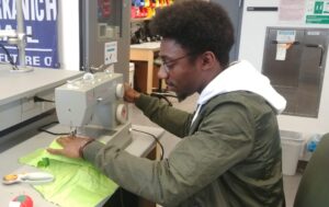
 To help others get started, the Penn BE Labs staff have put a wide range of resources online, including extensive video and photo archives, FAQ’s, tutorials, information about student projects and startups, and equipment inventories. A 2019 post written for the BE Blog by BE alumna Sophie Burkholder (BSE ‘20 & MSE ‘21) gives the reader tips on “how to build your own MakerSpace for under $1500.”